. 2022 Nov 9;96(21):e0099022.
doi: 10.1128/jvi.00990-22. Epub 2022 Oct 18.

Cryo-EM Structure of Gokushovirus ΦEC6098 Reveals a Novel Capsid Architecture for a Single-Scaffolding Protein, Microvirus Assembly System

Affiliations

Cryo-EM Structure of Gokushovirus ΦEC6098 Reveals a Novel Capsid Architecture for a Single-Scaffolding Protein, Microvirus Assembly System

Hyunwook Lee et al. J Virol. .

Abstract

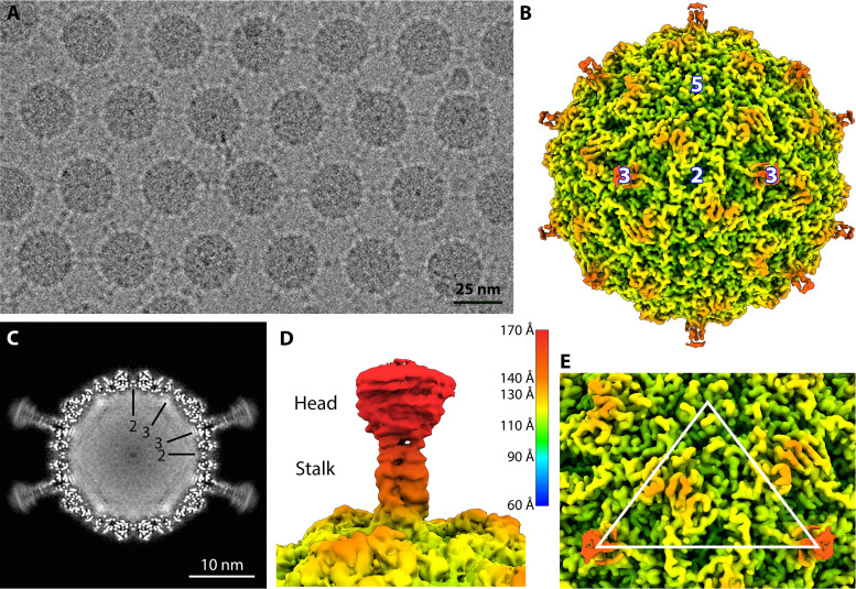
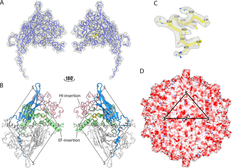
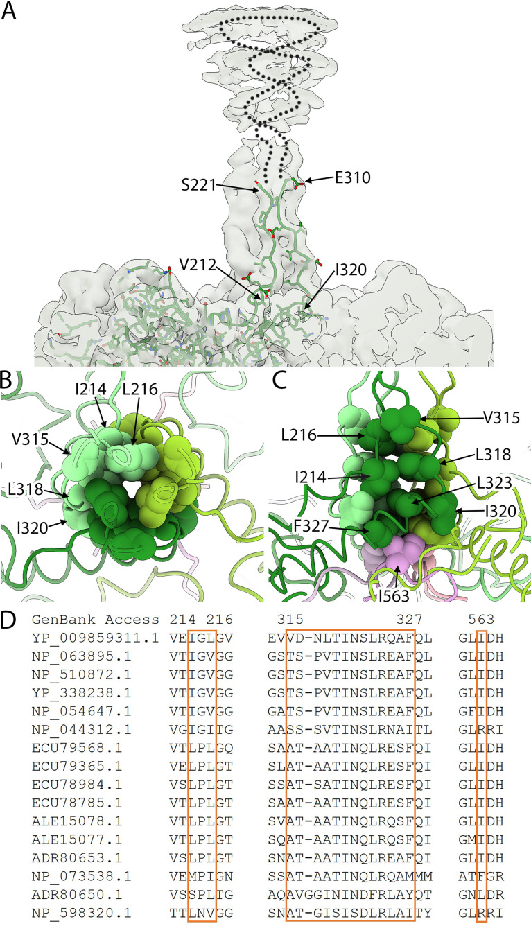
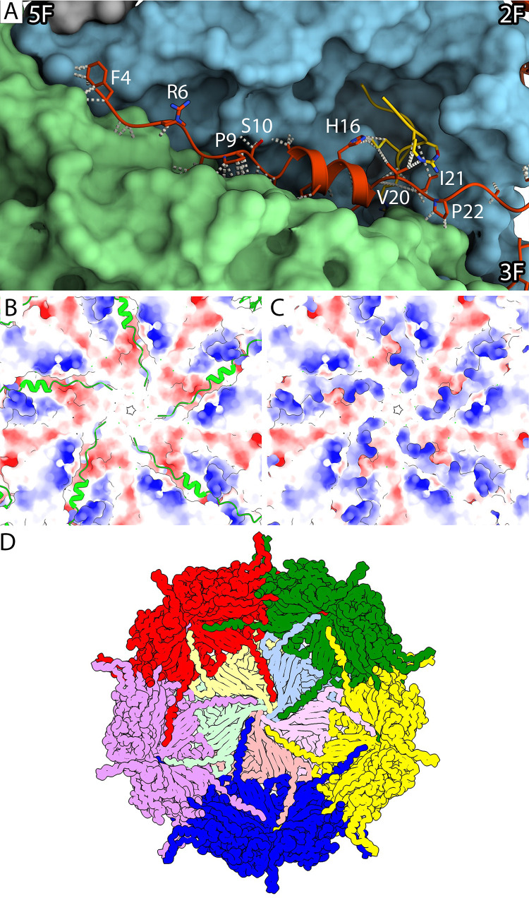
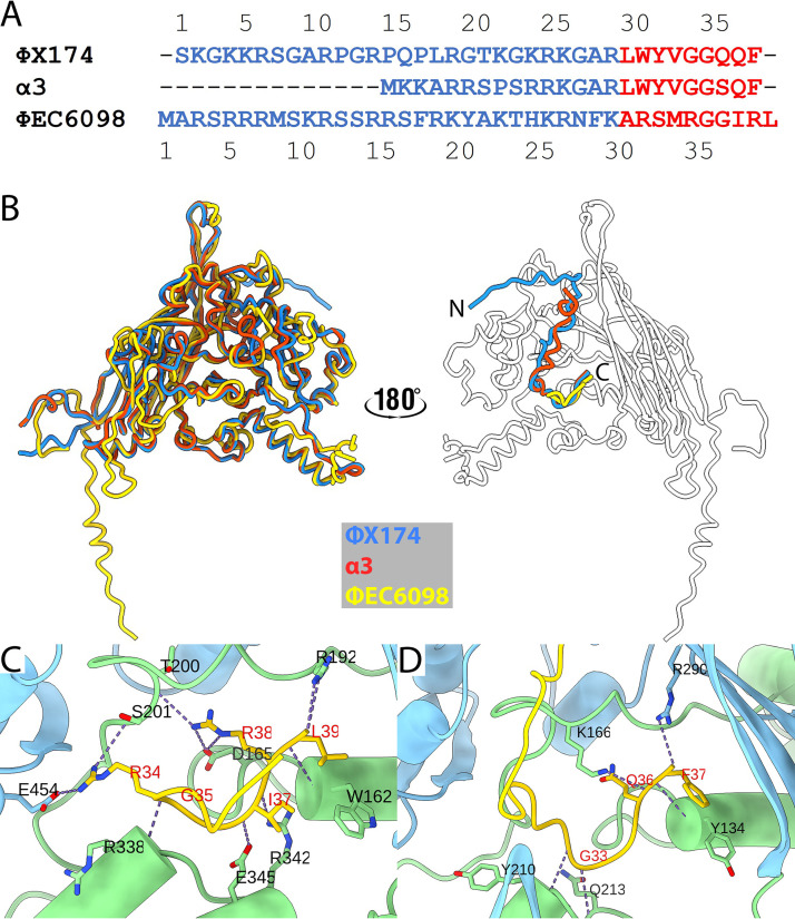
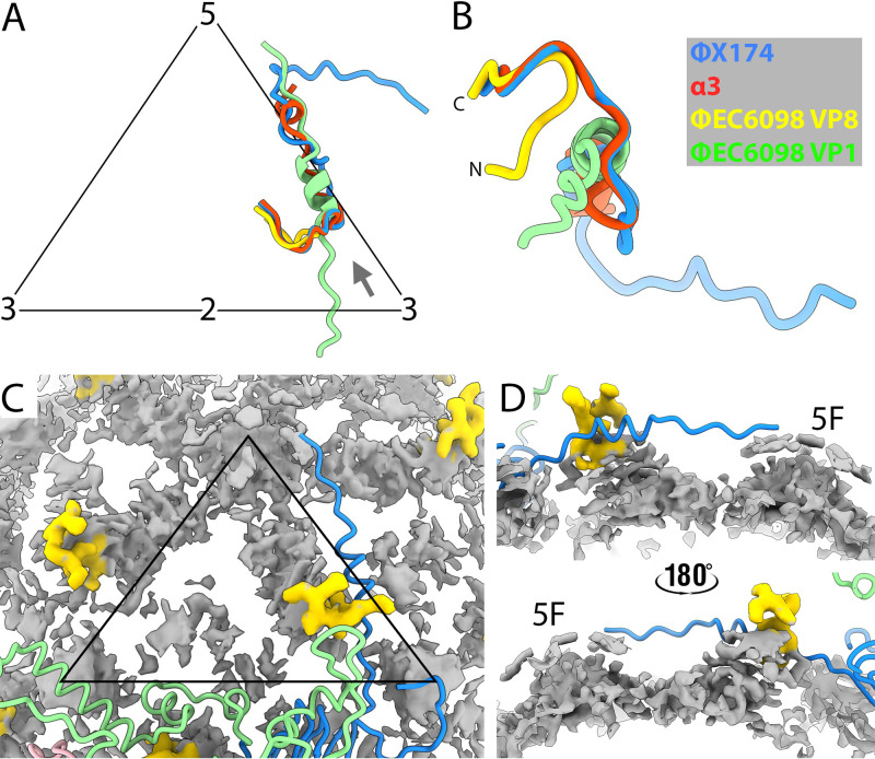
Ubiquitous and abundant in ecosystems and microbiomes, gokushoviruses constitute a Microviridae subfamily, distantly related to bacteriophages ΦX174, α3, and G4. A high-resolution cryo-EM structure of gokushovirus ΦEC6098 was determined, and the atomic model was built de novo. Although gokushoviruses lack external scaffolding and spike proteins, which extensively interact with the ΦX174 capsid protein, the core of the ΦEC6098 coat protein (VP1) displayed a similar structure. There are, however, key differences. At each ΦEC6098 icosahedral 3-fold axis, a long insertion loop formed mushroom-like protrusions, which have been noted in lower-resolution gokushovirus structures. Hydrophobic interfaces at the bottom of these protrusions may confer stability to the capsid shell. In ΦX174, the N-terminus of the capsid protein resides directly atop the 3-fold axes of symmetry; however, the ΦEC6098 N-terminus stretched across the inner surface of the capsid shell, reaching nearly to the 5-fold axis of the neighboring pentamer. Thus, this extended N-terminus interconnected pentamers on the inside of the capsid shell, presumably promoting capsid assembly, a function performed by the ΦX174 external scaffolding protein. There were also key differences between the ΦX174-like DNA-binding J proteins and its ΦEC6098 homologue VP8. As seen with the J proteins, C-terminal VP8 residues were bound into a pocket within the major capsid protein; however, its N-terminal residues were disordered, likely due to flexibility. We show that the combined location and interaction of VP8's C-terminus and a portion of VP1's N-terminus are reminiscent of those seen with the ΦX174 and α3 J proteins. IMPORTANCE There is a dramatic structural and morphogenetic divide within the Microviridae. The well-studied ΦX174-like viruses have prominent spikes at their icosahedral vertices, which are absent in gokushoviruses. Instead, gokushovirus major coat proteins form extensive mushroom-like protrusions at the 3-fold axes of symmetry. In addition, gokushoviruses lack an external scaffolding protein, the more critical of the two ΦX174 assembly proteins, but retain an internal scaffolding protein. The ΦEC6098 virion suggests that key external scaffolding functions are likely performed by coat protein domains unique to gokushoviruses. Thus, within one family, different assembly paths have been taken, demonstrating how a two-scaffolding protein system can evolve into a one-scaffolding protein system, or vice versa.

Keywords: DNA-binding protein; Microviridae; cryo-EM; gokushovirus; microvirus; mushroom spike.


Conflict of interest statement

The authors declare no conflict of interest.

Figures

FIG 1
FIG 1
Cryo-EM Analysis of φEC6098. (A) A representative micrograph of ΦEC6098 from the data collection shows an array of capsids with adjoining protrusions (scale bar, 25 nm). (B) A surface-rendered icosahedrally averaged map of ΦEC6098 (colored radially according to key) was reconstructed to 2.6-Å resolution. The 3-fold protrusions were visible only at low contour due to weaker density. Icosahedral symmetry axes are marked (white numbers). (C) The central section of the ΦEC6098 capsid shows the 3-fold protrusions. The capsid is filled with amorphous cryo-EM density corresponding to the icosahedrally averaged ssDNA genome. Icosahedral symmetry axes are shown by black lines. (D) A surface rendering of the 3-fold protrusion shown at low contour illustrates the globular head domain and narrow stalk domain, colored according to key. (E) The asymmetric unit of the icosahedral cryo-EM map is shown, marked by a white triangle.
FIG 2
FIG 2
Atomic model of ΦEC6098. (A) The protein structure built into one asymmetric unit, which consists of one copy each of VP1 (dodger blue) and VP8 (yellow), shown in a licorice representation within the cryo-EM density map (transparent surface). The left and right images show the external and internal surfaces flipped by 180°. (B) The atomic model of VP1 has a core anti-parallel β-barrel motif (BIDG and CHEF) with an N-terminal extension (blue). The EF- (green) and HI-insertion loops (pink) are also highlighted. The adjacent asymmetric unit across the 2-fold symmetry axis is colored in light gray. The left and right images show the external and internal surfaces of the coat protein dimer to illustrate the position of the VP1 N-terminal extension (blue) and VP8 (yellow). The two icosahedral asymmetric units are marked by triangles, and the symmetry axes are labeled. (C) The ordered region of VP8 encompassing residues 30 to 39 (yellow) was built into the map density (transparent gray). The VP8 region prior to residue 30 was too disordered to build. (D) The electrostatic potential is shown for the icosahedral capsid surface of ΦEC6098 and colored red and blue for negative and positive charges, respectively. The black triangle designates the asymmetric unit.
FIG 3
FIG 3
The 3-fold protrusion and hydrophobic core. (A) The 3-fold protrusion is depicted in a transparent surface rendering. A single VP1 built into one of the three copies of VP1 that form the trimeric stalk is shown in a stick representation (green). Rising from the capsid, residues were modeled up to residue 221, where the density became too disordered to continue. Extending from residue 221, a dotted line suggests a path to indicate that VP1 continues into the globular domain before tracing back toward the capsid surface. At residue 310, the build was resumed into the ordered stalk density. (B and C) Top and side views of the hydrophobic core at the center of the stalk region. (B) Three copies of VP1 (light, medium, and dark green) comprising the trimeric stalk are shown where the stalk adjoins the capsid surface. The residues contributing to the hydrophobic core are rendered as spheres and labeled. (C) At the bottom of the core are three I563 residues (light, medium, and dark pink). These residues are contributed to by three different, 2-fold symmetry related VP1 proteins, according to color. For example, the light green and light pink VP1 proteins are related by 2-fold symmetry. (D) Sequence comparison of the hydrophobic core (boxed residues) illustrates conservation among gokushoviruses. YP_009859311.1 is the accession code for the ΦEC6098 strain.
FIG 4
FIG 4
Characterization of the N-terminal extension of VP1. (A) A closeup view along a cleft in the capsid’s inner surface where each VP1 N-terminal extension interacts with two neighboring pentamers. The long N-terminal extension (red) and VP8 (yellow) are depicted in a ribbon diagram with side chains. The neighboring pentamers (blue and green) are shown surface rendered to visualize the cleft. Representative VP1 residues are labeled, and icosahedral symmetry axes are indicated. (B and C) The electrostatic potential of the inner capsid’s surface around the 5-fold symmetry axis is shown to illustrate that the positively charged N-terminal extensions (green) bind across the negatively charged clefts in the capsid inner surface (C). (D) A cartoon of the inner surface of the capsid demonstrates the N-terminal arm-interconnecting network. Coat protein pentamers (red, green, yellow, blue, purple) surround a pentamer illustrated with its five VP1s colored in tints (yellow, blue, pink, salmon, and green). Each N-terminal extension crosses over to an adjacent pentamer, where it makes connections in the cleft between two VP1 proteins, extending toward the 5-fold axis.
FIG 5
FIG 5
Comparison of the DNA-binding proteins. (A) Amino acid sequence alignment for the DNA-binding proteins from ΦX174, α3, and ΦEC6098. The residues for the N- and C-terminal domains are shown in blue and red, respectively. (B) Left, seen from the outside of the capsid, the coat and DNA-binding proteins are shown superimposed as ribbon diagrams colored according to the key. On the right, the ΦX174 (blue) and α3 (red) J proteins are superimposed onto VP8 (yellow) on the inner surface of the ΦEC6098 capsid protein (white). (C and D) ΦEC6098 VP8 (C) and ΦX174 J protein (D) bound into coat protein pockets formed by portions of the EF- (light green) and HI-insertion loops (light blue). H-bonds between the coat protein and DNA-binding protein are shown in dots, and the residues forming the H-bonds are labeled.
FIG 6
FIG 6
Superimposition of DNA-binding proteins and local disordered density. (A) Shown within the labeled asymmetric unit (black), ΦX174 and α3 J proteins are superimposed on the ΦEC6098 DNA-binding protein. The helical domain of the ΦEC6098 VP1 N-terminal extension (green) also maps to this region, where DNA is tethered to the inner surface of the capsid. (B) The superimposed DNA-binding proteins and N-terminal extension in panel A are rotated to view from the vantage point indicated by the gray arrow in panel A. The N-terminus of VP8 bends away from the superimposed J proteins because of the location of the VP1 N-terminal extension. (C) VP1 densities were subtracted from the cryo-EM map to show non-VP1 density in the interior (gray). Most of the density is amorphous and corresponds to genomic DNA plus the disordered region of VP8 that could not be built. However, the C-terminal VP8-ordered region (residues 30 to 39, yellow) was distinct. The icosahedral 2-fold symmetry related VP1 is shown in a ribbon diagram and colored as in Fig. 2B. Icosahedral symmetry axes are marked by a black triangle. (D) Side views of the amorphous density connected to the VP8 C-terminal domain. The 5-fold symmetry axis is labeled. The extra densities are likely the disordered portion of VP8 and associated ssDNA that do not conform to icosahedral symmetry.

Similar articles

Cited by

References

    1. McKenna R, Xia D, Willingmann P, Ilag LL, Krishnaswamy S, Rossmann MG, Olson NH, Baker TS, Incardona NL. 1992. Atomic structure of single-stranded DNA bacteriophage phi X174 and its functional implications. Nature 355:137–143. 10.1038/355137a0. - DOI - PMC - PubMed
    1. Bernal RA, Hafenstein S, Olson NH, Bowman VD, Chipman PR, Baker TS, Fane BA, Rossmann MG. 2003. Structural studies of bacteriophage α3 assembly. J Mol Biol 325:11–24. 10.1016/s0022-2836(02)01201-9. - DOI - PMC - PubMed
    1. Dokland T, McKenna R, Ilag LL, Bowman BR, Incardona NL, Fane BA, Rossmann MG. 1997. Structure of a viral procapsid with molecular scaffolding. Nature 389:308–313. 10.1038/38537. - DOI - PubMed
    1. McKenna R, Bowman BR, Ilag LL, Rossmann MG, Fane BA. 1996. Atomic structure of the degraded procapsid particle of the bacteriophage G4: induced structural changes in the presence of calcium ions and functional implications. J Mol Biol 256:736–750. 10.1006/jmbi.1996.0121. - DOI - PubMed
    1. Chipman PR, Agbandje-McKenna M, Renaudin J, Baker TS, McKenna R. 1998. Structural analysis of the spiroplasma virus, SpV4: implications for evolutionary variation to obtain host diversity among the Microviridae. Structure 6:135–145. 10.1016/s0969-2126(98)00016-1. - DOI - PMC - PubMed

Publication types

Substances