Review
. 2023 Feb;24(1):5-21.
doi: 10.1007/s11154-022-09756-3. Epub 2022 Oct 18.

Development and function of the fetal adrenal

Affiliations
Review

Development and function of the fetal adrenal

Emanuele Pignatti et al. Rev Endocr Metab Disord. 2023 Feb.

Abstract

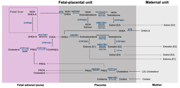
The adrenal cortex undergoes multiple structural and functional rearrangements to satisfy the systemic needs for steroids during fetal life, postnatal development, and adulthood. A fully functional adrenal cortex relies on the proper subdivision in regions or 'zones' with distinct but interconnected functions, which evolve from the early embryonic stages to adulthood, and rely on a fine-tuned gene network. In particular, the steroidogenic activity of the fetal adrenal is instrumental in maintaining normal fetal development and growth. Here, we review and discuss the most recent advances in our understanding of embryonic and fetal adrenal development, including the known causes for adrenal dys-/agenesis, and the steroidogenic pathways that link the fetal adrenal with the hormone system of the mother through the fetal-placental unit. Finally, we discuss what we think are the major open questions in the field, including, among others, the impact of osteocalcin, thyroid hormone, and other hormone systems on adrenal development and function, and the reliability of rodents as models of adrenal pathophysiology.

Keywords: Adrenal development; C11-oxy androgens; Cortisol; Fetal-placental unit; NR5A1; Sex differentiation; Steroidogenesis.


Conflict of interest statement

The authors declare no conflict of interest.

Figures

Fig. 1
Fig. 1
Schematic illustration of the cellular rearrangements during adrenal development in mice and humans. The adrenogonadal primordium (AGP) originates from cells localized between the coelomic epithelium and the dorsal aorta (blue arrow) at around 28–30 dpc (E9.5 in the mouse). At 32–40 dpc (E10.5 in the mouse), the adrenal primordium separates from the AGP. Between 48 and 52 dpc (E12.5), neural crest cells, precursors of the chromaffin cells of the medulla, invade the AGP. From 56 dpc (E14.5) onward, the fetal cortical cells are replaced by the definitive cortex, which gives rise to the adult zG and zF around the time of birth
Fig. 2
Fig. 2
Steroid biosynthesis in the fetal adrenal, and steroid metabolism in the fetal liver and placenta, marking steroid metabolic pathways in the fetal-placental-maternal unit. PREG, pregnenolone; 17OHPREG, 17α-hydroxypregnenolone; PROG, progesterone; DHEA, dehydroepiandrosterone; DHEA-S, dehydroepiandrosterone-sulfate; CYP11A1, cytochrome P450 cholesterol side chain cleavage; StAR, steroidogenic acute regulatory protein; CYP17A1, cytochrome P450 17α-hydroxylase/17,20-lyase; SULT2A1, sulfotransferase; CYP3A7, cytochrome P450 family 3 subfamily A member 7; CYP19A1, cytochrome P450 aromatase; HSD17B1, 17β-hydroxysteroid dehydrogenase type 1; HSD17B2, 17β-hydroxysteroid dehydrogenase type 2; HSD17B7, 17β-hydroxysteroid dehydrogenase type 7; HSD17B3, 17β-hydroxysteroid dehydrogenase type 3; AKR1C3, 17β-hydroxysteroid dehydrogenase type 5; HSD3B1, 3β-hydroxysteroid dehydrogenase type 1; HSD11B2, 11β-hydroxysteroid dehydrogenase type 2; STS, sulfatase
Fig. 3
Fig. 3
Steroid metabolic pathways in the fetal adrenal zones, A; with the developmental timeline of each zone during gestation, B. Steroidogenic acute regulatory protein (StAR), expressed in the fetal zone (FZ) (and transition zone [TZ]) from 6–8 wpc and in the definitive zone (DZ) only from 22–23 wpc; cytochrome P450 cholesterol side chain cleavage (CYP11A1), expressed in the FZ from 8–9 wpc and onwards, and in the TZ from 14–22 weeks, while the expression in the DZ appears only from 17–19 and 23 wpc; cytochrome P450 17α-hydroxylase/17,20-lyase (CYP17A1), expressed in the FZ (already from 8–9 wpc) and TZ from 14–24 wpc; 3β-hydroxysteroid dehydrogenase (3βHSD2) is expressed from 8–19 wpc in the DZ and from 24–41 wpc in the TZ; cytochrome P450 steroid 21-hydroxylase (CYP21A2) is sparsely expressed in the DZ between 13–24 wpc (only becoming prominent during late gestation) and detected in the FZ and TZ from 14 wpc (while detection in the FZ at 9–19 wpc has been shown); cytochrome P450 aldosterone synthase (CYP11B2), becomes prominent in the DZ during late gestation; and cytochrome P450 11β-hydroxylase (CYP11B1) is expressed in the fetal adrenal, in both the FZ and TZ by 13–24 wpc (limited weak staining in the FZ between 8–12 wpc has also been shown) [39, 43, 63, 64, 66, 90]. Steroids boxed in orange, steroid substrates for the backdoor pathway; steroids boxed in grey, steroid substrates for placental aromatase; steroid boxed in blue, steroid substrate for placental HSD11B2; steroids boxed in black, steroid end-products of each zone of the fetal adrenal

References

    1. Sucheston ME, Cannon MS. Development of zonular patterns in the human adrenal gland. J Morphol. 1968;126:477–91. - PubMed
    1. Hornsby PJ. Aging of the human adrenal cortex. Sci Aging Knowledge Environ. 2004;2004:re6. - PubMed
    1. Orentreich N, Brind JL, Rizer RL, Vogelman JH. Age changes and sex differences in serum dehydroepiandrosterone sulfate concentrations throughout adulthood. J Clin Endocrinol Metab. 1984;59:551–5. - PubMed
    1. Miller WL, Flück CE, Breault DT, Feldman BJ. Adrenal cortex and its disorders. In: Sperling MA, editor. Sperling - Pediatric Endocrinology. 5th ed.: Elsevier; 2020.
    1. Morel Y, Roucher F, Plotton I, Goursaud C, Tardy V, Mallet D. Evolution of steroids during pregnancy: Maternal, placental and fetal synthesis. Ann Endocrinol (Paris) 2016;77:82–9. - PubMed

Publication types

Substances