Imaging features of immune checkpoint inhibitor-related nephritis with clinical correlation: a retrospective series of biopsy-proven cases
- PMID: 36255488
- PMCID: PMC9957799
- DOI: 10.1007/s00330-022-09158-8
Imaging features of immune checkpoint inhibitor-related nephritis with clinical correlation: a retrospective series of biopsy-proven cases
Abstract
Objectives: Imaging appearances of immune checkpoint inhibitor-related nephritis have not yet been described. The primary objective of this study is to describe the appearances of immunotherapy-related nephritis on computerized tomography (CT) and positron emission tomography (PET). The secondary objectives are to investigate the association of radiologic features with clinical outcomes.
Methods: CT and PET-CT scans before the initiation of immunotherapy (baseline), at nephritis, and after resolution of pathology-proven nephritis cases were reviewed. Total kidney volume, renal parenchymal SUVmax, renal pelvis SUVmax, and blood pool SUVmean were obtained.
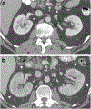
Results: Thirty-four patients were included. The total kidney volume was significantly higher at nephritis compared to baseline (464.7 ± 96.8 mL vs. 371.7 ± 187.7 mL; p < 0.001). Fifteen patients (44.1%) had > 30% increase in total kidney volume, which was associated with significantly higher renal toxicity grade (p = 0.007), higher peak creatinine level (p = 0.004), and more aggressive medical treatment (p = 0.011). New/increasing perinephric fat stranding was noted in 10 patients (29.4%) at nephritis. Among 8 patients with contrast-enhanced CT at nephritis, one (12.5%) developed bilateral wedge-shaped hypoenhancing cortical. On PET-CT, the renal parenchymal SUVmax-to-blood pool ratio was significantly higher at nephritis compared to baseline (2.13 vs. 1.68; p = 0.035). The renal pelvis SUVmax-to-blood pool SUVmean ratio was significantly lower at nephritis compared to baseline (3.47 vs. 8.22; p = 0.011).
Conclusions: Bilateral increase in kidney size, new/increasing perinephric stranding, and bilateral wedge-shaped hypoenhancing cortical foci can occur in immunotherapy-related nephritis. On PET-CT, a diffuse increase in radiotracer uptake throughout the renal cortex and a decrease in radiotracer activity in the renal pelvis can be seen.
Key points: • CT features of immune checkpoint inhibitor-related nephritis include an increase in kidney volume, new/increasing perinephric stranding, and bilateral ill-defined wedge-shaped hypoenhancing cortical foci. • FDG-PET features of immune checkpoint inhibitor-related nephritis include an increase in FDG uptake throughout the renal cortex and a decrease in FDG activity/excretion in the collecting system. • > 30% increase in total kidney volume is associated with worse toxicity grade and more aggressive medical management.
Keywords: Immunotherapy; Nephropathy; Radiographic; Radiology; Toxicity.
© 2022. The Author(s), under exclusive licence to European Society of Radiology.
Conflict of interest statement
Conflicts of interest:
N.A. has received honoraria for serving on a scientific advisory board as a consultant for ChemoCentryx. A.D. has received honoraria from Nektar and he has served as a consultant for Nektar, Memgen and Pfizer.
Figures


References
MeSH terms
Substances
Grants and funding
LinkOut - more resources
Full Text Sources
Medical

