. 2023 Jan;270(1):82-100.
doi: 10.1007/s00415-022-11399-y. Epub 2022 Oct 18.

Vestibular paroxysmia entails vestibular nerve function, microstructure and endolymphatic space changes linked to root-entry zone neurovascular compression

Affiliations

Vestibular paroxysmia entails vestibular nerve function, microstructure and endolymphatic space changes linked to root-entry zone neurovascular compression

Emilie Kierig et al. J Neurol. 2023 Jan.

Abstract

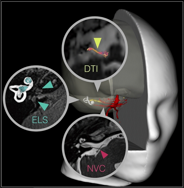
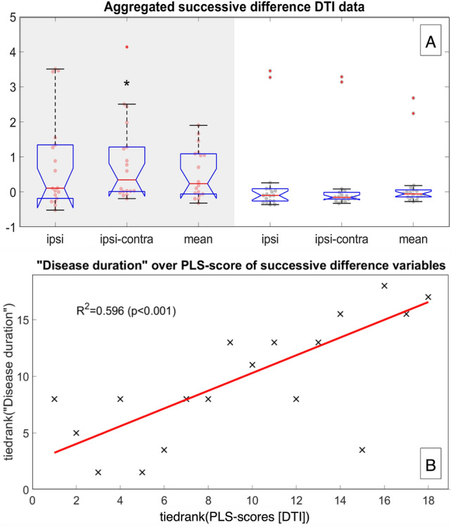
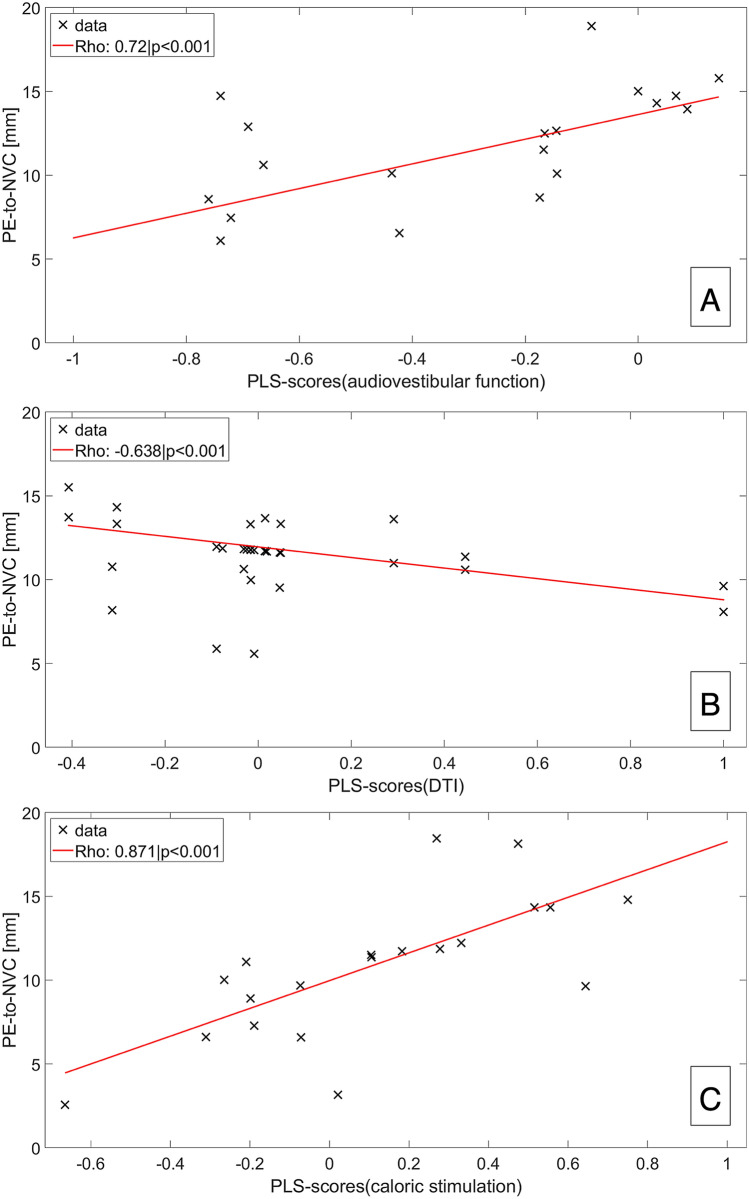
Combining magnetic resonance imaging (MRI) sequences that permit the determination of vestibular nerve angulation (NA = change of nerve caliber or direction), structural nerve integrity via diffusion tensor imaging (DTI), and exclusion of endolymphatic hydrops (ELH) via delayed gadolinium-enhanced MRI of the inner ear (iMRI) could increase the diagnostic accuracy in patients with vestibular paroxysmia (VP). Thirty-six participants were examined, 18 with VP (52.6 ± 18.1 years) and 18 age-matched with normal vestibulocochlear testing (NP 50.3 ± 16.5 years). This study investigated whether (i) NA, (ii) DTI changes, or (iii) ELH occur in VP, and (iv) to what extent said parameters relate. Methods included vestibulocochlear testing and MRI data analyses for neurovascular compression (NVC) and NA verification, DTI and ELS quantification. As a result, (i) NA increased NVC specificity. (ii) DTI structural integrity was reduced on the side affected by VP (p < 0.05). (iii) 61.1% VP showed mild ELH and higher asymmetry indices than NP (p > 0.05). (iv) "Disease duration" and "total number of attacks" correlated with the decreased structural integrity of the affected nerve in DTI (p < 0.001). NVC distance within the nerve's root-entry zone correlated with nerve function (Roh = 0.72, p < 0.001), nerve integrity loss (Roh = - 0.638, p < 0.001), and ELS volume (Roh = - 0.604, p < 0.001) in VP. In conclusion, this study is the first to link eighth cranial nerve function, microstructure, and ELS changes in VP to clinical features and increased vulnerability of NVC in the root-entry zone. Combined MRI with NVC or NA verification, DTI and ELS quantification increased the diagnostic accuracy at group-level but did not suffice to diagnose VP on a single-subject level due to individual variability and lack of diagnostic specificity.

Keywords: DTI; Diffusion imaging; Endolymphatic hydrops; Endolymphatic space; Gadolinium-based contrast agent; Inner ear; Intravenous; MRI; Root-entry zone; Vestibular nerve; Vestibular paroxysmia.


Conflict of interest statement

There are no conflicts of interest.

Figures

Fig. 1
Fig. 1
MR imaging approach tailored to VP. This study investigated the vestibular nerve with a MR imaging approach tailored to vestibular paroxysmia (VP). A 3D rendered T1-image visualizes the spatial relationship between overlaid blood vessels (red), the eighth cranial nerve (8 CN, yellow), and the vestibulocochlear end organ (white shell). These key structures are further shown magnified in a circle within a transversal slice of the cerebellopontine angle in the respective raw data with color-coded arrowheads pointing towards them. Particular emphasis was put on the site of neurovascular compression (NVC, behind red arrowhead in the lower circle), and the DTI tractography quantification of the cisternal segment of the eighth cranial nerve (behind yellow arrowhead in the upper circle) between the point of entry (PE: point of entry for afferent fibers, point of exit for efferent fibers into/out of the brainstem) and the internal auditory canal (IAC). In addition, the study focused on the volumetric quantification of the endolymphatic space (ELS, behind the turquoise arrowheads in the middle circle) within the membranous and bony labyrinth of the inner ear. The upper arrowhead points toward the cochlea, and the lower arrowhead points towards the vestibulum. The corresponding volumetric visualization of these structures (ELS in turquopoise) is depicted on the left, next to these structures. Visualization tools were a combination of 3D Slicer (https://www.slicer.org/, version 4.11 [36]) and DSI Studio (http://dsi-studio.labsolver.org/, version 2021-02-12).
Fig. 2
Fig. 2
DTI tractography results. Compiled results of DTI group differences (A) and correlations (B): The upper graphic (A) shows the aggregated group difference data for the ipsilateral, the difference between ipsilateral and contralateral sides, and the average (mean) of both sides as boxplots with overlaid data points. The VP data is shown in red with a gray background, and the NP data is shown in black with a white background. Significant differences in the median between the groups are marked by a black star “*”, with a significance level p < 0.05. The lower graphic (B) depicts PLS rank-correlations between disease parameter “disease duration” and the 1st order of successive differences. The scatter plot of the ranked “disease duration” data and ranks of the 1st order successive difference PLS scores are shown as black crosses, with an overlayed trendline in red. The explained variance r-squared was 0.596 with significance p < 0.001 (no failures in 1000 permutation tests)
Fig. 3
Fig. 3
Interparametric correlations. Scatter plot depiction with trendlines of the correlations of “distance between PE and NVC (PE-to-NVC [mm])” and the eighth cranial nerve (A) audiovestibular function, i.e. neurophysiological testing (caloric stimulation, HIT and PTA), (B) structural nerve integrity (DTI SSPs, or deviations of successive DTI differences), and inner ear ELS volumes (C). A decreasing distance between PE to NVC can be translated into the transition zone of the eighth nerve (6-15 mm as measured from the PE, where oligodendrocytes change into Schwann cells [89]), and the root-entry zone from 6 mm downwards [88]. In consequence, the positive correlation of PE-to-NVC to audiovestibular function (A, Roh 0.72, p < 0.001) indicates better function the more the NVC lies outside the root-entry area of the intracisternal part of the eighth cranial nerve, and not covered by central myelin (oligodendrocytes), but peripheral myelin (Schwann cells). Conversely, the negative correlation of PE-to-NVC to nerval structural microstructure DTI SSPs (B, Roh − 6.638, p < 0.001), or inner ear ELS volumes (C, Roh − 6.604, p < 0.001) indicates less nerve deterioration, or less ELS volume the higher the PE-to-NVC distance. Altogether, these correlations link the suttle clinical, diagnostic, DTI-proxy for nerve microstructure, and ELS volume findings in VP to NVC, and in particular to NVC being in the root-entry zone. Abbreviations: ELS endolymphatic space, NVC neurovascular compression, PE point of entry/exit for afferent/efferent fibers out of/into the brainstem, PE-to-NVC distance between PE and NVC [mm], SSP summary statistic parameters, or deviations in successive differences.

References

    1. Brandt T, Dieterich M, Strupp M. Vertigo and dizziness: common complaints. 2. London: Springer-Verlag; 2013.
    1. Brandt T, Dieterich M. Vestibular paroxysmia: vascular compression of the eighth nerve? Lancet. 1994;343:798–799. doi: 10.1016/s0140-6736(94)91879-1. - DOI - PubMed
    1. Jannetta PJ, Møller MB, Møller AR. Disabling positional vertigo. N Engl J Med. 1984;310:1700–1705. doi: 10.1056/NEJM198406283102604. - DOI - PubMed
    1. Møller MB, Møller AR. Vascular compression syndrome of the eighth nerve. Clinical correlations and surgical findings. Neurol Clin. 1990;8:421–439. doi: 10.1016/S0733-8619(18)30365-7. - DOI - PubMed
    1. Strupp M, Lopez-Escamez JA, Kim J-S, et al. Vestibular paroxysmia: diagnostic criteria. J Vestib Res. 2016;26:409–415. doi: 10.3233/VES-160589. - DOI - PMC - PubMed