. 2022 Oct 18;15(756):eabn4948.
doi: 10.1126/scisignal.abn4948. Epub 2022 Oct 18.

Fusobacterium nucleatum induces proliferation and migration in pancreatic cancer cells through host autocrine and paracrine signaling

Affiliations

Fusobacterium nucleatum induces proliferation and migration in pancreatic cancer cells through host autocrine and paracrine signaling

Barath Udayasuryan et al. Sci Signal. .

Abstract

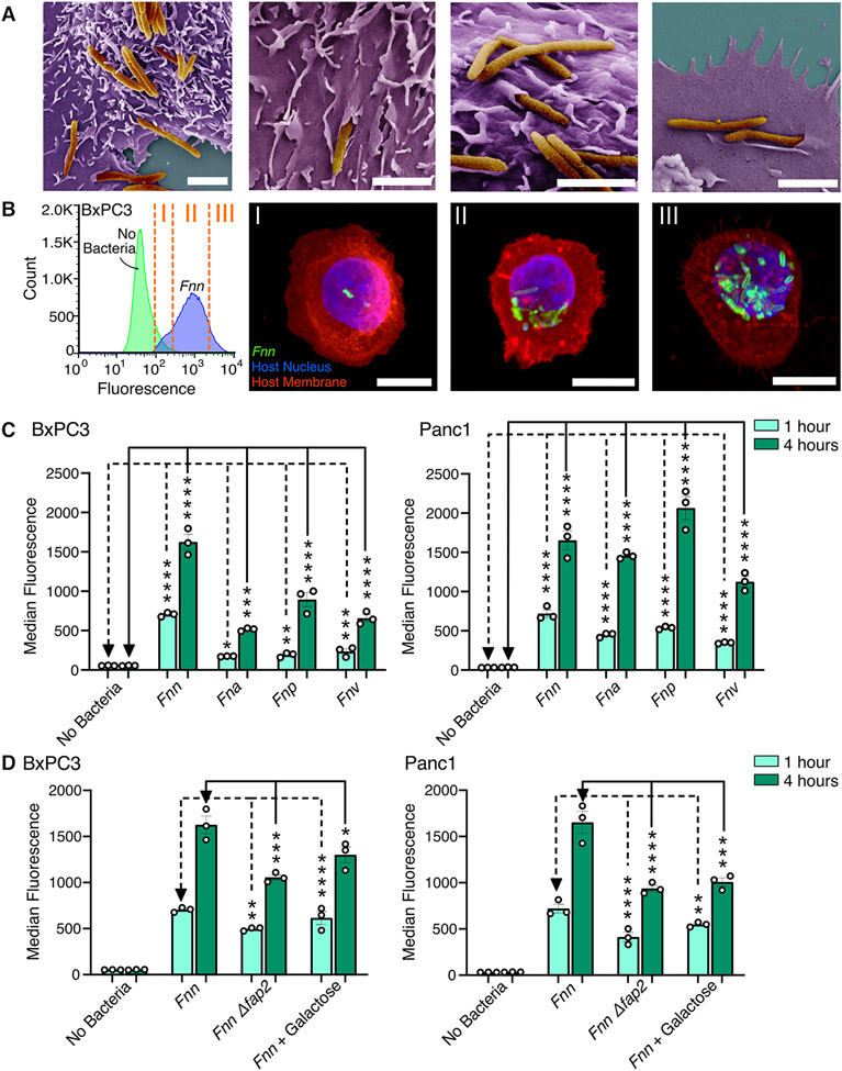
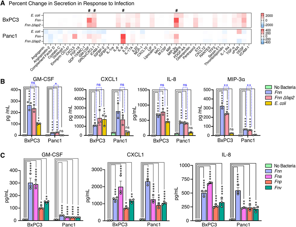
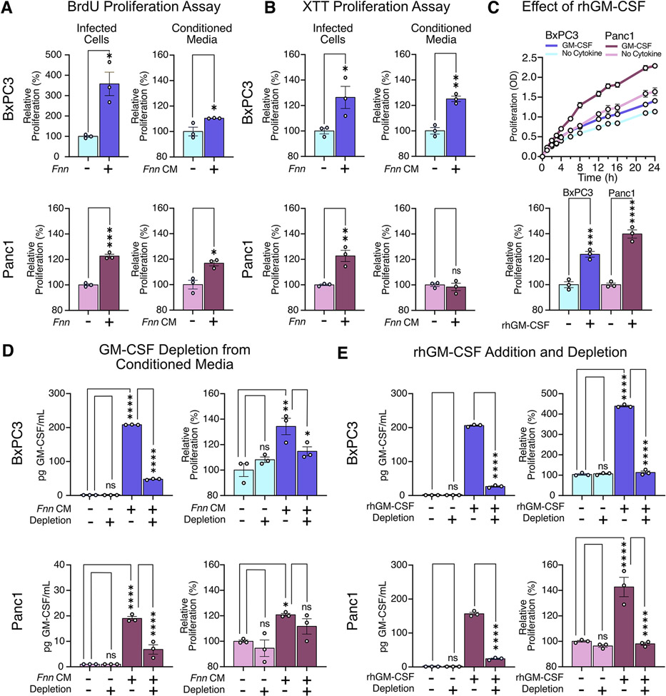
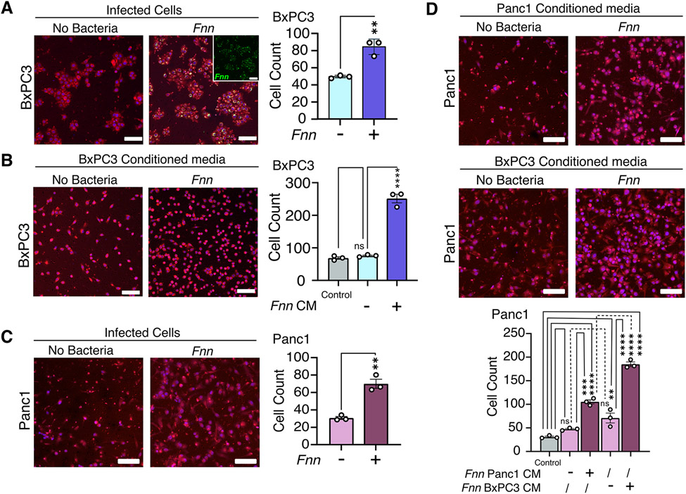
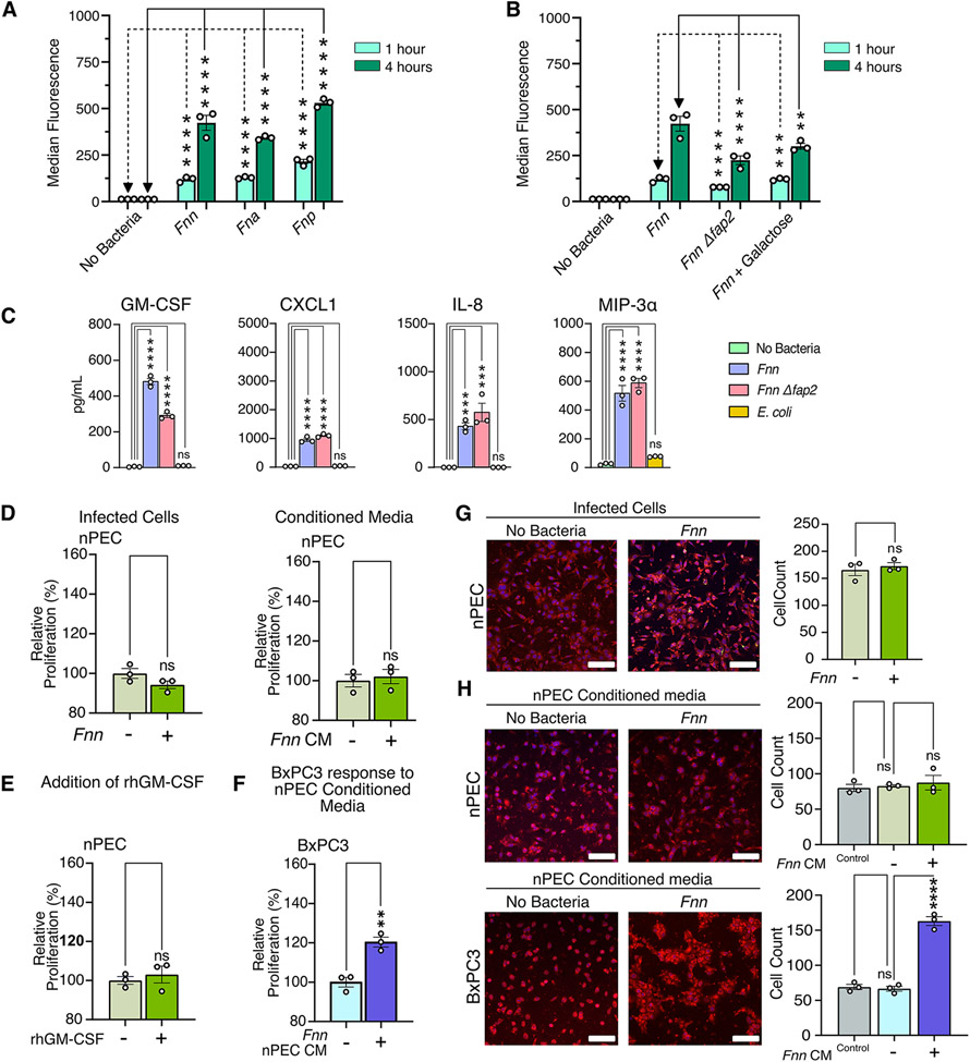
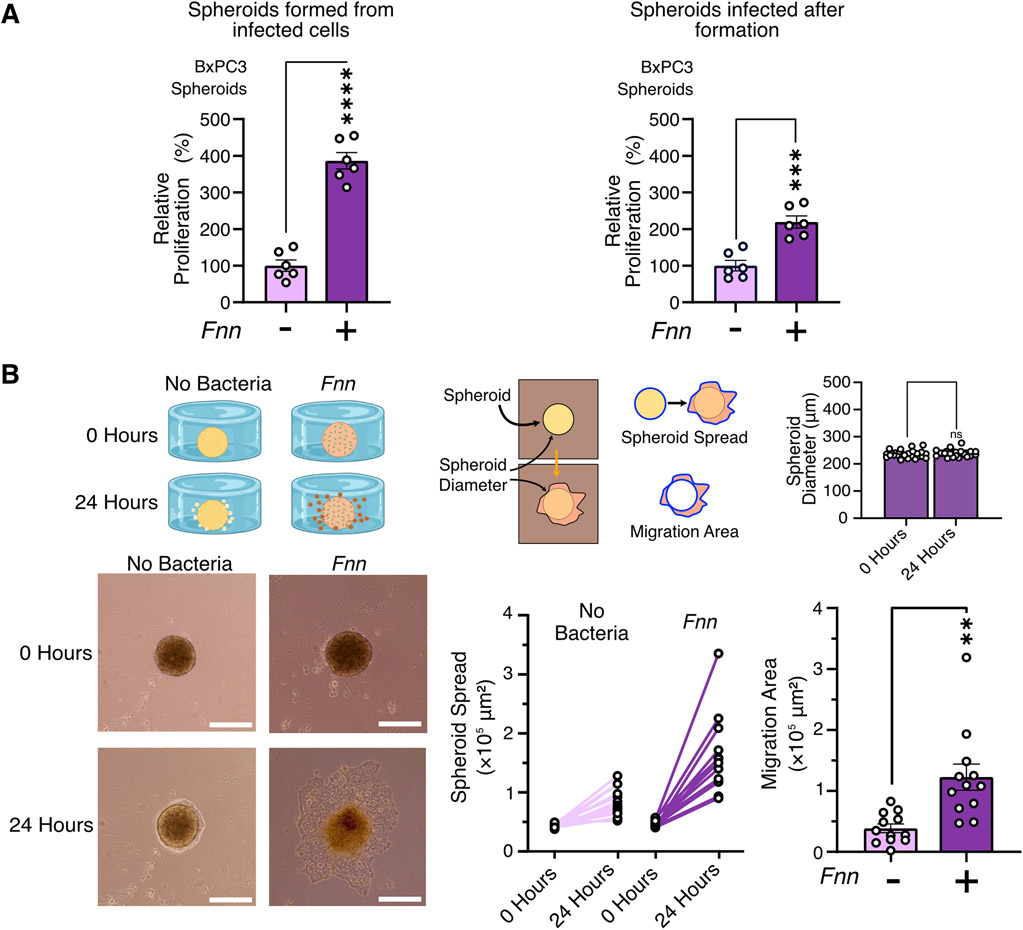
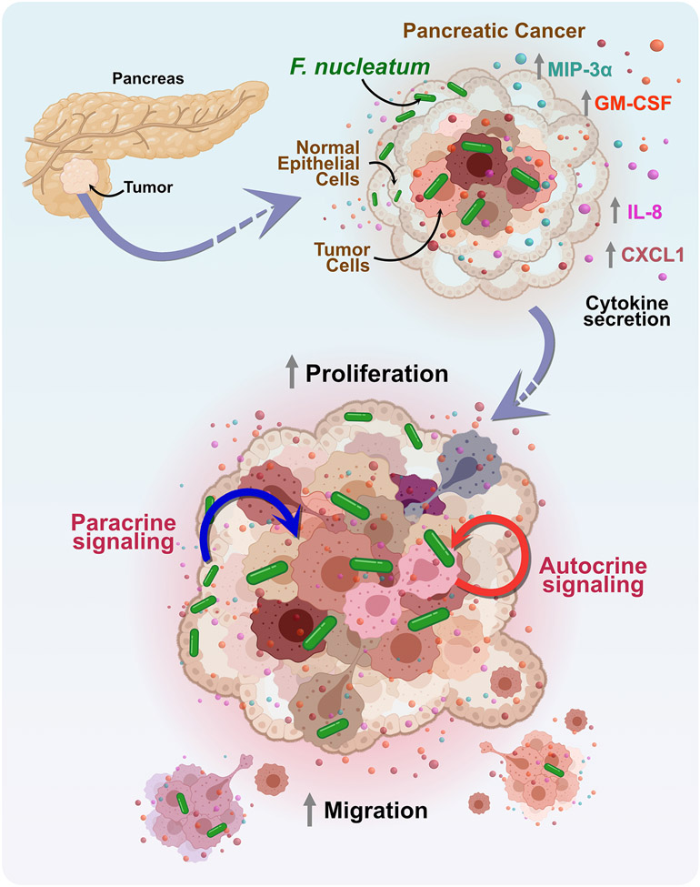
The tumor microbiome is increasingly implicated in cancer progression and resistance to chemotherapy. In pancreatic ductal adenocarcinoma (PDAC), high intratumoral loads of Fusobacterium nucleatum correlate with shorter survival in patients. Here, we investigated the potential mechanisms underlying this association. We found that F. nucleatum infection induced both normal pancreatic epithelial cells and PDAC cells to secrete increased amounts of the cytokines GM-CSF, CXCL1, IL-8, and MIP-3α. These cytokines increased proliferation, migration, and invasive cell motility in both infected and noninfected PDAC cells but not in noncancerous pancreatic epithelial cells, suggesting autocrine and paracrine signaling to PDAC cells. This phenomenon occurred in response to Fusobacterium infection regardless of the strain and in the absence of immune and other stromal cells. Blocking GM-CSF signaling markedly limited proliferative gains after infection. Thus, F. nucleatum infection in the pancreas elicits cytokine secretion from both normal and cancerous cells that promotes phenotypes in PDAC cells associated with tumor progression. The findings support the importance of exploring host-microbe interactions in pancreatic cancer to guide future therapeutic interventions.


Figures

Figure 1.
Figure 1.. Fusobacterium nucleatum binds to and invades pancreatic cell lines.
(A) Scanning electron microscopy images of F. nucleatum subsp. nucleatum 23726 (Fnn) invading BxPC3 pancreatic cancer cells. Pseudocolored with orange for bacteria and purple for host cells. Scale bar: 2μm. (B) Flow cytometry analysis of BxPC3 cells cultured without bacteria or with Fnn at 50:1 MOI. Cells sorted based on fluorescence intensity correspond to intracellular bacterial loads (I: low, II: medium, III: high). These cells were plated and imaged on a confocal microscope to identify relative levels of infection. Host cell nuclei were stained with DAPI (blue), Fnn membranes were stained with FM 1-43 FX (green), and host cell membranes were stained with MemBrite 568 (red). Scale bar: 10 μm. (C) Flow cytometry analysis of infection of BxCP3 and Panc1 cells cultured with Fnn, Fna, Fnp, and Fnv at 50:1 MOI after 1 and 4 hours. (D) Flow cytometry analysis of infection of BxpC3 and Panc1 cells cultured with Fnn, Fnn Δfap2, or Fnn in the presence of Fap2-binding inhibitor galactose, at 50:1 MOI for 1 and 4 hours. Data in (B to D) are means ± SEM from N = 3 independent experiments per condition; comparisons by ordinary one-way ANOVA followed by Dunnett’s multiple comparisons test (C) or Šídák's multiple comparisons test (D): * P≤0.05, ** P≤0.01, *** P≤0.001, and **** P≤0.0001; ns, not significant.
Figure 2:
Figure 2:. Fusobacterium infection elicits secretion of specific cytokines from host cells.
(A) Heat maps depicting the percent change in cytokine secretion from BxPC3 and Panc1 cells in response to E. coli, Fnn, or Fnn Δfap2 infection in comparison to non-infected cells using the Human XL Cytokine Array. # denotes increases in secretion of GM-CSF, CXCL1, IL-8, and MIP-3α. (B) Quantitative analysis of select cytokine secretion from BxPC3 and Panc1 cells upon infection with E. coli, Fnn, or Fnn Δfap2, using ELISA. (C) Quantitative analysis of select cytokine secretion from BxPC3 and Panc1 cells upon infection with other F. nucleatum strains: animalis (Fna), polymorphum (Fnp), and vincentii (Fnv). Data in (B and C) are means ± SEM from N = 3 independent experiments per condition; comparisons by ordinary one-way ANOVA followed by Tukey’s multiple comparisons test (B) or Dunnett’s multiple comparisons test (C): * P≤0.05, ** P≤0.01, *** P≤0.001, and **** P≤0.0001; ns, not significant.
Figure 3:
Figure 3:. GM-CSF induces pancreatic cell proliferation.
(A and B) Results of BrdU and XTT assays to assess proliferation of BxPC3 and Panc1 cells upon Fnn infection and when cultured in conditioned medium from infected cells from the same line (Fnn CM). (C) Effect of recombinant human GM-CSF (rhGM-CSF, 200 pg/mL) on the proliferation of BxPC3 and Panc1 cells, measured using the XTT assay. (D) Effect of GM-CSF depletion from conditioned media obtained from Fnn infected cells on the proliferation of BxPC3 and Panc1 cells, measured using the XTT assay. (E) Proliferation of BxPC3 and Panc1 cells upon GM-CSF depletion from the regular culture medium, upon supplementation of the regular culture medium with rhGM-CSF, and upon depletion of the supplemented rhGM-CSF, measured using the XTT assay. Data in (A to E) are means ± SEM from N = 3 independent experiments per condition; comparisons by unpaired t-test (A, B) or ordinary one-way ANOVA followed by Šídák's multiple comparisons test (C, D, E): * P≤0.05, ** P≤0.01, *** P≤0.001, **** P≤0.0001; ns, not significant.
Figure 4:
Figure 4:. Increased cell migration observed for both F. nucleatum-infected cells and non-infected cells in response to conditioned media obtained from F. nucleatum infected cells.
(A) Representative images and results from Transwell assays to measure BxPC3 cell migration over 16 hours in response to Fnn infection. Staining by Cell Tracker Red, DAPI (blue), and FM 1-43X (inset; Fnn, green). (B) Transwell assays assessing BxPC3 cell migration in response to conditioned, concentrated media obtained from Fnn-infected BxPC3 cells, stained as described in (A). Control: 0.5% FBS. (C) Transwell assays assessing the migration of Panc1 cells upon infection with Fnn. (D) Transwells assays assessing migration in Panc1 cells cultured with conditioned, concentrated medium from Fnn-infected Panc1 or BxPC3 cells. Data in (A to D) are means ± SEM from N = 3 independent experiments; comparisons by unpaired t-tests (A and C) or ordinary one-way ANOVA followed by Tukey's multiple comparisons test (B) or Šídák's multiple comparisons test (D): * P≤0.05, ** P≤0.01, *** P≤0.001, and **** P≤0.0001; ns, not significant. Scale bars = 100μm.
Figure 5:
Figure 5:. Normal pancreatic cells secrete cytokines upon Fusobacterium infection.
(A) Flow cytometry analysis of nPEC infection with Fnn, Fna, and Fnp and Fnn Δfap2 at 1 and 4 hours to assess bacterial invasion. (B) Flow cytometry analysis of nPEC infection with Fnn, Fnn Δfap2, and Fnn + Fap2-binding inhibitor galactose at 1 and 4 hours to assess bacterial invasion. (C) Secretion of GM-CSF, CXCL1, IL-8, and MIP-3α from nPECs upon infection with Fnn, Fnn Δfap2, or E. coli compared with each from uninfected nPECs, measured using ELISA. (D) XTT proliferation assays assessing proliferation of nPECs upon infection or culture in conditioned media from infected nPECs. (E) Proliferation of nPECs in response to rhGM-CSF, measured using the XTT assay. (F) Proliferation of BxPC3 cells cultured in conditioned media from infected nPECs, measured using the XTT assay. (G and H) Transwell migration assays to assess cell migration of infected nPECs (G) and of nPECs and BxPC3 cells cultured in conditioned, concentrated media from infected nPECs (H), stained with Cell Tracker Red. Scale bar: 100 μm. Data in (A to H) are means ± SEM from N = 3 independent experiments; comparisons by unpaired t-test (D, E, F, and G), and ordinary one-way ANOVA followed by Dunnett’s multiple comparisons test (A and C), Tukey’s multiple comparisons test (H), or Šídák's multiple comparisons test (B): * P≤0.05, ** P≤0.01, *** P≤0.001, and **** P≤0.0001; ns, not significant.
Figure 6:
Figure 6:. F. nucleatum infection induces proliferation of BxPC3 spheroids and migration of BxPC3 cells from spheroids.
(A) XTT proliferation assays measure proliferation of BxPC3 spheroids (5000 cells) when infected with F. nucleatum at 50:1 MOI both before spheroid formation and after spheroid formation. Data are representative of means ± SEM from N = 6 independent experiments per condition. (B) Representative images of the migration of BxPC3 cells from 5000 cell spheroids in the custom designed chamber. Spheroid diameters were quantified at the start and at the end of the experiment to confirm no expansion and contraction of the spheroids during the duration of the experiment. Migration over 24 hours is quantified by measuring extent of spheroid spread and overall area of migration after normalization to spheroid sizes. Data are representative of means ± SE from N=4 with 3 biological replicates per condition; comparisons by unpaired t-test: * P≤0.05, ** P≤0.01, *** P≤0.001, and **** P≤0.0001; ns, not significant.
Figure 7:
Figure 7:. A model of pancreatic cancer response to F. nucleatum infection induced cytokine signaling.
Model of the mechanism, depicting that F. nucleatum binds to pancreatic cancer cells in a Fap2-driven mechanism and induces the specific secretion of cytokines, GM-CSF, CXCL1, IL-8, and MIP-3α. These cytokines play a role to enhance proliferation and migration of pancreatic cancer cells through paracrine and autocrine signaling to adversely impact cancer progression.

Similar articles

Cited by

References

    1. Adamska A, Domenichini A & Falasca M Pancreatic Ductal Adenocarcinoma: Current and Evolving Therapies. Int. J. Mol. Sci 18, 1338 (2017). - PMC - PubMed
    1. Buscail L, Bournet B & Cordelier P Role of oncogenic KRAS in the diagnosis, prognosis and treatment of pancreatic cancer. Nat. Rev. Gastroenterol. Hepatol 17, 153–168 (2020). - PubMed
    1. Singh K et al. Kras mutation rate precisely orchestrates ductal derived pancreatic intraepithelial neoplasia and pancreatic cancer. Lab. Invest 101, 177–192 (2021). - PMC - PubMed
    1. Waters AM & Der CJ KRAS: The Critical Driver and Therapeutic Target for Pancreatic Cancer. Cold Spring Harb. Perspect. Med 8, a031435 (2018). - PMC - PubMed
    1. Ho WJ, Jaffee EM & Zheng L The tumour microenvironment in pancreatic cancer - clinical challenges and opportunities. Nat. Rev. Clin. Oncol (2020) doi:10.1038/s41571-020-0363-5. - DOI - PMC - PubMed

Publication types

Substances