. 2022 Nov;119(44):e2206509119.
doi: 10.1073/pnas.2206509119. Epub 2022 Oct 18.

Structural basis for mouse receptor recognition by SARS-CoV-2 omicron variant

Affiliations

Structural basis for mouse receptor recognition by SARS-CoV-2 omicron variant

Wei Zhang et al. Proc Natl Acad Sci U S A. 2022 Nov.

Abstract

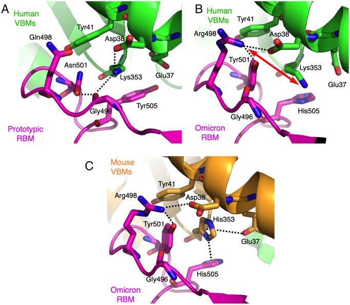
The sudden emergence and rapid spread of the severe acute respiratory syndrome coronavirus 2 (SARS-CoV-2) omicron variant has raised questions about its animal reservoir. Here, we investigated receptor recognition of the omicron's receptor-binding domain (RBD), focusing on four of its mutations (Q493R, Q498R, N501Y, and Y505H) surrounding two mutational hotspots. These mutations have variable effects on the RBD's affinity for human angiotensin-converting enzyme 2 (ACE2), but they all enhance the RBD's affinity for mouse ACE2. We further determined the crystal structure of omicron RBD complexed with mouse ACE2. The structure showed that all four mutations are viral adaptations to mouse ACE2: three of them (Q493R, Q498R, and Y505H) are uniquely adapted to mouse ACE2, whereas the other one (N501Y) is adapted to both human ACE2 and mouse ACE2. These data reveal that the omicron RBD was well adapted to mouse ACE2 before omicron started to infect humans, providing insight into the potential evolutionary origin of the omicron variant.

Keywords: COVID-19; X-ray crystallography; mouse angiotensin-converting enzyme 2; omicron variant; receptor-binding domain (RBD).


Conflict of interest statement

The authors declare no competing interest.

Figures

Fig. 1.
Fig. 1.
Binding interactions between SARS-CoV-2 RBD (from prototypic strain or omicron strain) and ACE2 (from human or mouse). (A) Structure of the interface between prototypic RBM and hACE2 (PDB ID: 6VW1). RBM is in magenta. hACE2 is in green. RBD residues that have undergone mutations from the prototypic strain to the omicron variant (strain BA.2) are shown as sticks. Three mutational hotspots are highlighted: hotspot-353 centers on Lys353 in hACE2, hotspot-31 centers on Lys31 in hACE2, and hotspot-ridge centers on the receptor-binding ridge in hACE2. (B and C) SPR assay for the binding of RBD (from prototypic strain or omicron strain) to ACE2 (from human or mouse). ACE2-Fc was coated to a protein A chip in a fixed direction, and individual RBDs flowed through. Data in B are from one of our recent studies (17), except that the omicron variant (strain BA.2) in this study replaced strain BA.1 in the previous study. Data in C are from the current study. The data in B and C are presented as mean ± SEM (n = 3 or n = 4) on a log scale. A Student’s two-tailed t test was performed to analyze the statistical difference between the RBD on the Left in either panel and each of the other RBDs in the same panel; the results are labeled on top of each bar. The statistical difference between the R493Q mutation and the N477S/R493Q double mutations was also analyzed in C; the result was labeled between the two bars. The horizontal dashed lines represent the measurements for the prototypic RBD in B or the omicron RBD in C and are used for comparison with other measurements in the respective panel. ***P < 0.001; **P < 0.01; *P < 0.05. n.s., statistically not significant, N.D., not detected.
Fig. 2.
Fig. 2.
Overall structures of chimeric omicron RBD complexed with chimeric mACE2 or hACE2. (A) Overall structure of chimeric omicron RBD complexed with chimeric mACE2. The chimeric omicron RBD contains the core structure (in cyan) from SARS-CoV-1 RBD and RBM (in magenta) from the omicron RBD (strain BA.2). The chimeric mACE2 contains the core structure (in green) from hACE2 and three VBMs (in orange) from mACE2. (B) Sequence alignment between hACE2 and mACE2 in three VBMs (shaded). The centers for hotspot-31 and hotspot-353 (i.e., residues 31 and 353, respectively) are both labeled in red. Residues that directly contact SARS-CoV-2 RBD are labeled in blue. Asterisks indicate positions that have a single, fully conserved residue. Colons indicate positions that have strongly conserved residues. Periods indicate positions that have weakly conserved residues. (C) Overall structure of chimeric omicron RBD complexed with hACE2. hACE2 is in green.
Fig. 3.
Fig. 3.
Structural details at mutational hotspot-31. (A) Interface between the prototypic RBM and human VBMs. PDB ID: 6VW1. (B) Interface between the omicron RBM and human VBMs. (C) Interface between the omicron RBM and mouse VBMs. Dotted lines indicate hydrogen bonds. Side chain of 490rbm is not shown for the sake of clarity. Double-ended red arrow indicates repulsion.
Fig. 4.
Fig. 4.
Structural details at mutational hotspot-353. (A) Interface between the prototypic RBM and human VBMs. PDB ID: 6VW1. (B) Interface between the omicron RBM and human VBMs. (C) Interface between the omicron RBM and mouse VBMs. Dotted lines indicate salt bridges, hydrogen bonds, hydrogen-π interaction, or anion-π interaction. Double-ended red arrow indicates repulsion.

References

    1. Karim S. S. A., Karim Q. A., Omicron SARS-CoV-2 variant: A new chapter in the COVID-19 pandemic. Lancet 398, 2126–2128 (2021). - PMC - PubMed
    1. Maslo C., et al. , Characteristics and outcomes of hospitalized patients in South Africa during the COVID-19 Omicron wave compared with previous waves. JAMA 327, 583–584 (2022). - PMC - PubMed
    1. Saxena S. K., et al. Characterization of the novel SARS-CoV-2 Omicron (B.1.1.529) variant of concern and its global perspective. J. Med. Virol. 94, 1738–1744 (2022). - PubMed
    1. Gu H., et al. , Probable transmission of SARS-CoV-2 Omicron variant in quarantine hotel, Hong Kong, China, November 2021. Emerg. Infect. Dis. 28, 460–462 (2022). - PMC - PubMed
    1. Mallapaty S., Where did Omicron come from? Three key theories. Nature 602, 26–28 (2022). - PubMed

Publication types

Substances

Supplementary concepts