. 2022 Oct 25;119(43):e2205492119.
doi: 10.1073/pnas.2205492119. Epub 2022 Oct 18.

Lysosomal positioning regulates Rab10 phosphorylation at LRRK2+ lysosomes

Affiliations

Lysosomal positioning regulates Rab10 phosphorylation at LRRK2+ lysosomes

Jillian H Kluss et al. Proc Natl Acad Sci U S A. .

Abstract

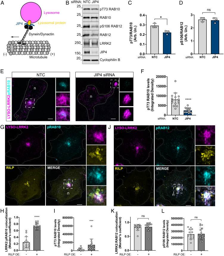
Genetic variation at the leucine-rich repeat kinase 2 (LRRK2) locus contributes to an enhanced risk of familial and sporadic Parkinson's disease. Previous data have demonstrated that recruitment to various membranes of the endolysosomal system results in LRRK2 activation. However, the mechanism(s) underlying LRRK2 activation at endolysosomal membranes and the cellular consequences of these events are still poorly understood. Here, we directed LRRK2 to lysosomes and early endosomes, triggering both LRRK2 autophosphorylation and phosphorylation of the direct LRRK2 substrates Rab10 and Rab12. However, when directed to the lysosomal membrane, pRab10 was restricted to perinuclear lysosomes, whereas pRab12 was visualized on both peripheral and perinuclear LRRK2+ lysosomes, suggesting that lysosomal positioning provides additional regulation of LRRK2-dependent Rab phosphorylation. Anterograde transport of lysosomes to the cell periphery by increasing the expression of ARL8B and SKIP or by knockdown of JIP4 blocked the recruitment and phosphorylation of Rab10 by LRRK2. The absence of pRab10 from the lysosomal membrane prevented the formation of a lysosomal tubulation and sorting process we previously named LYTL. Conversely, overexpression of RILP resulted in lysosomal clustering within the perinuclear area and increased LRRK2-dependent Rab10 recruitment and phosphorylation. The regulation of Rab10 phosphorylation in the perinuclear area depends on counteracting phosphatases, as the knockdown of phosphatase PPM1H significantly increased pRab10 signal and lysosomal tubulation in the perinuclear region. Our findings suggest that LRRK2 can be activated at multiple cellular membranes, including lysosomes, and that lysosomal positioning further provides the regulation of some Rab substrates likely via differential phosphatase activity or effector protein presence in nearby cellular compartments.

Keywords: JIP4; LLOMe; LYTL; Parkinson's disease; kinase.


Conflict of interest statement

The authors declare no competing interest.

Figures

Fig. 1.
Fig. 1.
Activation of LRRK2 and subsequent Rab phosphorylation is achieved using FKBP/FRB complex and chimera-LRRK2 constructs regardless of membrane identity. HEK293FT cells transfected with either LAMP1-FRB-CFP and FKBP-LRRK2 plasmids (specified as “lyso-trap”; Top) or LYSO-LRRK2 plasmid (Bottom). Endogenous LAMP1 is also shown in the bottom image of LYSO-LRRK2. Construct designs are shown to the left of the respective ICC images in (A). Scale bar, 10 μm. (B–E) Western blot analyses of LRRK2 S1292 autophosphorylation, Rab10 and Rab12 phosphorylation under conditions of NT-LRRK2 or lyso-trap–expressing cells with or without rapamycin treatment, or LYSO-LRRK2 chimera only. (F) Cells transfected with iRFP-FRB-RAB5 and FKBP-LRRK2 plasmids treated with rapamycin (specified as “EE-trap”; Top) or the EE-LRRK2 chimera (Bottom). Endogenous EEA1 is also shown in the bottom image of EE-LRRK2. Construct designs are shown on the left of the images. Scale bar, 10 μm. DAPI is shown in blue. (G–J) Western blot analysis from densitometry measurements of LRRK2 S1292 autophosphorylation, Rab10 and Rab12 phosphorylation under conditions of NT-LRRK2, or EE-trap–expressing cells with and without rapamycin treatment or the EE-LRRK2 chimera alone. (C–E, H–J) One-way ANOVA with Tukey’s post hoc; n = 3; SD bars shown; (C) F(4, 10) = 19.67, (D) F(4, 10) = 16.87, (E) F(4, 10) = 81.71, (H) F(4, 10) = 25.86, (I) F(4, 10) = 25.22, and (J) F(4, 10) = 16.00. ns = not significant.
Fig. 2.
Fig. 2.
Rab phosphorylation is LRRK2 dependent and pRab colocalization patterns differ between Rab10 and Rab12 at the lysosomal membrane. HEK293FT cells were transfected with either NT-LRRK2 or LYSO-LRRK2 chimera, and pT73 Rab10 and pS106 Rab12 integrative densities were measured (A–D). MLi-2 treatment was compared to untreated cells expressing LYSO-LRRK2 (A–D). Scale bar, 10 μm. (E–I) Cells were transfected with either NT-LRRK2 or EE-LRRK2 chimera, and pT73 Rab10 and pS106 Rab12 integrative densities were measured (A–D). MLi-2 treatment was compared to untreated cells expressing EE-LRRK2 (E–I). Scale bar, 10 μm. In all merged images, “n” denotes the nucleus. (B, D, F, and H) One-way ANOVA with Tukey’s post hoc; (I) two-way ANOVA with Tukey’s post hoc; n = 3, n = 13 cells; SD bars shown; (B) F(2, 32) = 33.11, (D) F(2, 32) = 34.92, (F) F(2, 32) = 26.67, (H) F(2, 32) = 52.36, and (I) chimera: F(1, 36) = 93.49, P < 0.0001, Rab: F(1, 36) = 73.37, P < 0.0001.
Fig. 3.
Fig. 3.
ARL8B- and SKIP-expressing cells promote lysosomal repositioning to the periphery of cells. Representative schematic describing lysosomal translocation to the cell periphery via expression of ARL8B and SKIP proteins along the plus end of microtubules (A). Airyscan microscopy showing peripheral LYSO-LRRK2 lysosomes, colocalizing with ARL8B and SKIP (B). Cell edges are outlined. Scale bars, 10 μm. LYSO-LRRK2 was transfected in cells alone or cotransfected with ARL8B and SKIP (C). Scale bars, 10 μm. Quantification of the ratio of peripheral to total LRRK2+ lysosomes such as those represented in (C) are shown in (D). Peripheral fluorescence refers to the presence of LRRK2 within 2 μm from the cell vertices. Horizontal lines indicate the mean ± SD from 3 independent experiments.
Fig. 4.
Fig. 4.
ARL8B- and SKIP-expressing cells prevent pRab10 colocalization on peripherally positioned LRRK2+ lysosomes. Representative confocal microscopy images show pT73 Rab10 signal, LYSO-LRRK2 with endogenous LAMP2 (Top), or ARL8B and SKIP (Bottom) (A). Integrative density and Mander’s correlation coefficient were used to measure pRab10 staining intensity (B) and LRRK2:pRab10 colocalization (C). (D) Cells with and without ARL8B/SKIP transfection were treated with 1 mM LLOMe for 2 h. Integrative density and Mander’s correlation coefficient were used to measure pRab10 staining intensity (E) and LRRK2:pRab10 colocalization (F). LYSO-LRRK2 expressing cells with and without ARL8B/SKIP cotransfection and LLOMe treatment were probed for S1292 LRRK2 and pT73 Rab10 for western blot densitometry analysis (G–J). In all merged images, “n” denotes the nucleus. (B, C, E, and F) Two-tailed unpaired t test; n = 13 cells across 2 independent experiments; error bars represent SDs; (B) ***P = 0.0004, (C) **P = 0.0071, (E) ****P < 0.0001, and (F) ****P < 0.0001. (H–J) One-way ANOVA with Tukey’s post hoc; n = 3 to 4; SD bars shown. (H) F(3, 12) = 59.39, (I) F(3, 12) = 4.624, and (J) F(3, 12) = 19.42. Scale bars, 10 μm. ns = not significant.
Fig. 5.
Fig. 5.
Phosphorylated Rab12 is found at peripheral lysosomes in cells transiently transfected with ARL8B and SKIP plasmids. A superresolution confocal microscopy image shows pS106 Rab12 staining at LRRK2+ lysosomes when ARL8B and SKIP are coexpressed (A). In the merged image, “n” denotes the nucleus. Scale bar, 10 μm. Mander’s coefficient was used to measure LRRK2:pRab12 colocalization in conditions in which ARL8B and SKIP are coexpressed with LYSO-LRRK2 as well as when only LYSO-LRRK2 is expressed alone (B). Western blot analysis from densitometry measurements of Rab12 phosphorylation are shown (C and D). (B) Unpaired, two-tailed t test; n = 3, n = 12 cells each; SD error bars are shown; P = 0.6045. (D–F) One-way ANOVA with Tukey’s post hoc; n = 3; SD bars shown. (D) F(3, 8) = 87.73.
Fig. 6.
Fig. 6.
Knockdown of endogenous JIP4 is sufficient to prevent peripheral lysosomes from returning to the perinuclear area, while RILP-expressing cells promote clustering of LRRK2+ lysosomes within the perinuclear region and significantly increase pRab10 signal. Simplified schematic of lysosomal movement into the perinuclear area via JIP4 adaptor binding to a lysosomal protein and the dynein/dynactin complex (A). Western blot analysis showing NTC (nontargeting control siRNA) versus JIP4 siRNA conditions in HEK293FT cells and densitometry measurements of pRab10 and pRab12 proteins (B–D). Representative confocal microscopy images of HEK293FT cells stained for LYSO-LRRK2 and pRab10 under conditions of NTC and JIP4 siRNA transfection (E), in which knockdown of JIP4 significantly reduces pRab10 signal (F). Cells expressing LYSO-LRRK2 and RILP were stained for pRab10 or pRab12 and colocalization with LRRK2 and integrated densities were measured, respectively (G–L). In all of the merged images, “n” denotes the nucleus. (C, D, F, H, I, K, and L) Two-tailed unpaired Student t test; SD error bars are shown. (C) P = 0.0132, n = 3, (F) P < 0.0001, n = 13 to 15 cells, (H) P < 0.0001, (I) P < 0.0001, (K) P = 0.6045, and (L) P = 0.8273, n = 12 to 15 cells.
Fig. 7.
Fig. 7.
ARL8B- and SKIP- expressing cells treated with LLOMe recruit NT-LRRK2-expressing HEK293FT and endogenous LRRK2 in primary mouse astrocytes but not pRab10. Representative Airyscan microscopy images of NT-LRRK2 with and without ARL8B/SKIP coexpressing cells under conditions of DMSO or LLOMe treatment, and “n” denotes the nuclei. (A and B) LRRK2+ lysosomes are counted in each condition followed by pT73 Rab10 (C and D), and lastly, JIP4 (E and F). Scale bars, 10 μm. (G–I) Western blot analysis from densitometry measurements of pRab10 from LYSO-LRRK2-expressing cells ± ARL8B/SKIP and ± LLOMe. (J) Primary astrocytes were treated with DMSO or LLOMe and probed for endogenous LAMP1 and pRab10 at endogenous levels of LRRK2. In the image shown with ARL8B, “periph.” and “perinuc.” abbreviations are shown for the insets that are peripheral and perinuclear, respectively. (K) pRab10+ lysosomes were counted in these treatment conditions ± ARL8B/SKIP. Scale bars, 10 μm. (B, D, F, H, I, and K) One-way ANOVA with Tukey’s post hoc; (B, D, F, and K) n = 32 to 40 cells counted. (H and I) n = 3; SD error bars are shown. (B) F(2, 105) = 15.91, (D) F(2, 107) = 141.1, (F) F(2, 94) = 116.4, (H) F(3, 8) = 14.54, (I) F(3, 8) = 0.5329, and (K) F(2, 99) = 27.14.
Fig. 8.
Fig. 8.
PPM1H knockdown affects pRab10-mediated lysosomal tubulation. Representative confocal images showing LYSO-LRRK2 and pT73 Rab10 after siRNA knockdown of NTC or PPM1H, where “n” denotes the nuclei (A). The integrated density of pRab10 signal was measured in ICC (B) and western blot densitometry measurements, with additional probing for Rab12 (C–E). Representative confocal images of JIP4 are shown accompanied by integrated density measurements in cells transfected with NTC or PPM1H siRNA (F and G). Airyscan images are shown of pT73 Rab10+ and JIP4+ tubules, and the number of tubules was counted (H–K). (B, D, E, G, J, and K) Two-tailed unpaired t test; n = 16 (B and G); (D and E) n = 3; n = 35 (J and K); SD bars shown. (B) P = 0.0013, (D) P = 0.0141, (E) P = 0.6540, (G) P = 0.0066, (J) P = 0.0055, and (K) P = 0.0038.

References

    1. Paisán-Ruíz C., et al. , Cloning of the gene containing mutations that cause PARK8-linked Parkinson’s disease. Neuron 44, 595–600 (2004). - PubMed
    1. Zimprich A., et al. , The PARK8 locus in autosomal dominant parkinsonism: Confirmation of linkage and further delineation of the disease-containing interval. Am. J. Hum. Genet. 74, 11–19 (2004). - PMC - PubMed
    1. Nalls M. A., et al. ; 23andMe Research Team; System Genomics of Parkinson’s Disease Consortium; International Parkinson’s Disease Genomics Consortium, Identification of novel risk loci, causal insights, and heritable risk for Parkinson’s disease: A meta-analysis of genome-wide association studies. Lancet Neurol. 18, 1091–1102 (2019). - PMC - PubMed
    1. Roosen D. A., Cookson M. R., LRRK2 at the interface of autophagosomes, endosomes and lysosomes. Mol. Neurodegener. 11, 73 (2016). - PMC - PubMed
    1. Greggio E., et al. , Kinase activity is required for the toxic effects of mutant LRRK2/dardarin. Neurobiol. Dis. 23, 329–341 (2006). - PubMed

Publication types

MeSH terms

Substances

LinkOut - more resources