. 2022 Oct 26;289(1985):20221126.
doi: 10.1098/rspb.2022.1126. Epub 2022 Oct 19.

Footedness for scratching itchy eyes in rodents

Affiliations

Footedness for scratching itchy eyes in rodents

Yukitoshi Katayama et al. Proc Biol Sci. .

Abstract

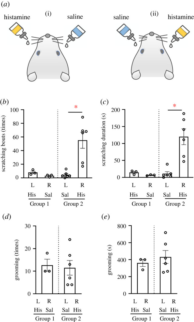
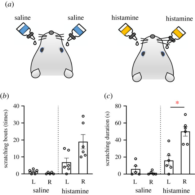
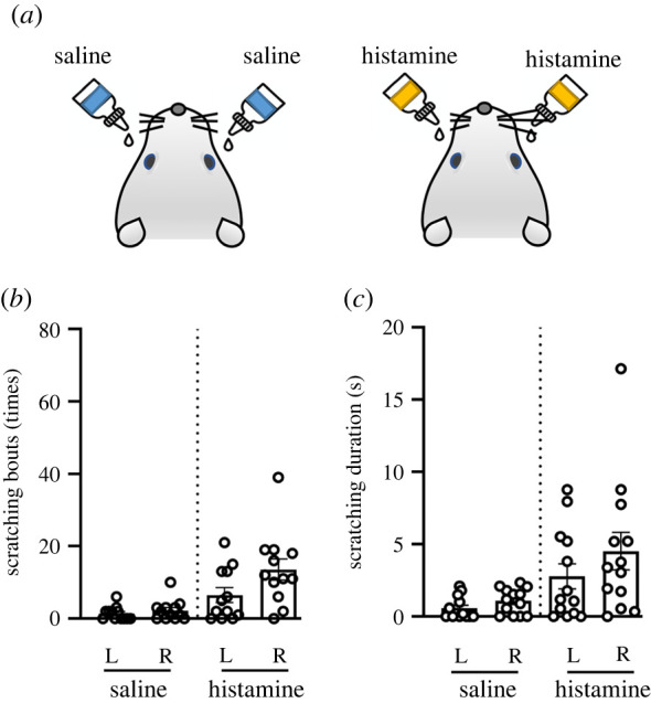
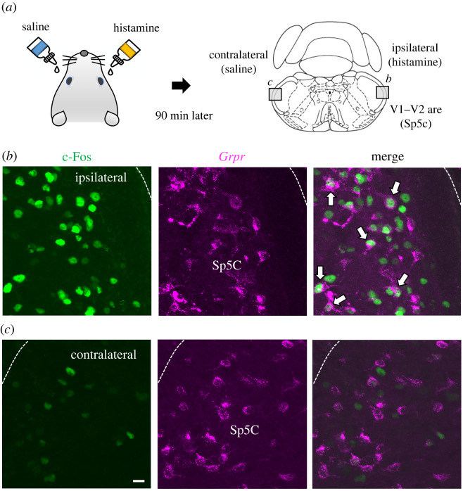
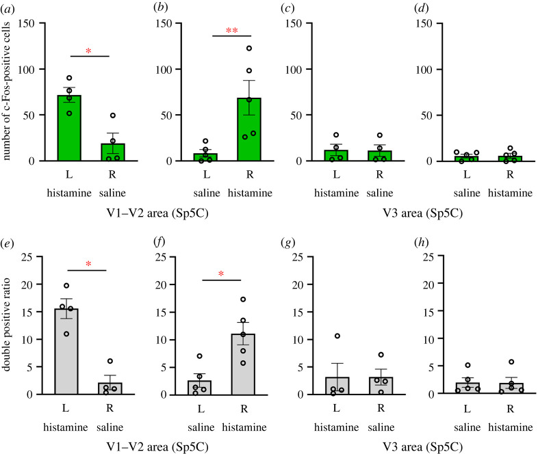
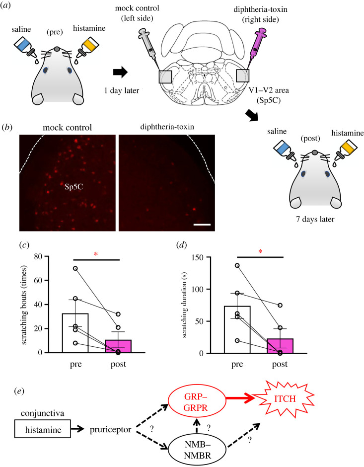
The neural bases of itchy eye transmission remain unclear compared with those involved in body itch. Here, we show in rodents that the gastrin-releasing peptide receptor (GRPR) of the trigeminal sensory system is involved in the transmission of itchy eyes. Interestingly, we further demonstrate a difference in scratching behaviour between the left and right hindfeet in rodents; histamine instillation into the conjunctival sac of both eyes revealed right-foot biased laterality in the scratching movements. Unilateral histamine instillation specifically induced neural activation in the ipsilateral sensory pathway, with no significant difference between the activations following left- and right-eye instillations. Thus, the behavioural laterality is presumably due to right-foot preference in rodents. Genetically modified rats with specific depletion of Grpr-expressing neurons in the trigeminal sensory nucleus caudalis of the medulla oblongata exhibited fewer and shorter histamine-induced scratching movements than controls and eliminated the footedness. These results taken together indicate that the Grpr-expressing neurons are required for the transmission of itch sensation from the eyes, but that foot preference is generated centrally. These findings could open up a new field of research on the mechanisms of the laterality in vertebrates and also offer new potential therapeutic approaches to refractory pruritic eye disorders.

Keywords: footedness; gastrin-releasing peptide receptor; histamine; itchy eyes.


Conflict of interest statement

We declare we have no competing interests.

Figures

Figure 1.
Figure 1.
Scratching behaviour evoked by unilateral instillation of histamine into the conjunctival sac of the eyes of rats. (a) Schematic showing the instillation of either 60 µmol histamine (His) (n = 6) or saline (Sal) (n = 3) unilaterally into the conjunctival sac of either the left (L) eye (i) or right (R) eye (ii). The number (b) and duration (c) of histamine- or saline-evoked hindfoot scratches during 60 min. The number and duration of scratches with the right foot was significantly greater than with that with the left foot ((b) p = 0.028, (c) p = 0.042). The number (d) and duration (e) of grooming events did not differ significantly in either Group 1 or Group 2 rats. *p < 0.05 with Wilcoxon signed-rank test. Data are shown as mean ± s.e.m. (Online version in colour.)
Figure 2.
Figure 2.
Scratching behaviour evoked by bilateral instillation of histamine into the conjunctival sacs of the eyes in rats. (a) Schematic showing the bilateral instillation of either saline (n = 5) or 60 µmol histamine (n = 6) into the conjunctival sac of the eyes of rats. The number (b) and duration (c) of saline- or histamine-evoked hindfoot scratches during 60 min. The duration of scratches was significantly greater on the right side (right foot to right eye) compared with the left side after histamine but not saline instillation ((b) p = 0.065, (c) p = 0.031). *p < 0.05 with Wilcoxon signed-rank test. Data are shown as mean ± s.e.m. (Online version in colour.)
Figure 3.
Figure 3.
Laterality of scratching behaviour evoked by bilateral instillation of histamine into the conjunctival sacs of the eyes in mice. (a) Schematic showing the bilateral instillation of saline or histamine into the conjunctival sacs in mice (n = 12). The number (b) and duration (c) of saline- or histamine-evoked hindfoot scratches during 30 min. The number of scratches induced by 60 µmol histamine tended to be increased in the right-eye group (p = 0.084) compared with left-eye group. There were no significant differences in scratching duration between the right side and the left side in mice (p = 0.140). *p < 0.05 with Wilcoxon signed-rank test. Data are shown as mean ± s.e.m. (Online version in colour.)
Figure 4.
Figure 4.
Histamine instillation induced the activation of Grpr-expressing neurons in the lateral part of the trigeminal nucleus of the medulla oblongata in rats. (a) Schematic showing the lateral part (V1 and V2 input area) of the spinal trigeminal nucleus caudalis (Sp5C) in rats. (b,c) Double-fluorescence images showing Grpr-expression (RFP, magenta) and c-Fos immunoreactivity (green) in the Sp5C (V1 and V2 area). Histamine instillation into an eye increased c-Fos expression in the ipsilateral side of Sp5C (b), compared to that in the contralateral side of Sp5C (control) (c). White arrows indicate neurons double-positve for c-Fos and Grpr. Scale bar, 10 µm. (Online version in colour.)
Figure 5.
Figure 5.
Quantitation of c-Fos-immunoreactive neurons in the trigeminal nucleus of the medulla oblongata in rats. Histamine instillation increased the number of c-Fos-immunoreactive neurons in the V1 and V2 areas of the ipsilateral side in Sp5C [(a) left-side instillation of histamine (L), n = 4; p = 0.028, (b) right-side instillation of histamine (R), n = 5; p = 0.008]. The number of c-Fos-immunoreactive neurons in the V3 area was not changed (c,d). The number of cells double positive for c-Fos and Grpr-expressed as a proportion of the total number of Grpr-expressing neurons was significantly higher in the histamine-instilled side than in the control side in the V1 and V2 area of ipsilateral side [(e) left-side instillation of histamine (L); p = 0.028; (f) right-side instillation of histamine (R); p = 0.015]. The number of c-Fos-immunoreactive neurons was not changed in the V3 area (g,h). *p < 0.05 with Wilcoxon signed-rank test. Data are shown as mean ± s.e.m. (Online version in colour.)
Figure 6.
Figure 6.
Scratching behaviour was reduced by the depletion of Grpr-expressing neurons in the medulla oblongata in rats. (a) Schematic showing the initial instillation of 60 µmol histamine into the conjunctival sac of the right eye and saline into the left eye of Grpr-RFP transgenic rats (pre). One day later, saline (n = 3) or DT (n = 5) was injected locally into the right trigeminal nucleus caudalis (Sp5C). Seven days after the toxin injection, 60 µmol histamine was again instilled in the conjunctival sac of the right eye and saline was instilled in the conjunctival sac of the left eye to examine the scratching behaviour (post). (b) RFP (Grpr-expression) fluorescent signals in the right Sp5C of the saline-injected rats (left, vehicle) and right Sp5C of the DT-injected rats (right, DT-lesioned). Scale bar, 100 µm. The number (c) and duration (d) of scratches with the right hindfoot were significantly decreased in the post-DT injection group in compared with the pre-DT injection group at 60 min (n = 5) [(c) p = 0.027, (d) p = 0.038]. *p < 0.05 with t-test. Data are shown as mean ± s.e.m. (e) Hypothetical model of NMB–NMBR and GRP–GRPR pathways for histaminergic itch transmission in the eye. Histamine in the eye activates GRP–GRPR and NMB–NMBR systems via pruriceptors. As suggested in the spinal sensory pathway of the body itch [6,28], Grpr-expressing neurons in the Sp5C might integrate multiple lines of information and transmit histaminergic itch information to the brain. Possible interaction between NMB–NMBR and GRP–GRPR systems should be investigated in future studies of ocular itch. Broken lines indicate possible actions in the itch transmission. (Online version in colour.)

Similar articles

Cited by

References

    1. Galor A, Batawi H, Felix ER, Margolis TP, Sarantopoulos KD, Martin ER, Levitt RC. 2016. Incomplete response to artificial tears is associated with features of neuropathic ocular pain. Br. J. Ophthalmol. 100, 745-749. (10.1136/bjophthalmol-2015-307094) - DOI - PubMed
    1. Hay M, Thomas DW, Craighead JL, Economides C, Rosenthal J. 2014. Clinical development success rates for investigational drugs. Nat. Biotechnol. 32, 40-51. (10.1038/nbt.2786) - DOI - PubMed
    1. Weisshaar E, Dalgard F. 2009. Epidemiology of itch: adding to the burden of skin morbidity. Acta Derm. Venereol. 89, 339-350. (10.2340/00015555-0662) - DOI - PubMed
    1. Yosipovitch G, Bernhard JD. 2013. Chronic pruritus. New Engl. J. Med. 368, 1625-1634. (10.1056/NEJMcp1208814) - DOI - PubMed
    1. Andersen HH, Yosipovitch G, Galor A. 2017. Neuropathic symptoms of the ocular surface: dryness, pain, and itch. Curr. Opin. Allergy Clin. Immunol. 17, 373. (10.1097/ACI.0000000000000389) - DOI - PMC - PubMed

Publication types