Review
. 2023 Sep;44(3):217-223.
doi: 10.1007/s10974-022-09629-x. Epub 2022 Oct 19.

John squire and endothelial glycocalyx structure: an unfinished story

Affiliations
Review

John squire and endothelial glycocalyx structure: an unfinished story

Kenton P Arkill et al. J Muscle Res Cell Motil. 2023 Sep.

Abstract

John Squire did not only produce leading works in the muscle field, he also significantly contributed to the vascular permeability field by ultrastructural analysis of the endothelial glycocalyx. Presented here is a review of his involvement in the field by his main collaborator C.C. Michel and his last postdoctoral researcher KP Arkill. We end on a reinterpretation of his work that arguably links to our current understanding of endothelial glycocalyx structure and composition predicting 6 glycosaminoglycans fibres per syndecan core protein, only achieved in the endothelium by dimerization.

Keywords: Albumin; Glycosaminoglycan; Proteoglycan; Reflection coefficient; Vascular permeability.


Conflict of interest statement

The authors declare no competing interests.

Figures

Fig. 1
Fig. 1
Freeze fracture measurement on the endothelial glycocalyx of a frog mesenteric capillary Taken from (Squire et al. 2001). A Micrograph from the technically challenging (including due to determining regions of interest, depth of freezing) untreated slam frozen and freeze fractured. B The auto-correlation of the (A) which reveals a striking quasi-hexagonal arrangement within the glycocalyx with spacing of between 80 and 120 nm as indicated by the white lines and arrows. Smaller periodicities can also be found within these major repeats. White scale bar is 200 nm
Fig. 2
Fig. 2
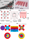
Relating the fibre spacing to the reflection coefficient. A From(Arkill et al. 2011) Rostgard-Qvortrup staining of the endothelial glycocalyx (eGlx) in the rat kidney glomerulus showing short clumps. B Proteoglycan cores (blue) with example of glycoasaminoglycan (GAG) fibres attached mid rotation to plan view (See Supplemental video 1). Ci The Squire filtration model (exampled on a square lattice for ease of display) (Squire et al. ; Arkill et al. 2011). The GAG fibres (red) are collapsed on the core, which can be expanded (Cii). Ciii represents the new expanded model in cross-section where there is a new inter-fibre spacing. D shows how the free space for a solute to travel is dependent on the relative molecular size (rf= fibre radius; ra= solute radius). Di The solute cannot fit freely around the fibres so can be approximated to a tube. The reflection coefficient can be determined via Anderson and Malone’s calculations (Anderson and Malone 1974). Dii If the solute can fit around the fibres the available space can be approximated to an annulus and the reflection coefficient can be determined via Zhang et al.’s calculations (Zhang et al. 2006). Ci–iii from and (D) adapted from (Arkill 2021)
Fig. 3
Fig. 3
The unraveled glycosaminoglycan (GAG) model predicts a mean of ≈ 6 fibres per proteoglycan core. The graph depicts the model for a spacing of 19.5 nm between proteoglycan cores in a hexagonal arrangement of cores and unraveled glycosaminoglycans with a fibre radius (rf) of 0.75 nm. Parameter justification for albumin (r3.5nm) using the tubular model (open red triangles) are in (Arkill 2021). The red filled triangles are using the annular Zhang model (Zhang et al. 2006) to determine the reflection coefficients, these are always higher than the tubular model for the same number of fibres per core and less applicable to albumin. The blue filled triangles are the annular Zhang model for r2nm to approximate myoglobin and ⍺-lactalbumin

References

    1. Anderson JL, Malone DM. Mechanism of osmotic flow in porous membranes. Biophys J. 1974;14(12):957–982. doi: 10.1016/S0006-3495(74)85962-X. - DOI - PMC - PubMed
    1. Arkill KP. A reinterpretation of evidence for the endothelial glycocalyx filtration structure. Front Cell Dev Biol. 2021 doi: 10.3389/fcell.2021.734661. - DOI - PMC - PubMed
    1. Arkill KP, Knupp C, Michel CC, Neal CR, Qvortrup K, Rostgaard J, Squire JM. Similar endothelial glycocalyx structures in microvessels from a range of mammalian tissues: Evidence for a common filtering mechanism? Biophys J. 2011;101(5):1046–1056. doi: 10.1016/j.bpj.2011.07.036. - DOI - PMC - PubMed
    1. Arkill KP, Neal CR, Mantell JM, Michel CC, Qvortrup K, Rostgaard J, Bates DO, Knupp C, Squire JM. 3d reconstruction of the glycocalyx structure in mammalian capillaries using electron tomography. Microcirculation. 2012;19(4):343–351. doi: 10.1111/j.1549-8719.2012.00168.x. - DOI - PMC - PubMed
    1. Arkill KP, Qvortrup K, Starborg T, Mantell JM, Knupp C, Michel CC, Harper SJ, Salmon AH, Squire JM, Bates DO, Neal CR. Resolution of the three dimensional structure of components of the glomerular filtration barrier. BMC Nephrol. 2014;15(1):1–13. doi: 10.1186/1471-2369-15-24. - DOI - PMC - PubMed

Publication types

Substances

LinkOut - more resources