

Evaluation of Prefrontal γ-Aminobutyric Acid and Glutamate Levels in Individuals With Major Depressive Disorder Using Proton Magnetic Resonance Spectroscopy
- PMID: 36260322
- PMCID: PMC9582968
- DOI: 10.1001/jamapsychiatry.2022.3384
Evaluation of Prefrontal γ-Aminobutyric Acid and Glutamate Levels in Individuals With Major Depressive Disorder Using Proton Magnetic Resonance Spectroscopy
Abstract
Importance: Major depressive disorder (MDD) is one of the most prevalent illnesses worldwide. Perturbations of the major inhibitory and excitatory neurotransmitters, γ-aminobutyric acid (GABA) and glutamate (Glu), respectively, as well as Glx (Glu or glutamine [Gln]) have been extensively reported in a multitude of brain areas of individuals with depression, but few studies have examined changes in Gln, the metabolic counterpart of synaptic Glu.
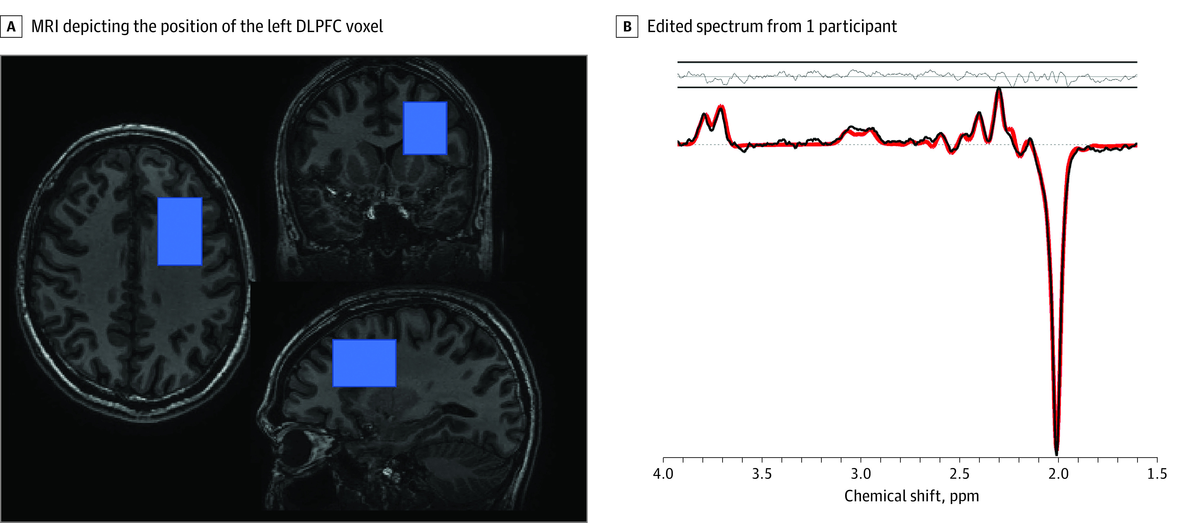
Objective: To investigate changes in GABA, Glx, Glu, and Gln levels in a voxel in the left dorsolateral prefrontal cortex of participants with no, past, and current MDD using proton magnetic resonance spectroscopy (1H-MRS).
Design, setting, and participants: This community-based study used a cross-sectional design using 3-T 1H-MRS in participants not taking MDD medication recruited from the community. The sample consisted of 251 healthy controls, 98 participants with a history of past MDD, and 47 participants who met the diagnostic criteria for current MDD. Diagnostic groups were comparable regarding age, education, income, and diet. Data were collected from March 2014 to October 2021, and data were analyzed from October 2021 to June 2022.
Main outcomes and measures: GABA, Glx, Glu, and Gln concentrations in the left dorsolateral prefrontal cortex.
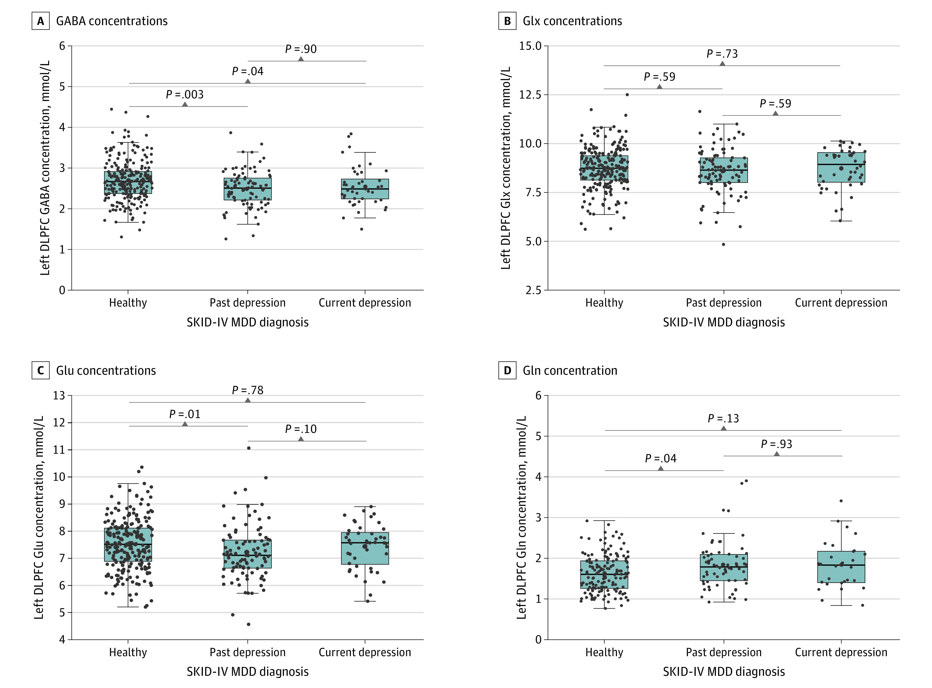
Results: Of 396 included participants, 258 (65.2%) were female, and the mean (SD) age was 25.0 (4.7) years. Compared with healthy controls, those with past MDD and current MDD had lower GABA concentrations (mean [SEM] concentration: healthy controls, 2.70 [0.03] mmol/L; past MDD, 2.49 [0.05] mmol/L; current MDD, 2.54 [0.07] mmol/L; 92 with past MDD vs 236 healthy controls: r = 0.18; P = .002; 44 with current MDD vs 236 healthy controls: r = 0.13; P = .04). Compared with healthy controls, those with past MDD also had lower Glu concentrations (mean [SEM] concentration: healthy controls, 7.52 [0.06] mmol/L; past MDD, 7.23 [0.11] mmol/L; 93 with past MDD vs 234 healthy controls: r = 0.16; P = .01) and higher Gln concentrations (mean [SEM] concentration: healthy controls, 1.63 [0.04] mmol/L; past MDD, 1.84 [0.07] mmol/L; 66 with past MDD 153 healthy controls: r = 0.17; P = .04).
Conclusions and relevance: In a large, mostly medication-free community sample, reduced prefrontal GABA concentrations were associated with past MDD, consistent with histopathologic studies reporting reduced glial cell and GABA cell density in the prefrontal cortex in individuals with depression. Patients with MDD also demonstrated increased Gln levels, indicative of increased synaptic Glu release, adding to previous evidence for the Glu hypothesis of MDD.
Conflict of interest statement
Figures
References
Publication types
MeSH terms
Substances
LinkOut - more resources
Full Text Sources
Miscellaneous
