. 2023 Feb 1;34(2):258-272.
doi: 10.1681/ASN.2022040477. Epub 2022 Oct 19.

Clinical Implications of a New DDX58 Pathogenic Variant That Causes Lupus Nephritis due to RIG-I Hyperactivation

Affiliations

Clinical Implications of a New DDX58 Pathogenic Variant That Causes Lupus Nephritis due to RIG-I Hyperactivation

Jiahui Peng et al. J Am Soc Nephrol. .

Abstract

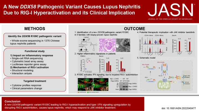
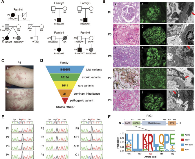
Background: Lupus nephritis (LN) is one of the most severe complications of systemic lupus erythematosus, with heterogeneous phenotypes and different responses to therapy. Identifying genetic causes of LN can facilitate more individual treatment strategies.

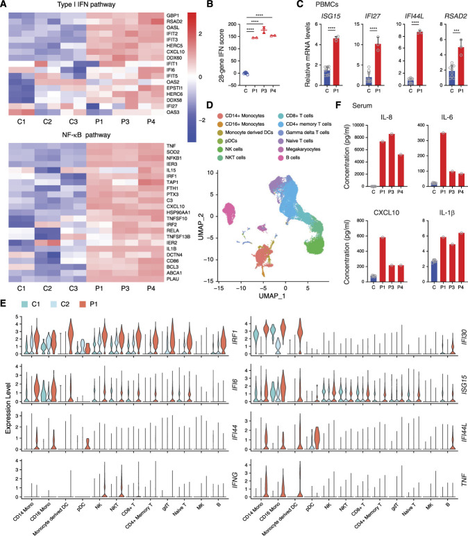
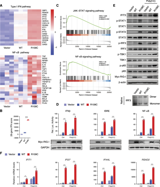
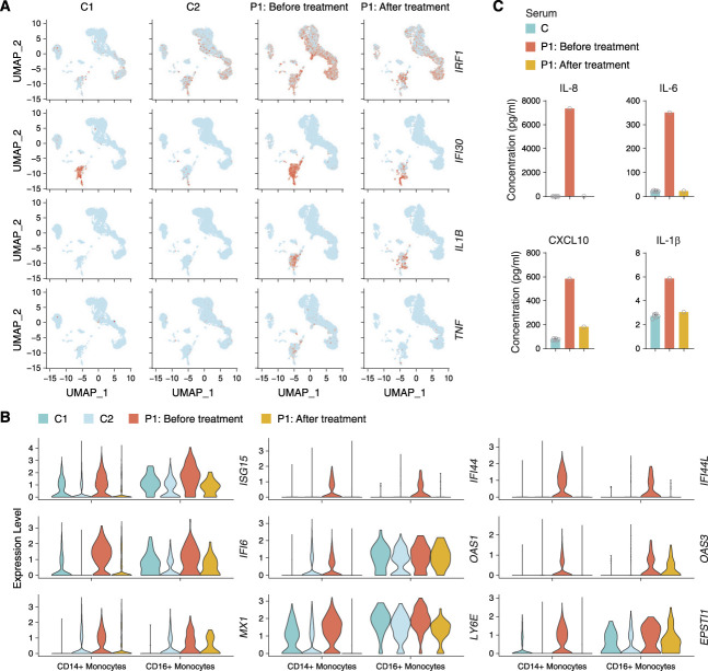
Methods: We performed whole-exome sequencing in a cohort of Chinese patients with LN and identified variants of a disease-causing gene. Extensive biochemical, immunologic, and functional analyses assessed the effect of the variant on type I IFN signaling. We further investigated the effectiveness of targeted therapy using single-cell RNA sequencing.

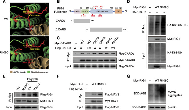
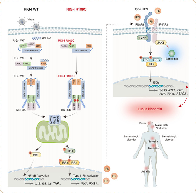
Results: We identified a novel DDX58 pathogenic variant, R109C, in five unrelated families with LN. The DDX58 R109C variant is a gain-of-function mutation, elevating type I IFN signaling due to reduced autoinhibition, which leads to RIG-I hyperactivation, increased RIG-I K63 ubiquitination, and MAVS aggregation. Transcriptome analysis revealed an increased IFN signature in patient monocytes. Initiation of JAK inhibitor therapy (baricitinib 2 mg/d) effectively suppressed the IFN signal in one patient.

Conclusions: A novel DDX58 R109C variant that can cause LN connects IFNopathy and LN, suggesting targeted therapy on the basis of pathogenicity.

Podcast: This article contains a podcast at.


Conflict of interest statement

All authors have nothing to disclose.

Figures

None
Graphical abstract
Figure 1
Figure 1
Identification of a heterozygous DDX58 R109C variant in patients with LN. (A) Pedigrees of the five unrelated families carrying the DDX58 variant. The proband is indicated by an arrow. Black indicates patients with SLE or LN, dark gray indicates patients with suspected SLE, and light gray indicates patients with psoriasis. (B) Renal histopathologic lesions of patients P1, P5, P6, P7, and P8. Light microscopy: (a) Diffuse mesangial and endocapillary proliferation in the glomerulus (PAS, ×400). (b) A fibrocellular crescent compressing the tuft of glomerulus (HE, ×400). (c) The prominent mesangial hypercellularity and segmental endocapillary proliferation in the glomerulus. It also demonstrates acute tubular injury and patchy interstitial inflammation (PAS, ×200). (d) The enlarged glomerulus, which shows a membranoproliferative pattern of injury, with mesangial hypercellularity and glomerular basement membrane duplication (PASM-Masson, ×400). (e) The segmental endocapillary proliferation and a cellular crescent formation in the glomerulus (HE, ×400). Immunofluorescence: (f, g, i, and j) The granular IgG deposits in the mesangial area and along the capillary wall (IF, ×400). (h) The granular C3 deposits in the mesangium and along the glomerular basement membrane (IF, ×400). Electromicroscopy: (k, m, n, and o) Tubuloreticular inclusions in the glomerular endothelial cells (red arrows). (l) Abundant electron-dense deposits in the subendothelial area with wrinkling of the glomerular basement membrane. (C) Psoriasiform skin rash on (a) the arm and (b) the back of P3. (D) Schematic representation of the WES data-filtering approach used to identify dominant inherited pathogenic variant in DDX58 in family 1. (E) Confirmation of the DDX58 R109C variant for patients and family members by Sanger sequencing. (F) Schematic illustration of the structure of human RIG-I protein, and evolutionary conservation of the site R109 in DDX58 across various species. AP, asymptomatic patient; CTD, Carboxy-terminal domain; HE, hematoxylin and eosin; IF, immunofluorescence; NA, not available; PAS, periodic acid–Schiff; PASM, periodic Schiff–methenamine silver.
Figure 2
Figure 2
Hyperactivation of type I IFN and NF-κB signaling in patients with the DDX58 R109C variant. (A) RNA sequencing analysis of type I IFN and NF-κB pathways in P1’s, P3’s, and P4’s PBMCs compared with three unaffected controls (C1–C3). Analysis of each sample was performed in duplicate. (B) Quantification of 28-gene IFN score of RNA sequencing data from (A). Data are presented as the mean±SD; ****P<0.0001, t test. (C) qPCR analysis of the expression of the IFN-stimulated genes in PBMCs from P1 compared with three unaffected controls. Data are presented as the mean±SD; n=3 independent experiments; ***P<0.001, ****P<0.0001, t test. (D) Marker-based annotation on UMAP plot of single-cell RNA sequencing data from two unaffected controls (C1 and C2) and patient P1. (E) Visualization of upregulated IFN-stimulated and inflammatory genes among all 12 clusters of P1 compared with two unaffected controls (C1 and C2) in violin plots. (F) CBA analysis of serum proinflammatory cytokines and chemokines (IL-6, IL-8, IL-1β, and CXCL10) levels in patients (P1, P3, and P4), compared with six unaffected controls. DCs, dendritic cells; gdT, gamma deta T cells; MK, megakaryocytes; Mono, monocytes; NK, natural killer cells; pDCs, plasmacytoid dendritic cells; UMAP, uniform manifold approximation and projection.
Figure 3
Figure 3
R109C mutation constitutively activates the IFN pathway in HEK293T cells. (A) RNA sequencing analysis of type I IFN and NF-κB pathways in HEK293T cells overexpressing with or without RIG-I-WT or R109C for 24 hours. Analysis of each sample was performed in triplicate. (B) Quantification of 28-gene IFN score of RNA sequencing data from (A). Data are presented as the mean±SD; ****P<0.0001, t test. (C) The gene set enrichment analysis plot of differential expression gene sets of RNA sequencing data from (A) enriched on JAK-STAT and NF-κB signaling pathways in RIG-I-WT and R109C overexpressing cells. (D) Luciferase reporter gene assay with IFN-β, ISRE, and NF-κB in HEK293T cells overexpressing with or without RIG-I-WT or R109C upon 5 μg/ml poly(I:C) stimulation for 12 hours or not. Data are presented as the mean±SD; n=3 independent experiments; ***P<0.001, t test. (E) Western blotting analysis of the activation of type I IFN and NF-κB signaling by using the indicated antibodies and IRF3 dimer detected by native PAGE in HEK293T cells overexpressing with or without RIG-I-WT or R109C for 24 hours upon 5 μg/ml poly(I:C) stimulation for 12 hours or not. (F) qPCR analysis of the expression of the IFN-stimulated genes in HEK293T cells treated as in (E). Data are presented as the mean±SD; n=3 independent experiments; **P<0.01, ***P<0.001, t test. NES, normalized enrichment score.
Figure 4
Figure 4
R109C mutation activates IFN signaling by disrupting the autorepressed conformation of RIG-I. (A) Molecular dynamics snapshot of intramolecular interaction in the CARD2 (brown): DEAD helicase (green) interface of duck RIG-I WT and R109C. The corresponding residues in human RIG-I are shown in parentheses. The polar bonds are shown by yellow lines. Models were generated using Pymol v2.3.5 and Protein Data Bank accession 4a2w. (B) Schematic of human RIG-I full length domains and the truncated fragments (CARDs and ΔCARD) constructed in this study. (C) Co-precipitation of Myc-tagged WT ΔCARD or mutated ΔCARD with Flag-tagged WT CARDs or R109C mutated CARDs, respectively, in HEK293T cells overexpressing with indicated plasmids for 24 hours. Immunoprecipitation was carried out with anti-Myc beads, and the precipitates were analyzed using anti-Flag antibody. (D) K63-linked ubiquitination of RIG-I-WT or R109C detected by SDS-PAGE. HEK293T cells were transfected with Myc-tagged RIG-I-WT or R109C, together with HA-tagged K63-Ub for 24 hours. Cell lysates were immunoprecipitated with anti-Myc beads, followed by immunoblotting analysis with the indicated antibodies. (E) Co-precipitation of Flag-tagged RIG-I-WT or R109C with Myc-tagged RIG-I-WT or R109C, respectively, in HEK293T cells overexpressing with indicated plasmids for 24 hours, with or without 5 μg/ml poly(I:C) stimulation for 12 hours. Immunoprecipitation was carried out with anti-Myc beads, and the precipitates were analyzed using anti-Flag antibody. (F) Co-precipitation of Myc-tagged RIG-I-WT or R109C with Flag-tagged MAVS in HEK293T cells overexpressing with indicated plasmids for 24 hours. Immunoprecipitation was carried out with anti-Myc beads, and the precipitates were analyzed using anti-Flag antibody. (G) MAVS aggregates formation detected by SDD-AGE in HEK293T cells overexpressing with RIG-I-WT or R109C for 24 hours. Ub, ubiquitination.
Figure 5
Figure 5
Treatment with baricitinib suppresses type I IFN signature. (A) Visualization of expression of IRF1, IFI30, IL1B, and TNF on UMAP plot from P1 before (n=8596 cells) and after baricitinib treatment for 3 months (n=5933 cells) and two unaffected controls (C1 [n=11,116 cells] and C2 [n=13,925 cells]). Colored dots indicate single cells, and cells with high expression level are highlighted in red. (B) Violin plots showing the expression of IFN-stimulated genes in the CD14+ and CD16+ monocytes of P1 before and after baricitinib treatment for 3 months compared with two unaffected controls (C1 and C2). (C) CBA analysis of serum proinflammatory cytokines and chemokines (IL-6, IL-8, IL-1β, and CXCL10) levels in P1 before and after baricitinib treatment for 3 months compared with six unaffected controls.
Figure 6
Figure 6
Schematic model of RIG-I R109C mutation leading to spontaneous activation of RIG-I and upregulation of type I IFN signaling and then causing LN.

References

    1. Demirkaya E, Sahin S, Romano M, Zhou Q, Aksentijevich I: New horizons in the genetic etiology of systemic lupus erythematosus and lupus-like disease: Monogenic lupus and beyond. J Clin Med 9: 712, 2020 - PMC - PubMed
    1. Wu YL, Brookshire BP, Verani RR, Arnett FC, Yu CY: Clinical presentations and molecular basis of complement C1r deficiency in a male African-American patient with systemic lupus erythematosus. Lupus 20: 1126–1134, 2011 - PubMed
    1. Al-Mayouf SM, Sunker A, Abdwani R, Abrawi SA, Almurshedi F, Alhashmi N, et al. : Loss-of-function variant in DNASE1L3 causes a familial form of systemic lupus erythematosus. Nat Genet 43: 1186–1188, 2011 - PubMed
    1. Bilginer Y, Düzova A, Topaloğlu R, Batu ED, Boduroğlu K, Güçer Ş, et al. : Three cases of spondyloenchondrodysplasia (SPENCD) with systemic lupus erythematosus: A case series and review of the literature. Lupus 25: 760–765, 2016 - PubMed
    1. Belot A, Kasher PR, Trotter EW, Foray AP, Debaud AL, Rice GI, et al. : Protein kinase cδ deficiency causes mendelian systemic lupus erythematosus with B cell-defective apoptosis and hyperproliferation. Arthritis Rheum 65: 2161–2171, 2013 - PMC - PubMed

Publication types