. 2022 Nov;611(7934):161-166.
doi: 10.1038/s41586-022-05336-2. Epub 2022 Oct 19.

Mechanism of an intramembrane chaperone for multipass membrane proteins

Affiliations

Mechanism of an intramembrane chaperone for multipass membrane proteins

Luka Smalinskaitė et al. Nature. 2022 Nov.

Abstract

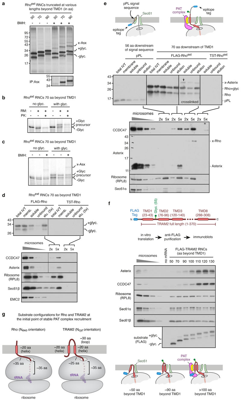
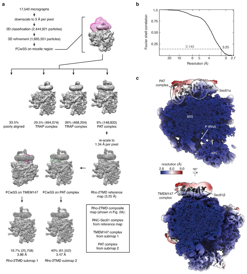
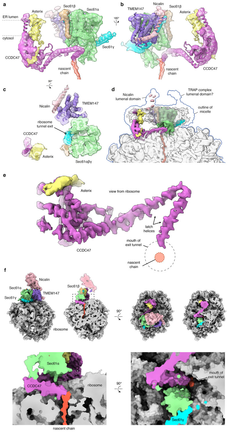
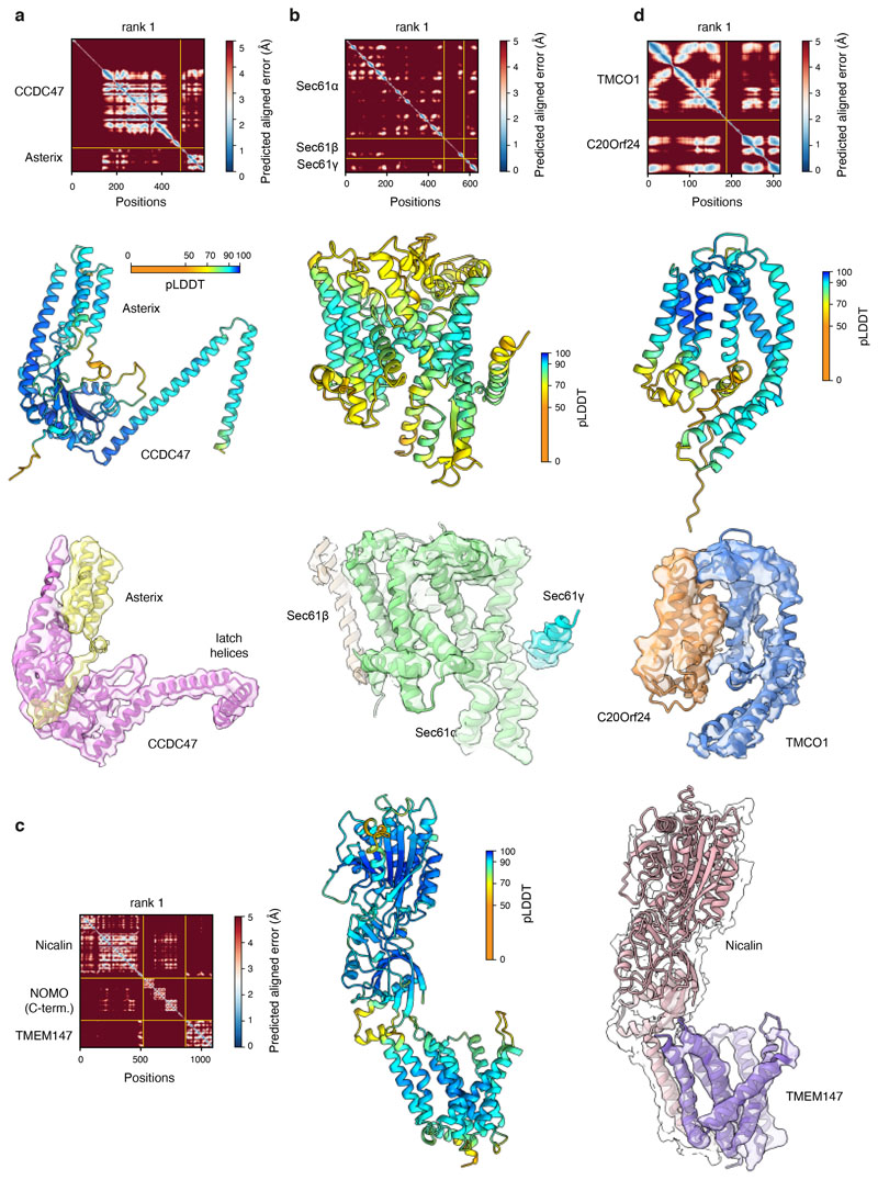
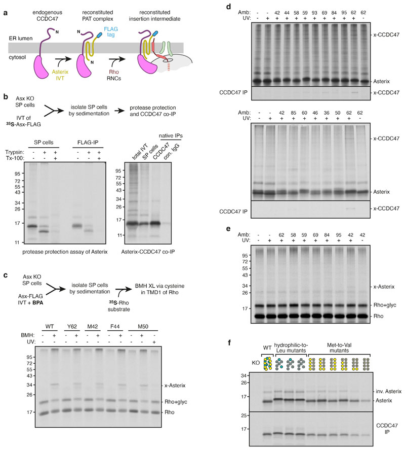
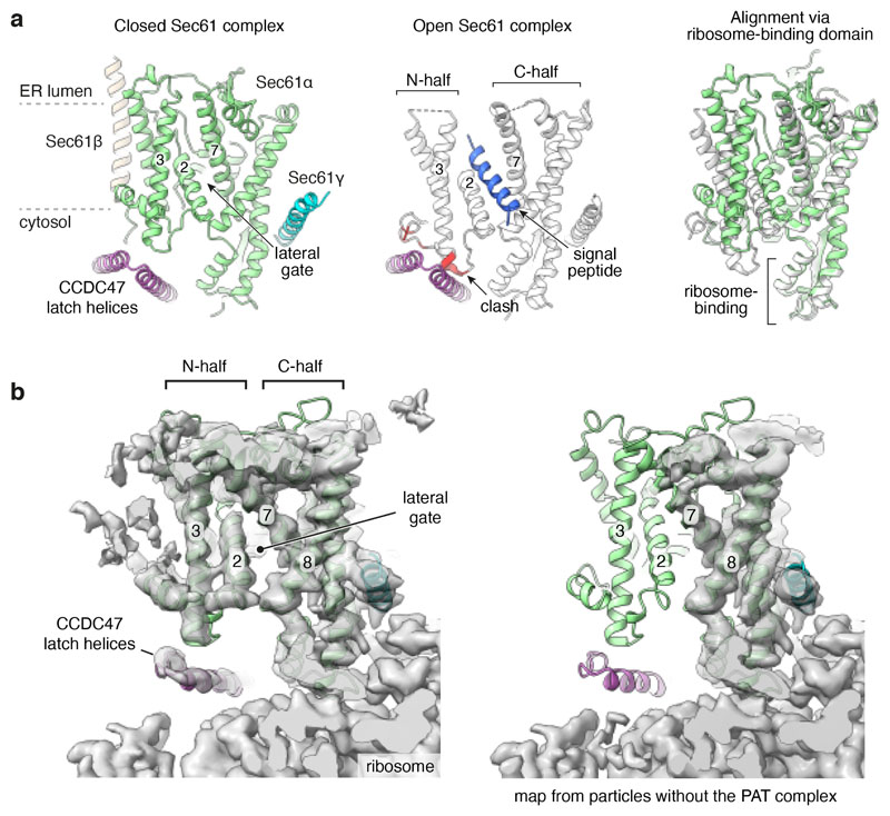
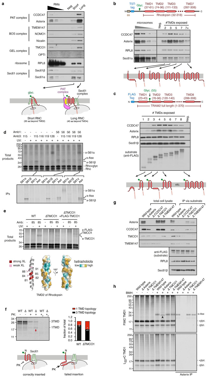
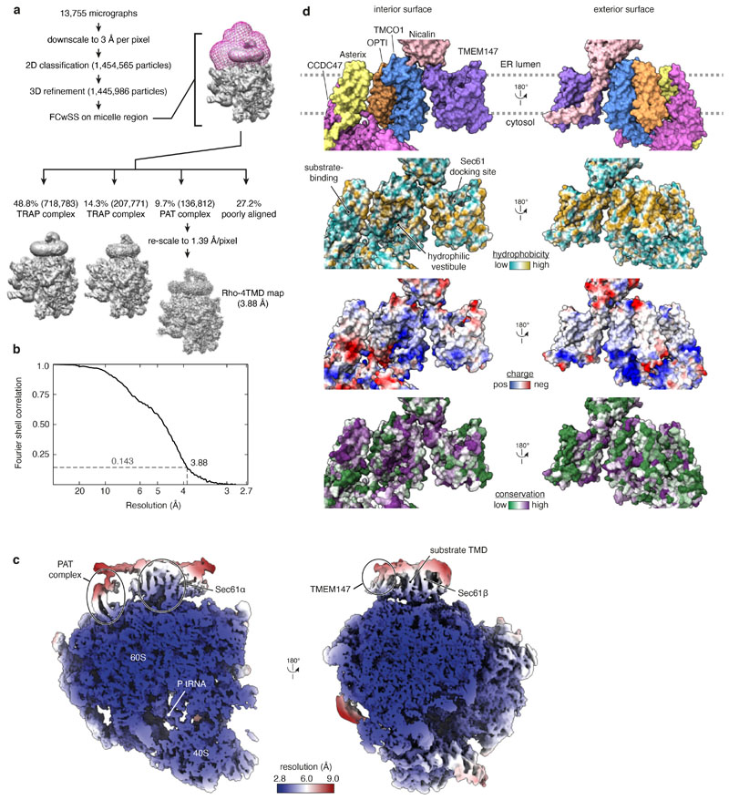
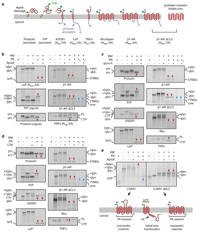
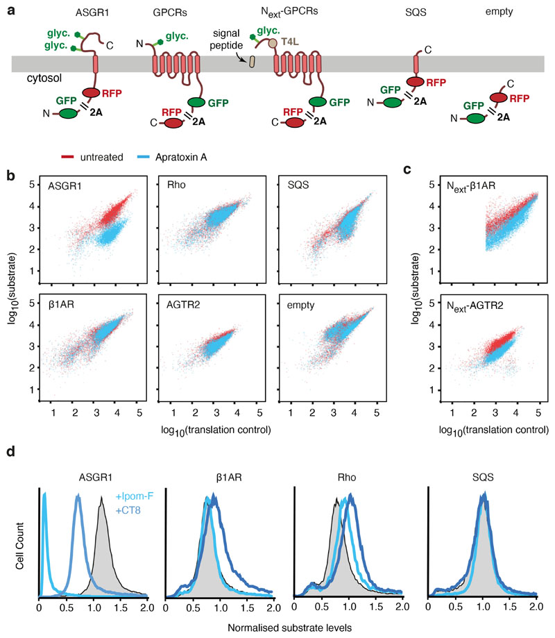
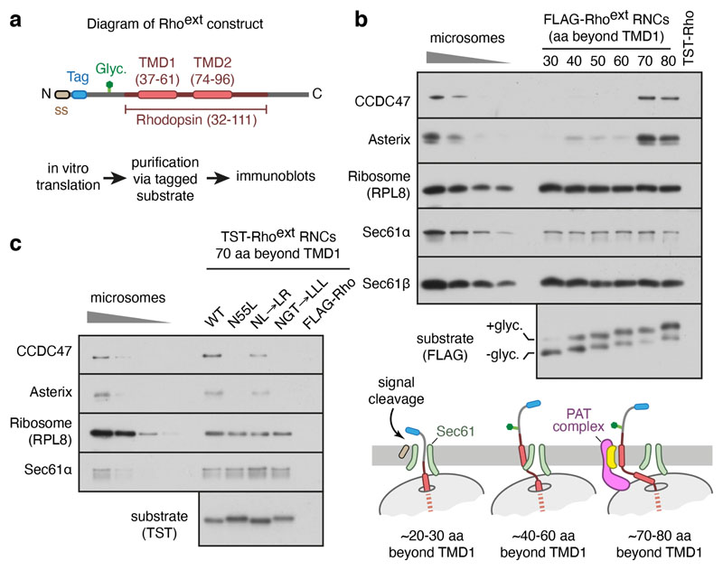
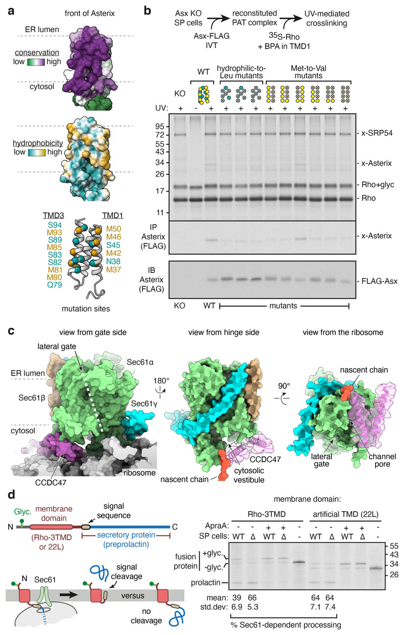
Multipass membrane proteins play numerous roles in biology and include receptors, transporters, ion channels and enzymes1,2. How multipass proteins are co-translationally inserted and folded at the endoplasmic reticulum is not well understood2. The prevailing model posits that each transmembrane domain (TMD) of a multipass protein successively passes into the lipid bilayer through a front-side lateral gate of the Sec61 protein translocation channel3-9. The PAT complex, an intramembrane chaperone comprising Asterix and CCDC47, engages early TMDs of multipass proteins to promote their biogenesis by an unknown mechanism10. Here, biochemical and structural analysis of intermediates during multipass protein biogenesis showed that the nascent chain is not engaged with Sec61, which is occluded and latched closed by CCDC47. Instead, Asterix binds to and redirects the substrate to a location behind Sec61, where the PAT complex contributes to a multipass translocon surrounding a semi-enclosed, lipid-filled cavity11. Detection of multiple TMDs in this cavity after their emergence from the ribosome suggests that multipass proteins insert and fold behind Sec61. Accordingly, biogenesis of several multipass proteins was unimpeded by inhibitors of the Sec61 lateral gate. These findings elucidate the mechanism of an intramembrane chaperone and suggest a new framework for multipass membrane protein biogenesis at the endoplasmic reticulum.


Conflict of interest statement

Competing interests: The authors declare that they have no competing interests.

Figures

Extended Data Fig. 1
Extended Data Fig. 1. Characterisation of the Rhoext construct.
a, 35S-methionine labelled Rhoext ribosome nascent chain complexes (RNCs) of indicated length were synthesised in rabbit reticulocyte lysate (RRL) in the presence of canine pancreas-derived rough microsomes (RMs). Where indicated, RNCs were chemically crosslinked using bismaleimidohexane (BMH). The positions of the glycosylated (+glyc.) and non-glycosylated (-glyc.) translation products are indicated. The crosslink to Asterix (indicated by x-Asx) was verified by immunoprecipitation under denaturing conditions using anti-Asterix antibody (bottom panel). The pattern of PAT complex recruitment for Rhoext construct is similar to the non-extended Rho construct construct described previously. b, 35S-methionine labelled Rhoext RNC of the indicated length, lacking or containing the glycosylation site, was synthesised in RRL containing or lacking RMs. The translation products were then digested with proteinase K (PK). The population of polypeptides inserted into the membrane is protected from PK. c, Translation products were produced as in panel b (lanes 1 and 4), after which the RMs were isolated by sedimentation (lanes 2 and 5) and subjected to crosslinking with BMH (lanes 3 and 6). The crosslink to Asterix is indicated. d, FLAG or twin-Strep tag (TST) containing membrane-inserted RNCs of Rhoext truncated 70 amino acids (aa) beyond TMD1 were subjected to fractionation and affinity purification. The total IVT products were centrifuged to obtain a membrane fraction, which was then solubilised under non-denaturing conditions (soluble fraction). The soluble fraction was then subjected to anti-FLAG affinity purification. Aliquots of the purification (with five-fold more loaded for the elution fraction) were analysed by anti-FLAG immunoblot (top panel). The bottom panel shows the elution fractions of the two purifications immunoblotted for CCDC47, Asterix, RPL8, Sec61b, and EMC2. Serial dilutions of ER microsomes were analysed in parallel. RNCs of Rhoext can be efficiently affinity-purified, recovering the associated PAT complex. Note that the left and right lanes of the CCDC47 and Asterix blots are from the same gel and taken from the same exposure, with the vertical line indicating the point where the lanes were spliced together. e, Membrane-inserted RNCs of bovine preprolactin (pPL) truncated 56 residues beyond the signal sequence and Rhoext truncated 70 residues beyond TMD1 were subjected to anti-FLAG affinity purification as in panel d. The TST-tagged Rhoext served as a specificity control. Where indicated, the sample was crosslinked with BMH just prior to solubilisation of RMs. The top panel shows the anti-FLAG immunoblot of the steps of affinity purification (as in panel d), and the bottom panel shows the elution fractions of each purification immunoblotted for CCDC47, Asterix, RPL8, and Sec61α. The blots indicate that the pPL translation intermediate does not associate with the PAT complex, while the PAT complex is recovered with Rhoext with comparable efficiency without or with crosslinking, which proved to be nearly quantitative as judged by the near absence of non-crosslinked Asterix. This indicates that association of the PAT complex with Rhoext RNCs is salt- and detergent-resistant under the purification conditions employed. The faint band in the final lane of the CCDC47 is IgG heavy chain contamination. f, The top panel shows a diagram depicting the construct encoding full-length N-terminal FLAG-tagged TRAM2, whose topology is opposite to that of Rho. The bottom panel shows an experiment analogous to that for Rhoext shown in Fig. 1b. The purified samples were analysed by immunoblot adjacent to serial dilutions of RMs. Equal translation levels and recovery of RNCs is reflected by both the substrate blot and by similar levels of Sec61 subunits and the ribosome. A reaction lacking mRNA served as a negative control for non-specific binding to the affinity resin. Note that as for Rhoext (Fig. 1b), recruitment of the PAT complex to TRAM2 translation intermediates is dependent on nascent chain length. Although a small amount of recruitment is detectable at lengths of 70-100 aa beyond TMD1, the initial point of stable maximal recruitment is observed at 110 aa beyond TMD1. g, Diagrams showing the substrate configurations, approximately to scale, for Rho and TRAM2 at the point of initial stable recruitment of the PAT complex. This corresponds to ~70 aa beyond TMD1 for Rho and ~110 aa beyond TMD1 for TRAM2. The difference in length can be accounted for by the different topology of TMD1. The translocon is not shown for simplicity.
Extended Data Fig. 2
Extended Data Fig. 2. Processing scheme and resolution information of the Rho-2TMD map.
a, Flowchart depicting the classification scheme and analysis of cryo-EM micrographs of the Rho-2TMD RNC sample. The classes denoted as “TRAP complex” appear to only contain the core translocon comprised of the Sec61 and TRAP complexes, evidently in somewhat different conformations, as characterised previously in cryo-EM and cryo-electron tomography work,–. These were not pursued further. Note that model building relied almost entirely on the Rho-2TMD reference map, with a few regions employing the two submaps for verification. The composite map shown in Fig. 2a is solely for illustration purposes as a means of simultaneously displaying the best regions contributed by each map. FCwSS indicates focused classification with signal subtraction. b, Fourier shell correlation (FSC) curve for the Rho-2TMD reference map illustrating an overall resolution of 3.25 Å by the gold-standard method. c, Two views of the Rho-2TMD reference map coloured by local resolution. Key structural elements are indicated.
Extended Data Fig. 3
Extended Data Fig. 3. Features of the Rho-2TMD map and model.
a, b, Two views from the plane of the membrane of the Rho-2TMD reference map, low-pass filtered to 7 Å resolution, fitted with the model. The approximate position of the membrane is indicated. The detergent micelle and ribosome are omitted for clarity. c, View from the ER lumen of the low-pass filtered Rho-2TMD reference map fitted with the model. The clipping plane is within the membrane close to the ER lumen. The detergent micelle and ribosome are omitted for clarity, with the position of the ribosome tunnel exit indicated. d, Overview of the low-pass filtered reference map in the context of the detergent micelle and ribosome. The map is shown at two contour levels. The blue line shows a contour level at which lumenal density and the complete micelle is visible. The opaque density is shown at a lower contour level to visualise the translocon, coloured as in panels a-c. The ribosome and micelle are in transparent grey. e, The PAT sub-classified map (submap 2 in Extended Data Fig. 2) filtered by local resolution viewed from the ribosome to illustrate that the regions closest to (and stabilised by) the ribosome are particularly well resolved. The position of the nascent chain density and the mouth of the ribosome exit tunnel are indicated. f, Space-filling depictions of the Rho-2TMD model illustrating how the C-terminal region of CCDC47 enters the mouth of the ribosome exit tunnel, abuts the nascent chain, and narrows the exit tunnel dimensions. The top shows overviews before and after clipping, with dashed boxes indicating the regions shown at higher magnification below.
Extended Data Fig. 4
Extended Data Fig. 4. AlphaFold2 predictions of the PAT, Sec61, GEL and BOS complexes.
All AlphaFold2 predictions were obtained using ColabFold. For all complexes, predicted alignment error (PAE) matrices, structural models coloured by the predicted Local Distance Difference Test (pLDDT) and the final models fitted into respective densities are shown. Note that the PAE matrix scale is from 0-5 Å, not the default 0-30 Å output, to emphasise the very high-confidence interactions. a, Human PAT complex, comprised of CCDC47 and Asterix. The final model is fitted into the respective density taken from the PAT sub-classified map (submap 2 in Extended Data Fig. 2) filtered by local resolution. b, Canine Sec61 complex. The final model is fitted into the respective density taken from the Rho-2TMD reference map filtered by local resolution. c, Human BOS complex, comprised of TMEM147, Nicalin, and NOMO. The final model (which omitted NOMO because it was not visualised in the map) is fitted into the respective density taken from the TMEM147 sub-classified map (submap 1 in Extended Data Fig. 2) filtered by local resolution. The density for Nicalin is also shown at a very low threshold in transparent white to visualise the lumenal domain density. d, Human GEL complex, comprised of TMCO1 and OPTI (C20orf24). The final model is fitted into the respective density taken from the Rho-4TMD map filtered by local resolution.
Extended Data Fig. 5
Extended Data Fig. 5. Characterisation of PAT complex reconstitution in SP cells.
a, Diagram illustrating the strategy for PAT complex reconstitution with Asterix variants followed by analysis of substrate interaction. Asterix KO cells contain residual levels of endogenous CCDC47. When Asterix is in vitro translated in RRL, supplemented with Asterix KO semi-permeabilised (SP) cells, it is inserted and interacts with CCDC47 to reconstitute the PAT complex. When substrate RNCs are subsequently introduced, their interaction with the PAT complex can be tested. b,35S-labelled Asterix was translated in the presence of Asterix KO SP cells. The cells were then isolated by centrifugation and analysed by protease protection to check Asterix topology (left) and interaction with CCDC47 by co-immunoprecipitation (IP; right). The cytosolic N-terminus of Asterix is accessible to protease (trypsin), and the remaining protected fragment is recoverable by immunoprecipitation via the C-terminal FLAG-tag. The right panel shows the total in vitro translated products (lane 1), the sedimented SP cells (lane 2), native IP with CCDC47 antibody (lane 3) or native IP using control antibody (lane 4). c, Wild-type and amber codon-containing Asterix variants (at the indicated positions) were translated in RRL (with non-radioactive methionine) containing Asterix KO SP cells and amber suppression reagents for site-specific incorporation of the photocrosslinking amino acid BPA. The cells were isolated and incubated with 35S-labelled Rho RNCs truncated at 70 residues beyond TMD1. The samples were then crosslinked using bismaleimidohexane (BMH) where indicated. The only cysteine of the substrate is located in the first TMD. Note that all Asterix variants form BMH-mediated crosslinks with the substrate, indicating successful reconstitution of the substrate-Asterix interaction. For comparison, the sample containing BPA at M50 of Asterix was UV-irradiated, illustrating that photo-crosslinks between Asterix and substrate can also be visualised (see also Fig. 2d and panel e). As expected, the efficiency of photo-crosslinking is lower than chemical crosslinking. d, Autoradiographs of photocrosslinking experiments between Asterix and CCDC47. 35S-labelled Asterix variants with BPA at the indicated positions were reconstituted into Asterix KO SP cells as in panel c. The cells were isolated, irradiated with UV light, and analysed directly or after denaturing IP using anti-CCDC47 antibody. e, Non-radioactive asterix variants with BPA at indicated positions were reconstituted into Asterix KO SP cells as in panel c. The reconstituted SP cells were then incubated at 32°C for 10 min with isolated 35S-labelled Rho RNCs truncated 70 residues beyond TMD1. The samples were irradiated with UV light and analysed by SDS-PAGE and autoradiography. The positions of non-glycosylated and glycosylated Rho, and the crosslink to Asterix (x-Asterix) are indicated. Only position 42 showed a strong crosslink among those tested in this experiment. f, Native CCDC47 IPs for the Asterix mutants analysed in Fig. 3b. 35S-labelled Asterix variants were translated in RRL supplemented with Asterix KO SP cells. The SP cells were isolated by centrifugation, solubilised under native conditions, and subjected to immunoprecipitation using anti-CCDC47 antibody. Aliquots of the isolated SP cells (top panel) and products of CCDC47 IP (bottom panel) are shown. Note that a small population of Asterix is evidently inserted in the inverted orientation (“inv. Asterix”) and becomes glycosylated. This population does not co-IP with CCDC47, providing an internal specificity control. The scheme for depicting the positions that are mutated is the same as shown in Fig. 3.
Extended Data Fig. 6
Extended Data Fig. 6. CCDC47 latch helices disfavour Sec61 opening.
a, The Sec61 complex in the Rho-2TMD model (left) is in a closed conformation with the latch helices of CCDC47 abutting the cytosolic loop between TMD2 and TMD3 of Sec61α. The open Sec61 complex (middle, PDB 3JC2) would clash with CCDC47 because the N-half of Sec61 would need to rotate away from the ribosome-bound C-half to accommodate a substrate at the lateral gate (such as a signal peptide in this structure). The right panel shows an alignment of the two structures by their ribosome-binding domains, illustrating how the N-half of Sec61 needs to rotate during lateral gate opening. b, Experimental cryo-EM density of reconstructions from the Rho-2TMD dataset from particles containing (left) or lacking (right) the PAT complex. At comparable contour levels where the ribosome and the C-half of Sec61 are essentially the same between the two maps, the N-half density is markedly better in the CCDC47-containing map. At more generous contour levels, the N-half can be seen in the CCDC47-lacking map, albeit at lower resolution due to presumed heterogeneity of positions. The differences in Sec61 density in particles without and with the PAT complex cannot be ascribed to refinement differences. The particle alignments during refinement are dominated by the strong signal provided by the ribosome, which does not differ between the two classes. No realignment was performed after focused classification. The fact that the density for the ribosome and many parts of Sec61 is very similar between the two maps provides post hoc validation that the refinement procedure was consistent. Differences in overall flexibility of the translocon-micelle complex relative to the ribosome can be excluded because significant density differences are seen only in the parts of Sec61 known to move during gating. By contrast, parts of Sec61 that are distal to the RBD but unmoved by gating remain unchanged. For these reasons, we ascribe the difference in Sec61 density between the PAT-containing and PAT-free maps to the former being stabilised in the closed state.
Extended Data Fig. 7
Extended Data Fig. 7. Analysis of the substrate-engaged multipass translocon.
a, RNCs of FLAG-TRAM2 truncated at the indicated lengths (see diagram) were synthesised in RRL supplemented with HEK293 SP cells. The RNCs were affinity-purified under native conditions via the N-terminal FLAG-tag (as in Extended Data Fig. 1f) and analysed by immunoblotting for the indicated proteins. The negative control (neg) is a translation reaction without mRNA. b, RNCs of Twin-Strep Tag (TST)-Rhoext (see diagram) truncated 40 residues downstream of TMD2, TMD3, TMD5 or TMD7 were analysed for PAT complex association as in Fig. 1b. The RNC of 7+ length represents a nascent chain truncated 70 residues beyond TMD7. c, RNCs of FLAG-TRAM2 (see diagram) truncated 40 residues downstream of indicated TMD were analysed for PAT complex association as in Extended Data Fig. 1f. d, Photo-crosslinking analysis of 35S-methionine-labelled Rho-4TMD translocation intermediate (as in Fig. 4d) via BPA installed at amber codons in TMD1 (Amb1), TMD3 (Amb3), or both. The top panel shows the autoradiograph of total RNCs after isolation by ultracentrifugation. Three of the key samples were also subjected to immunoprecipitation using the indicated antibodies and analysed in the bottom panel by autoradiography. Non-glycosylated and glycosylated substrate (Rho and Rho+glyc., respectively) and the adducts to Asterix (x-Asterix), Sec61β (x-S61β) and Sec61α (x-S61α) are indicated. e, Photo-crosslinking analysis of 35S-methionine-labelled Rho-4TMD translocation intermediate via BPA installed at TMD2 (position 85). Three types of semi-permeabilised cells were compared: wild type (WT), TMCO1 knockout (∆TMCO1) and ∆TMCO1 cells transiently transfected with FLAG-tagged TMCO1. The major crosslink seen with WT SP cells is lost in ∆TMCO1 SP cells, where other weak crosslinks to similar-sized unidentified proteins are seen. When a subset of the ∆TMCO1 cells now express FLAG-TMCO1, a new crosslink is seen that migrates slightly slower than the major product seen in WT cells. These results verify that the major crosslink in WT cells is TMCO1 and the slightly larger crosslink in the reconstituted ∆TMCO1 cells is FLAG-TMCO1. Crosslinking with BPA at different positions in TMD2 show that its more hydrophilic face interacts with TMCO1 (summarised in the diagram). f, Protease protection assay of Rho-4TMD RNC in WT or ∆TMCO1 SP cells. The autoradiograph shows equal glycosylation indicative of equal efficiencies of TMD1 insertion, but different amounts of fully-protected product indicative of successful 3-TMD insertion. In ∆TMCO1 cells, a larger population of proteins failed to insert TMDs 2 and 3, leading to protease-accessibility (see diagram). PK digestion in the presence of detergent (subscripted d) leads to complete digestion. Graph shows quantification of three independent experiments showing the mean and standard deviation. g, RNCs of Rhoext stalled 70 residues beyond TMD1 were assembled in WT, ∆Asterix, ∆CCDC47, ∆TMCO1 and ∆TMEM147 SP cells and affinity-purified via the FLAG-tag under native conditions as in Fig. 1b. Total cell lysates and purified RNCs were analysed by immunoblotting for the indicated proteins. h, Bismaleimidohexane (BMH) mediated crosslinking via cysteine in place of F56 in TMD1 of Rhodopsin stalled 70 residues downstream of TMD1 (top panel), or the equivalent RNC in which TMD1 is replaced with 22 leucine residues and a similarly positioned cysteine residue (bottom panel). 35S-methionine-labelled RNCs were assembled with WT, ∆Asterix, ∆TMCO1, and ∆TMEM147 SP cells, subjected to BMH crosslinking where indicated, and either directly analysed by SDS-PAGE and autoradiography or after denaturing anti-Asterix IP. Equal glycosylation efficiency in all samples without BMH crosslinking indicate TMD1 insertion is unaffected by the loss of MPT components. Asterix crosslinking (marked as x-Asx) is unimpaired in ∆TMCO1 and ∆TMEM147 SP cells. In ∆Asterix cells, TMD1 of Rho crosslinks to an unidentified product that migrates slightly faster than Asterix (upward arrowhead). The 22L TMD does not crosslink to Asterix or the unidentified product, suggesting these interactions need partial TMD hydrophilicity.
Extended Data Fig. 8
Extended Data Fig. 8. Features of the Rho-4TMD map and model.
a, Flowchart depicting the classification scheme and analysis of cryo-EM micrographs of the Rho-4TMD RNC sample. The classes denoted as “TRAP complex” appear to only contain the core translocon comprised of the Sec61 and TRAP complexes, evidently in somewhat different conformations, as characterised previously in cryo-EM and cryo-electron tomography work,–. These were not pursued further. b, Fourier shell correlation (FSC) curve for the Rho-4TMD map illustrating an overall resolution of 3.88 Å by the gold-standard method. c, Two views of the Rho-4TMD reference map coloured by local resolution. Key structural elements, including the density assigned to TMD3 of the substrate, are indicated. d, Space-filling models showing the multipass translocon’s highly conserved interior surface that faces the lipid-filled cavity (left) and poorly conserved exterior surface that faces the surrounding membrane (right). The models are coloured successively (from top to bottom) by protein, hydrophobicity, charge and conservation (calculated by ConSurf). The conserved amphiphilic substrate-binding domain of the PAT complex, the conserved positively-charged hydrophilic vestibule of the GEL complex, and the conserved Sec61-docking site on the BOS complex are each indicated.
Extended Data Fig. 9
Extended Data Fig. 9. Effect of Sec61 inhibitors on various substrates in vitro.
a, Diagram of substrates tested for their sensitivity to Sec61 inhibitors. Cleavable signal sequences (tan), glycosylation sites (green), and experimentally determined sites of protease accessibility (lavender) are indicated. The core of β1AR∆CL3 that is resistant to protease digestion is also indicated. PrP is prion protein; LeP is leader peptidase; ASGR1 is asialoglycoprotein receptor 1; TNFα is tumor necrosis factor alpha. b-d, 35S-methionine containing RRL translation reactions in the absence or presence of RMs and 2 μM of the indicated Sec61 inhibitor were analysed by protease protection. Panel b employed Apratoxin A (ApraA, as in Fig. 4e), panel c employed Ipomeassin F (Ipom-F) and panel d used cotransin-8 (CT8). An equivalent volume of DMSO was used as a negative control. The translation products were either left untreated or digested with proteinase K (PK) without or with detergent (subscripted d) as indicated. The samples were analysed directly by SDS-PAGE and autoradiography or immunoblotting via a C-terminal FLAG tag (in the case of TNFa, which lacks methionines in the protease-protected fragment). Red arrows represent the fragments protected from PK digestion that are indicative of successful translocation. The protease-resistant folded core of β1AR∆CL3 is marked by blue arrows. NTF and CTF stand for N-terminal fragment and C-terminal fragment, respectively. Asterisk in the TNFa blot marks a background band that is sometimes detected by the FLAG antibody. e, Protease-protection analysis of C3AR1 (a GPCR) or a variant in which the ~180 amino acid long second extracellular loop (EL2) was shortened by deleting 61 residues (from T166 to D326). For simplicity, the majority of the C-terminal cytosolic domain was omitted from the constructs. Translation reactions of C3AR1 (left) and C3AR1∆EL2 (right) were performed in the presence or absence of 2 μM ApraA and analyzed by protease protection. At the protease concentration used, all of the short cytosolic loops are resistant to digestion if the protein is inserted correctly, but become accessible if any insertion step fails (see diagram). Successfully inserted and PK-resistant products are indicated with red downward arrows. Failed insertion of TMD4 in the presence of ApraA has two consequences: the downstream loop is not glycosylated and the protein is protease-accessible downstream of TMD3. Thus, the doubly-glycosylated product is not observed and a protected N-terminal fragment (NTF, marked with dark blue upward arrow) representing the first three TMDs is generated upon PK digestion. Note that by these criteria, some failed insertion is seen even without ApraA. C3AR1∆EL2 is unaffected by ApraA and the majority of it is correctly inserted. The asterisk marks the position of the C3AR1∆EL2 fragment generated by PK digestion of the noninserted (and hence, non-glycosylated) population.
Extended Data Fig. 10
Extended Data Fig. 10. Effect of Sec61 inhibitors on various substrates in cells.
a, Diagram of fluorescent protein (FP)-tagged substrates tested for their sensitivity to Sec61 inhibitors in cells. 2A is the viral sequence at which peptide bond formation fails without disrupting elongation. Thus, each translation cycle generates two products: an FP-tagged substrate and a different coloured FP that serves as an internal control for translation levels. For RFP-tagged ASGR1 and SQS, GFP, which precedes the viral 2A sequence, serves as the translation control, while RFP fluorescence levels report on the substrate. The other substrates are GFP-tagged, with RFP serving as the translation control. b, Scatter plots of the indicated reporters, each expressed from an inducible promoter in stable cell lines. Cells treated with 200 nM Apratoxin A (ApraA) during reporter induction are shown in blue, while cells treated with vehicle are shown in red. The histograms corresponding to some of these constructs are shown in Fig. 4f. c, Scatter plots of N-terminally extended (Next) β1AR and AGTR2 (see diagram in panel a) from an experiment similar to that shown in panel b. Next-AGTR2 is expressed stably whereas Next-b1-AR was expressed for 24 h by transient transfection with ApraA included during the final 12 hours. d, Histograms of normalised substrate levels after 6 hours of expression in the presence of vehicle (grey), 200 nM Ipomoeassin F (Ipom-F, light blue) or 1 μM CT8 (dark blue).
Fig. 1
Fig. 1. Substrate-mediated recruitment of the PAT complex to Sec61.
a, Diagram of the Rhoext construct (not to scale) containing the first two TMDs and flanking regions of bovine rhodopsin (amino acids 32-111, Uniprot ID P02699). The Rho region is preceded by a signal sequence (ss) and a polypeptide of 52 amino acids containing a site for N-linked glycosylation (Glyc.) to monitor translocation. The experimental strategy to analyse intermediates of Rhoext is indicated. b, Ribosome nascent chain complexes (RNCs) of FLAG-tagged Rhoext truncated at the indicated lengths (in amino acids, aa) beyond TMD1 were assembled by in vitro translation in rabbit reticulocyte lysate (RRL) containing canine pancreas rough microsomes (RMs). The RNCs were affinity-purified via the FLAG tag and analysed by immunoblotting relative to serial two-fold dilutions of RMs. Rhoext tagged with the twin-Strep tag (TST) and truncated 70 amino acids beyond TMD1 served as a specificity control for anti-FLAG affinity purification. The diagram below (not to scale) depicts key stages of biogenesis as deduced from the results. c, Affinity purification of TST-tagged Rhoext RNCs similar to panel B for constructs in which TMD1 contains mutations: NL→LR stands for N55L and L47R mutations; NGT→LLL changes N55, G51, T58 to leucines. The FLAG-tagged wt Rhoext served as a specificity control for the purifications via the TST. The substrates in this experiment do not contain a glycosylation site. Source data for all gels in this and other figures can be found in Supplementary Fig. 1.
Fig. 2
Fig. 2. Structure of the PAT complex recruited to Sec61.
a, Composite map (top) and model (bottom) of the Rho-2TMD translocation intermediate stalled 70 amino acids beyond TMD1 (see Fig. 1b). The density for the PAT and BOS complexes derives from submaps obtained after focussed classification on each complex (see Extended Data Fig. 2). The PAT complex is shown at two contour levels to visualise the lower-resolution membrane domain simultaneously with the higher resolution ribosome-proximal regions. The GEL and TRAP complexes, visible only at very low contour levels, were not modelled. The detergent micelle has been removed for clarity. b, View from the ER lumen of the Rho-2TMD translocation intermediate model. c, Site-specific photo-crosslinking from benzoyl-phenylalanine (BPA) installed in Asterix by amber (Amb) suppression at residue 42 or 62. Semi-permeabilised (SP) Asterix knockout (KO) cells were reconstituted with 35S-methionine labelled Asterix-FLAG variants (see Extended Data Fig. 5d), irradiated with UV, and analysed directly (total products) or after immunoprecipitation (IP). The red arrow indicates the Asterix-CCDC47 adduct. d, Analysis of substrate-Asterix photo-crosslinking from BPA installed at amber codons (Amb) at the indicated positions in Asterix. Asterix KO cells are reconstituted with non-radioactive Asterix-FLAG variants as in panel c, then incubated with 35S-methionine labelled RNCs of Rho-2TMD. After UV irradiation, the samples were analysed directly or after IP. Non-glycosylated and glycosylated substrate (Rho and Rho+glyc., respectively), and the adduct to Asterix (x-Asterix) are indicated. e, Summary of all site-specific photo-crosslinking results (see Fig. 2d and Extended Data Fig. 5d-e) mapped onto the PAT complex structure shown in three different views.
Fig. 3
Fig. 3. The PAT complex binds the substrate and redirects it to the back of Sec61.
a, The substrate-binding surface of Asterix showing conservation (top), hydrophobicity (middle) and positions subject to mutagenesis (bottom). b, Photo-crosslinking from a BPA probe in place of F56 in TMD1 of the Rho-2TMD substrate. Semi-permeabilised (SP) Asterix KO cells were reconstituted with either wild-type or mutant Asterix-FLAG variants and tested for photo-crosslinking with 35S-methionine labelled Rho-2TMD RNCs similar to Fig. 2d. The adducts to Asterix (x-Asterix) and SRP54 (x-SRP54, from the non-translocated population of RNCs) are indicated. The bottom panel shows an immunoblot for Asterix to verify comparable levels of reconstitution for each mutant. The diagram above the gel indicates which positions were mutated (indicated by an ‘X’), using the positional scheme in the bottom diagram of panel a. Hydrophilic (teal) and methionine (yellow) residues were mutated to leucine and valine, respectively. c, Three views of the Rho-2TMD structure showing that CCDC47’s latch helices are wedged between the ribosome and a closed Sec61. d, Experimental strategy (left) to measure hydrophobic domain access to the Sec61 lateral gate downstream of a membrane domain. Either the first three TMDs of Rho (Rho-3TMD) or an artificial poly-leucine TMD (22L), is fused ahead of preprolactin. If the preprolactin signal sequence successfully engages Sec61, the glycosylated fusion protein is cleaved; otherwise, the glycosylated fusion protein remains intact. 35S-methionine labelled fusion proteins were translated with wild type (WT) or CCDC47 KO (∆) semi-permeabilised cells in the absence or presence of 2 μM Apratoxin A (ApraA, a Sec61 inhibitor). The pelleted cells were analysed by SDS-PAGE and autoradiography. Glycosylated (+glyc) and non-glycosylated (-glyc) fusion protein and the cleaved prolactin domain are marked. Quantification from three experiments (mean ± standard deviation) is shown below the gel.
Fig. 4
Fig. 4. Insertion of multipass proteins independent of Sec61 activity.
a, Tandem-mass-tag (TMT) mass spectrometry of proteins copurified with early and late Rhoext insertion intermediates (see diagram). The top four proteins differentially recovered with the late intermediate are indicated. The raw data are in Supplementary Table 1. b, Samples prepared as in panel a and a negative control lacking substrate were analysed by immunoblotting for the indicated proteins. c, Cryo-EM map (top) and map overlaid with model (bottom) of the Rho-4TMD translocation intermediate (stalled 40 amino acids beyond TMD3) containing the PAT, BOS, GEL and Sec61 complexes. The PAT complex is shown at two contour levels. The detergent micelle was removed for clarity. The position of the substrate is based on crosslinking data showing that TMD1, TMD2, and TMD3 are adjacent to Asterix, TMCO1 and the closed Sec61 complex, respectively. d, Photo-crosslinking of 35S-methionine-labelled Rho-4TMD translocation intermediates via BPA at the indicated positions of each TMD. Non-glycosylated and glycosylated substrate (Rho and Rho+glyc., respectively) and the adducts to Asterix (x-Asterix), TMCO1 (x-TMCO1), Sec61β (x-S61β) and Sec61α (x-S61α), verified in Extended Data Fig. 7d, and 7e, are indicated. e, In vitro translation reactions of ASGR1 and Rho in the absence or presence of RMs and 2 μM Apratoxin A were analysed by protease protection. The translation products were digested with proteinase K (PK) without or with detergent (subscripted d) where indicated. Red arrows represent PK-protected fragments indicative of successful translocation. f, Cell lines containing the indicated inducible fluorescent protein reporters (see Extended Data Fig. 10) were treated with control or Sec61A1-targeting siRNAs. Reporter expression was induced in the presence or absence of 200 nM Apratoxin A and the cells were analysed by flow cytometry. Histograms of normalised reporter expression are shown. SQS is a tail-anchored protein inserted by EMC.

Comment in

References

    1. von Heijne G. The membrane protein universe: what’s out there and why bother? J Intern Med. 2007;261:543–57. - PubMed
    1. Hegde RS, Keenan RJ. The mechanisms of integral membrane protein biogenesis. Nature Reviews Molecular Cell Biology. 2022;23:107–124. - PMC - PubMed
    1. Blobel G. Intracellular protein topogenesis. Proc Natl Acad Sci U S A. 1980;77:1496–500. - PMC - PubMed
    1. Heinrich SU, Mothes W, Brunner J, Rapoport TA. The Sec61p complex mediates the integration of a membrane protein by allowing lipid partitioning of the transmembrane domain. Cell. 2000;102:233–44. - PubMed
    1. Görlich D, Rapoport TA. Protein translocation into proteoliposomes reconstituted from purified components of the endoplasmic reticulum membrane. Cell. 1993;75:615–30. - PubMed

Publication types

MeSH terms