Review
. 2023 Jan;18(1):58-80.
doi: 10.1038/s41596-022-00753-z. Epub 2022 Oct 19.

Efficient and safe single-cell cloning of human pluripotent stem cells using the CEPT cocktail

Affiliations
Review

Efficient and safe single-cell cloning of human pluripotent stem cells using the CEPT cocktail

Carlos A Tristan et al. Nat Protoc. 2023 Jan.

Abstract

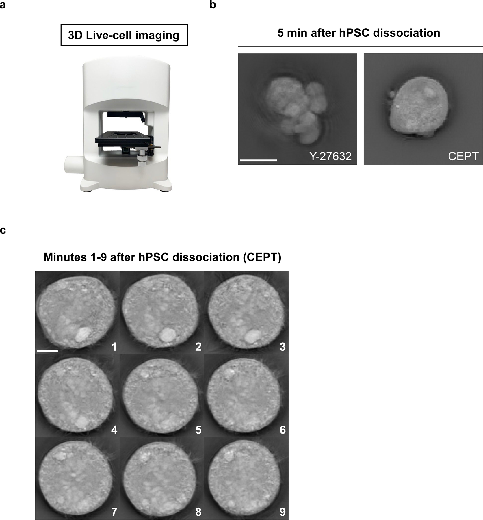
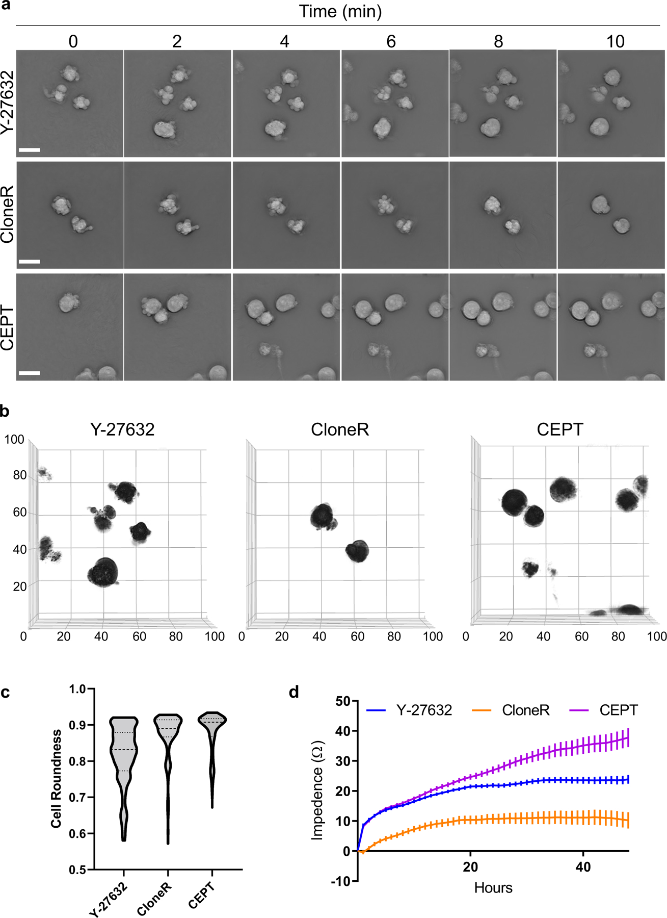
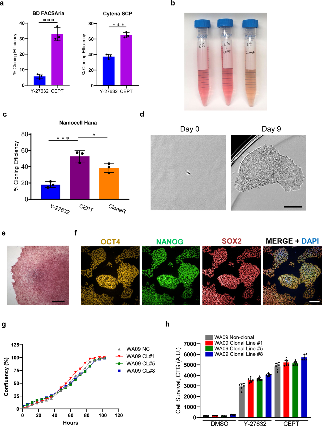
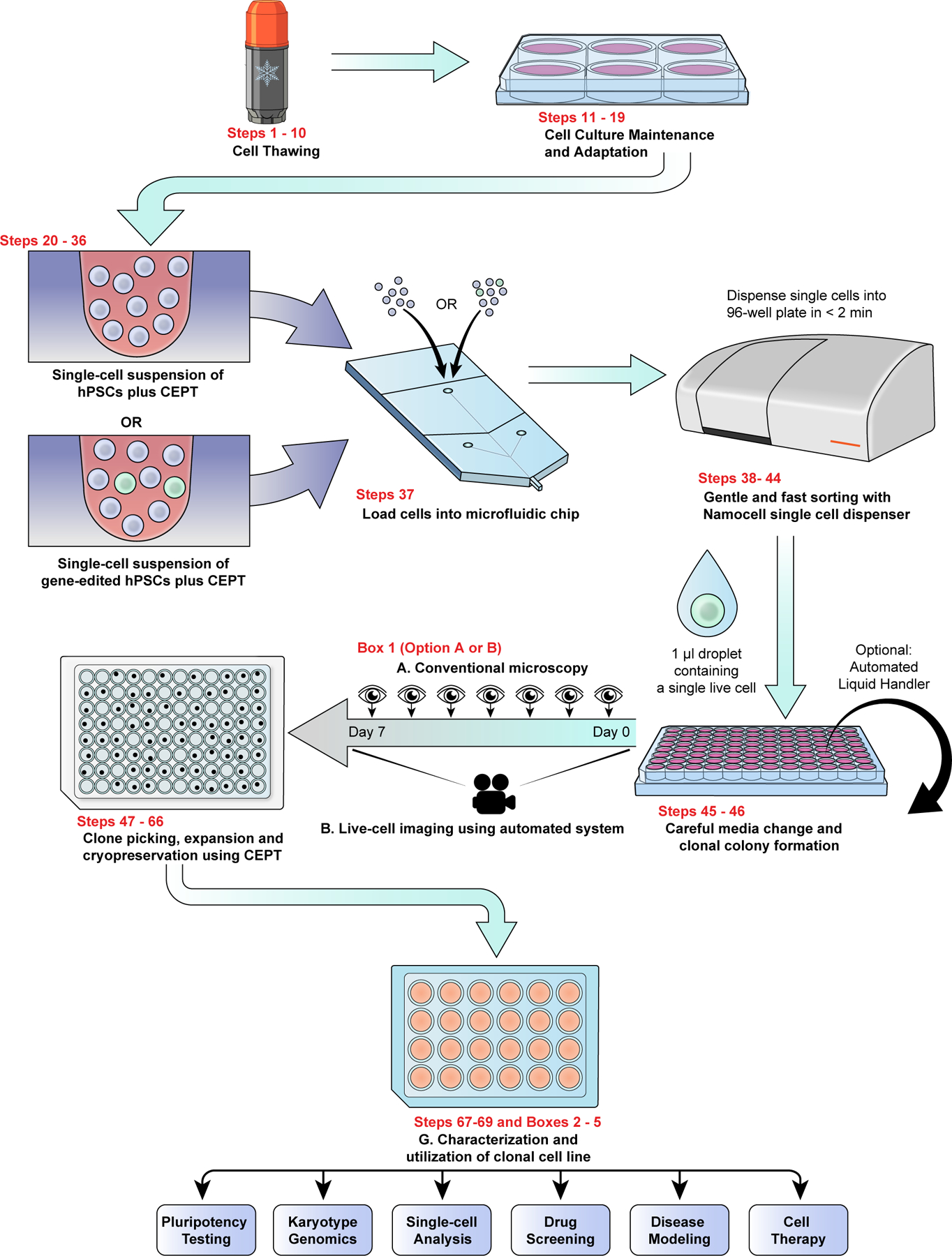
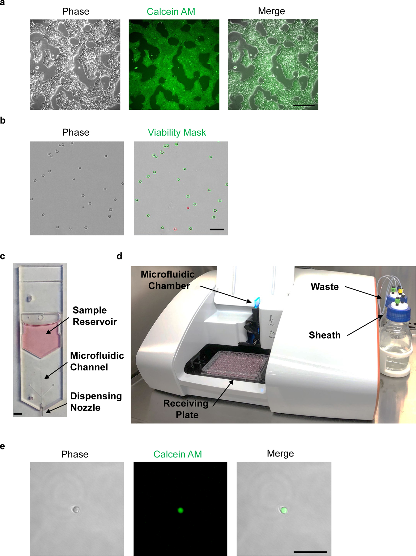
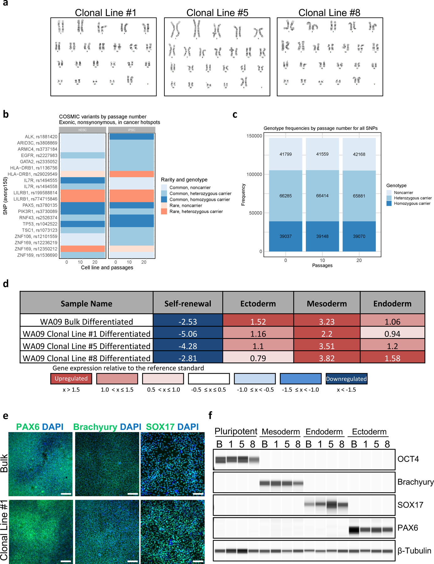
Human pluripotent stem cells (hPSCs) are inherently sensitive cells. Single-cell dissociation and the establishment of clonal cell lines have been long-standing challenges. This inefficiency of cell cloning represents a major obstacle for the standardization and streamlining of gene editing in induced pluripotent stem cells for basic and translational research. Here we describe a chemically defined protocol for robust single-cell cloning using microfluidics-based cell sorting in combination with the CEPT small-molecule cocktail. This advanced strategy promotes the viability and cell fitness of self-renewing stem cells. The use of low-pressure microfluidic cell dispensing ensures gentle and rapid dispensing of single cells into 96- and 384-well plates, while the fast-acting CEPT cocktail minimizes cellular stress and maintains cell structure and function immediately after cell dissociation. The protocol also facilitates clone picking and produces genetically stable clonal cell lines from hPSCs in a safe and cost-efficient fashion. Depending on the proliferation rate of the clone derived from a single cell, this protocol can be completed in 7-14 d and requires experience with aseptic cell culture techniques. Altogether, the relative ease, scalability and robustness of this workflow should boost gene editing in hPSCs and leverage a wide range of applications, including cell line development (e.g., reporter and isogenic cell lines), disease modeling and applications in regenerative medicine.


Conflict of interest statement

Ethics declarations

Competing interests

I.S., Y.C., and A.S. are co-inventors on a U.S. Department of Health and Human Services patent application covering the CEPT cocktail and its use.

Figures

Fig. 1:
Fig. 1:. Live-cell imaging of acutely dissociated hPSCs
a, Nanolive (3D Cell Explorer) microscope enabling label-free cell imaging. b, Fast-acting CEPT is cytoprotective and maintains cell morphology of WA09 dissociated cells, whereas ongoing cell contractions lead to abnormal cells in the presence of Y-27632. Representative image was taken 5 minutes after cell dissociation. c, Monitoring the morphology of dissociated hPSCs and treatment with CEPT. Images were taken every minute for the first 9 minutes after cell dissociation and plating into the well. Scale bars, 10 µm (b) and 5 µm (c).
Fig. 2:
Fig. 2:. Cell morphology and cell adhesion profile of dissociated hPSCs after treatment with Y-27632, CloneR, or CEPT.
a, Label-free live-cell imaging (Nanolive) showing the first 10 min after plating dissociated cells (WA09). Abnormal phenotypes are caused by cell contractions and membrane blebbing in the presence of Y-27632 (10 µM) and CloneR (1X), whereas treatment with fast-acting CEPT mitigates cell stress and ensures circularity of cell bodies. b, Analysis of membrane blebbing and cell roundness of single cells at 10 min after cell dissociation and treatment with Y-27632 (10 µM), CloneR (1X), or CEPT. Images represent 3D renditions of 60–70 z-planes stacked together. A customized fully automated digital image analysis algorithm segmented each cell as an individual object and measured the shape. Cell roundness is quantified per cell as deviation from perfect mathematical roundness. Six fields of view were analyzed for each condition. Note that CloneR has less cells (black circles) in the field of view because the addition of CloneR (1:10 dilution as recommended by the manufacturer) changes the density of the medium and the cells do not settle to the bottom of the plate as fast as they do with Y-27632 and CEPT. c, Violin plots of live-cell images (3D Cell Explorer) showing distribution of cell roundness and membrane blebbing at 10 min post-treatment with Y-27632, CloneR, or CEPT. Data are from n ≥ 230 for each condition. d, Cell adhesion time-course measured by impedance analysis of cells passaged with Y-27632 (10 µM), CloneR (1X), or CEPT. Data are mean ± s.e.m.; n = 16 wells for each group. Scale bars, 20 µm (a and b); x and y-axes in µm.
Fig. 3:
Fig. 3:. CEPT improves single-cell cloning efficiency and maintains pluripotency
a, Single-cell cloning efficiency in the presence of Y-27632 (10 µM) and CEPT as tested on two different platforms including single-cell printing with Cytena (Molecular Devices), and cell sorting with FACSAria (Becton Dickinson). Figure adapted with permission from ref. 21. b, E8 Medium after supplementation with CEPT or CloneR. Note that addition of CloneR (1:10 dilution according to manufacturer) changes the color of cell culture medium. c, Quantification of single-cell cloning efficiency (WA09) using microfluidics-based cell sorting (Namocell Hana) after treatment with Y-27632 (10 µM), CloneR (1X), or CEPT. d, Phase-contrast images showing a single cell (WA09) that survived dissociation and generated a clonal colony in 9 days. e, Representative image of a clonal colony (WA09) expressing pluripotency-associated marker alkaline phosphatase. f, Representative immunocytochemical images showing that clonal cell lines maintain pluripotency and express OCT4, NANOG and SOX2 (clonal cell line #1 from WA09). g, Image-based analysis comparing cell growth rate of non-clonal parental line (NC) and derived clonal cell lines 1, 5, and 8 (CL #1, 5 and 8). h, Luminescent cell viability assay showing that the sensitivity to enzymatic cell dissociation is comparable in non-clonal parental line and derived clonal cell lines 1, 5, and 8. Scale bars, 300 µm (d), 400 µm (e), and 100 µm (f). Data are mean ± s.d.; n = 3 96-well plates for each group (a, c), *P ≤0.05, **P ≤0.01, ***P ≤0.001, two-way ANOVA and Student’s t-test, respectively. Data are mean ± s.d.; n ≥ 3 (g, h).
Fig. 4:
Fig. 4:. Scalable single-cell cloning workflow for hPSCs.
Combined use of the CEPT cocktail and microfluidic cell-dispensing ensures that single-cell cloning is safe, efficient, and practical. Overview of workflow depicting the various steps to perform streamlined single-cell cloning in high-throughput fashion followed by recommended cell characterization and indication of potential downstream applications.
Fig. 5.
Fig. 5.. Live-cell staining and microfluidic single-cell dispensing of hPSCs.
a, Phase-contrast image of hPSCs before Calcein AM staining (left panel). Fluorescence microscopic analysis of hPSCs at 1 h post-staining with Calcein AM (center and right panels). b, Cell suspension of dissociated hPSCs should avoid clumps and yield low frequency of doublets (left panel). Trypan blue exclusion test can be used to measure cell viability; live and dead cells are shown with green or red outline, respectively (right panel). c, Disposable microfluidic cell cartridge depicting sample reservoir, microfluidic channel, and dispensing nozzle. d, Hana (Namocell) single-cell dispenser with microfluidic chamber, receiving plate, waste and sheath bottles. e, Fluorescence microscopic image of a single calcein-labeled hPSC after microfluidic dispensing in a 1 μl droplet. Scale bars, 400 µm (a), 200 µm (b and e) and 5 mm (c).
Fig. 6:
Fig. 6:. Karyotype analysis, whole-exome sequencing, and multi-lineage differentiation of clonal cell lines.
a, Three representative examples of clonal cell lines (WA09) that maintained normal karyotypes after 32 passages. Single-cell cloning, clone-picking, cell line establishment, cell expansion, and routine passaging were performed by using the CEPT cocktail. b, WES variant, genotype, and CNV analysis show genetic stability of WA09 and LiPSC-GR1.1 passaged with CEPT for 20 passages. COSMIC variants by passage number in cancer hotspots show no change in rarity or genotype over passaging. c, Genotype frequencies in WA09 cells by passage number for all SNPs are proportionally constant. Figures b and c were adapted with permission from ref. 21. d, EB differentiation potential of non-clonal parental line (WA09) and derived clonal cell lines measured by using Scorecard analysis. e, f, Immunocytochemistry and immunoblot analysis of non-clonal parental (N) and derived clonal cell lines (1, 5, and 8) after directed differentiation into ectoderm, mesoderm, and endoderm. Scale bar, 200 µm.

References

    1. Thomson JA et al. Embryonic stem cell lines derived from human blastocysts. Science 282, 1145–1147 (1998). - PubMed
    1. Takahashi K et al. Induction of Pluripotent Stem Cells from Adult Human Fibroblasts by Defined Factors. Cell 131, 861–872 (2007). - PubMed
    1. Shi Y, Inoue H, Wu JC & Yamanaka S Induced pluripotent stem cell technology: A decade of progress. Nat. Rev. Drug Discov. 16, 115–130 (2017). - PMC - PubMed
    1. Sharma A, Sances S, Workman MJ & Svendsen CN Multi-lineage Human iPSC-Derived Platforms for Disease Modeling and Drug Discovery. Cell Stem Cell 26, 309–329 (2020). - PMC - PubMed
    1. Evans MJ & Kaufman MH Establishment in culture of pluripotential cells from mouse embryos. Nature 292, 154–156 (1981). - PubMed

Key references using this protocol:

    1. Chen Y, Tristan CA, et al. A versatile polypharmacology platform promotes cytoprotection and viability of human pluripotent and differentiated cells. Nat Methods 18, 528–541 (2021). 10.1038/s41592-021-01126-2 - DOI - PMC - PubMed
    1. Tristan CA et al. Robotic high-throughput biomanufacturing and functional differentiation of human pluripotent stem cells Stem Cell Reports. 16, 1–17 (2021). 10.1016/j.stemcr.2021.11.004 - DOI - PMC - PubMed

Publication types