Review
. 2023 Jan;18(1):188-207.
doi: 10.1038/s41596-022-00752-0. Epub 2022 Oct 19.

Optimized single-nucleus transcriptional profiling by combinatorial indexing

Affiliations
Review

Optimized single-nucleus transcriptional profiling by combinatorial indexing

Beth K Martin et al. Nat Protoc. 2023 Jan.

Abstract

Single-cell combinatorial indexing RNA sequencing (sci-RNA-seq) is a powerful method for recovering gene expression data from an exponentially scalable number of individual cells or nuclei. However, sci-RNA-seq is a complex protocol that has historically exhibited variable performance on different tissues, as well as lower sensitivity than alternative methods. Here, we report a simplified, optimized version of the sci-RNA-seq protocol with three rounds of split-pool indexing that is faster, more robust and more sensitive and has a higher yield than the original protocol, with reagent costs on the order of 1 cent per cell or less. The total hands-on time from nuclei isolation to final library preparation takes 2-3 d, depending on the number of samples sharing the experiment. The improvements also allow RNA profiling from tissues rich in RNases like older mouse embryos or adult tissues that were problematic for the original method. We showcase the optimized protocol via whole-organism analysis of an E16.5 mouse embryo, profiling ~380,000 nuclei in a single experiment. Finally, we introduce a 'Tiny-Sci' protocol for experiments in which input material is very limited.


Conflict of interest statement

Ethics declarations

Competing interests

J.S. is a scientific advisory board member, consultant and/or cofounder of Cajal Neuroscience, Guardant Health, Maze Therapeutics, Camp4 Therapeutics, Phase Genomics, Adaptive Biotechnologies and Scale Biosciences. C.T. is a founder of Scale Biosciences. All other authors have no competing interests.

Figures

Figure 1.
Figure 1.. Summary of optimized sci-RNA-seq3 method.
Colors in plate wells represent 96 unique index sequences for each of the 3 rounds of indexing. RNA transcript in red, DNA in dark blue. a, Nuclei (in green) are isolated in lysis buffer with DEPC, then fixed with DSP and methanol. b, Nuclei are then distributed to a 96-well plate for reverse transcription, where the first index (in purple well) is introduced. If desired, nuclei from different samples can be deposited to different wells during this first round of indexing, facilitating multi-sample processing while minimizing batch effects. c, After indexed reverse transcription, the nuclei are pooled and split into a new plate to add a second index (in orange well) via ligation. d, The nuclei are pooled and split again. After second-strand synthesis (e), protease digestion, and tagmentation (f), the third index is added by PCR (in blue well), with an optional plate index (aqua) (g)., Finally, the library is purified and sequenced (h).
Figure 2.
Figure 2.. Detailed schematic of the sci-RNA-seq3 combinatorial indexing strategy.
a, The indexed primer sets used. “#” indicates the index sequence, “N” are random bases incorporated into the primer for the unique molecular identifier (UMI). There are 96–384 indexed reverse transcription (RT) primers, 96–384 indexed ligation primers, 96–384 indexed PCR P7 primers, and indexed PCR P5 primers. The P5 primer can be used as a plate index for multiple plates, or optionally a full set of 96–384 for fully dual indexed PCRs. b, In the first round of indexing, the oligodT of the RT primer binds to the polyA tail of mRNA, extending to make the complementary cDNA strand. c, After pooling and redistribution of the nuclei, the ligation primer, which can form a hairpin, anneals to the 6bp linker on the RT primer allowing ligation to the phosphorylated 5’ end of the RT primer. d, Nuclei are pooled and redistributed into the third and final plate, and during second strand synthesis the RNA is nicked and used as primers to create the second strand with DNA Polymerase I. e, Tn5 Tagmentation fragments the now double-stranded DNA and adds adaptors for the PCR primers. f, PCR adds the final indexes and sequencing adaptors. g, The final product can be sequenced on Illumina platforms with their standard primers.
Figure 3.
Figure 3.. Yoyo-1 stained nuclei from an E16.5 mouse embryo visualized on a Countess Cell Counter.
Nuclei are counted by hand in a 6cm × 6cm square. The method is perhaps inelegant, but in our hands fast and remarkably consistent.
Figure 4.
Figure 4.. Smashing tissue in a foil packet on a slab of dry ice with a hammer.
Foil packet with sample must stay on the dry ice until the powdered tissue is added to the lysis buffer.
Figure 5.
Figure 5.. Nuclear pellet size.
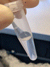
This is approximately the size of the nuclei pellet (~2 million nuclei) needed for 1 plate of reverse transcription. Extra fixed nuclei can be aliquoted and snap-frozen.
Figure 6.
Figure 6.. Visualizing protease digestion of the nuclei.
a, Nuclei after about 10min of protease digestion, swelling and starting to lose integrity. b, Nuclei after 30min of protease digestion; DNA has been released and now the protease can be heat-inactivated.
Figure 7.
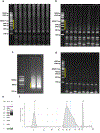
Figure 7.. Evaluating the libraries after PCR.
Yellow bars indicate the size range to look for. a, A good quality library - sampling of 8 wells from the pcr plate, 1.5 μl of each well is run on a 6% PAGE gel, 200V for 30 min. A bright, long smear of products above the 200 bp marker indicates a robust library. This one is from a 384×384 experiment with 4000 nuclei plated in each well of the last plate. b, A medium quality library with fainter smears. This was from a 96×96 experiment that underperformed - quality of the data was good and the number of UMIs per cell was as expected, but had less total cells overall, most likely due to counting estimates during the last round of plating. c, Gel size-selection. Libraries have been concentrated via ampure purification and run on a 1% agarose gel at 100V for about 1 hour. Most of the primer dimers are gone, and the libraries are cut out of the gel at the. This is a 384×384 experiment so these smears are very intense. d, A failed experiment. The smaller number of primer dimer bands suggests that the failure resulted from incomplete protease digestion. e,f, The final, gel-purified library fragment distribution as measured on an Agilent tapestation. Primer dimers have been removed and you should be left with a library ranging from 300–700bp in size.
Figure 8.
Figure 8.. High-quality data of E16.5 mouse embryo generated by application of the optimized sci-RNA-seq3 protocol.
2D UMAP visualization of the new E16.5 dataset. All nuclei colored by each of the 20 cell trajectories are shown on the left. Subview of global 2D UMAP visualization highlighting subpopulations of the white blood cells trajectory is shown on the right.

References

    1. Domcke S et al. A human cell atlas of fetal chromatin accessibility. Science 370, (2020). - PMC - PubMed
    1. Cusanovich DA et al. Multiplex single cell profiling of chromatin accessibility by combinatorial cellular indexing. Science 348, 910–914 (2015). - PMC - PubMed
    1. Cao J et al. Comprehensive single-cell transcriptional profiling of a multicellular organism. Science 357, 661–667 (2017). - PMC - PubMed
    1. Cao J et al. The single-cell transcriptional landscape of mammalian organogenesis. Nature 566, 496–502 (2019). - PMC - PubMed
    1. Rosenberg AB et al. Single-cell profiling of the developing mouse brain and spinal cord with split-pool barcoding. Science 360, 176–182 (2018). - PMC - PubMed

Publication types