Review
. 2022 Oct 3:10:995508.
doi: 10.3389/fcell.2022.995508. eCollection 2022.

Native valve, prosthetic valve, and cardiac device-related infective endocarditis: A review and update on current innovative diagnostic and therapeutic strategies

Affiliations
Review

Native valve, prosthetic valve, and cardiac device-related infective endocarditis: A review and update on current innovative diagnostic and therapeutic strategies

Joop J P Kouijzer et al. Front Cell Dev Biol. .

Abstract

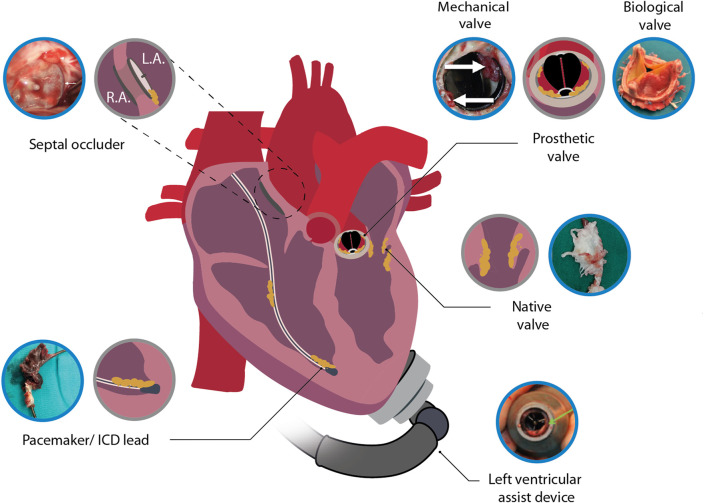
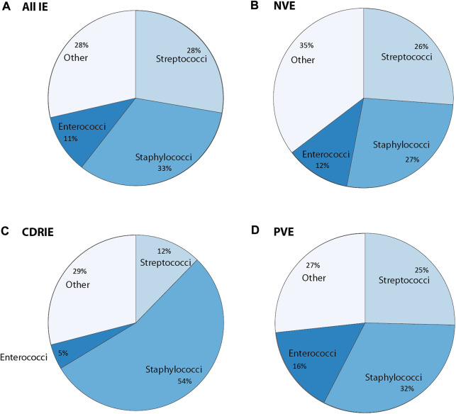
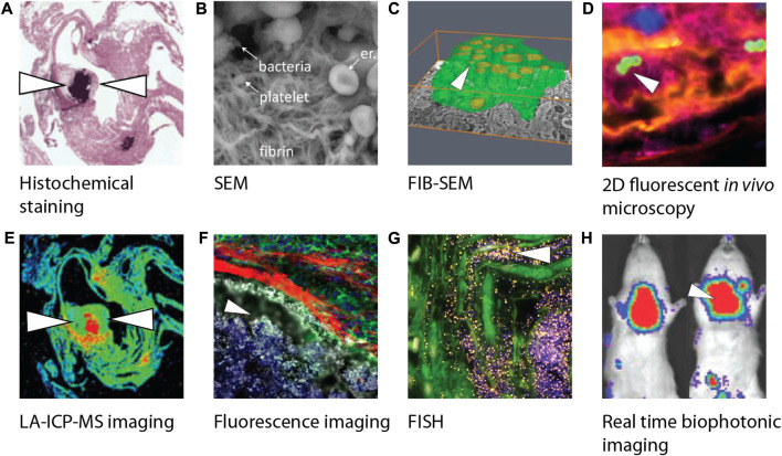
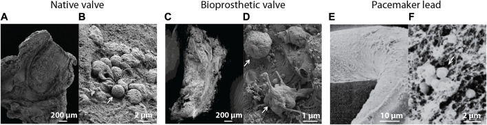
Infective endocarditis (IE) is a life-threatening microbial infection of native and prosthetic heart valves, endocardial surface, and/or indwelling cardiac device. Prevalence of IE is increasing and mortality has not significantly improved despite technological advances. This review provides an updated overview using recent literature on the clinical presentation, diagnosis, imaging, causative pathogens, treatment, and outcomes in native valve, prosthetic valve, and cardiac device-related IE. In addition, the experimental approaches used in IE research to improve the understanding of disease mechanisms and the current diagnostic pipelines are discussed, as well as potential innovative diagnostic and therapeutic strategies. This will ultimately help towards deriving better diagnostic tools and treatments to improve IE patient outcomes.

Keywords: biofilm; cardiac device; diagnosis; infection; infective endocarditis; native valve; prosthetic valve; treatment.


Conflict of interest statement

The authors declare that the research was conducted in the absence of any commercial or financial relationships that could be construed as a potential conflict of interest.

Figures

FIGURE 1
FIGURE 1
Illustration depicting infective endocarditis associated with native heart tissue and cardiac devices. Included images are examples of an infected septal occluder (Nguyen et al., 2016), mechanical heart valve (Pettersson et al., 2014), biological prosthetic heart valve (Jainandunsing et al., 2014), native heart valve (Li et al., 2022), LVAD (Akin et al., 2018) and pacemaker lead (Boljevic et al., 2019). Blue encircled images were adapted with permission from the original publishers and used as examples. L.A., left atrium; R.A., right atrium.
FIGURE 2
FIGURE 2
Overview of causative bacteria in infective endocarditis (IE) concerning (A) all IE cases, (B) native valve endocarditis (NVE), and (C) cardiac device related-infective endocarditis (CDRIE) that is further subdivided into (D) prosthetic valve endocarditis (PVE). Other refers to either other cultured or unidentified microorganisms. The values in this graph were obtained using the following references (Cahill and Prendergast, 2016; Wang A. et al., 2018; DeSimone and Sohail, 2018; DeSimone and Sohall, 2018, Babeș et al., 2021; Doring et al., 2018; Teoh and Hannan, 2018; Slawinski et al., 2019; Mateos Gaitán et al., 2020; Khalil and Soufi, 2022).
FIGURE 3
FIGURE 3
Overview of different biofilm visualization modalities used in infective endocarditis research. (A) Crystal violet staining visualizing gram-positive bacteria of paraffin-embedded female C57BL/6 mice heart tissue slices using light microscopy (Schwarz et al., 2022). (B) SEM image visualizing bacteria including a platelet-containing fibrin network (Hannachi et al., 2020). (C) Three-dimensional reconstruction of phagocytic cells (green) infected by bacteria (yellow) using FIB-SEM (Oberbach et al., 2017). (D) Confocal image of an early infective endocarditis lesions in mice visualizing platelets (pink), fibrin (yellow), endothelium (blue) and S. aureus (green) (Liesenborghs et al., 2019). (E) LA-ICP-MS imaging makes it possible to produce a quantitative distribution map (min to max, blue to red) of elemental distribution (Ca, Mg, Mn, Fe, Cu, or Zn) within female C57BL/6 mice heart tissue infected by bacteria (Schwarz et al., 2022). (F) Visualization of myeloid cells’ (green) inability to interact with bacteria (white) due to a fibrin barrier (red) in endocarditis in mice (Panizzi et al., 2020). (G) Visualization of S. epidermidis on aortic valve tissue with bacteria after incubation with a FISH probe (yellow) and DAPI (blue) (Lauten et al., 2021). (H) Real time imaging of rats infected by S. aureus using a bioluminescent in vivo imaging system (Xiong et al., 2005) showing high (red) to low (blue) bioluminescent signals. White arrowheads indicate the presence of bacteria. Images were adapted with permission from the original publishers and used as examples. SEM, Scanning electron microcopy; FIB-SEM, Focused ion beam scanning electron microscopy; LA-ICP-MS, Laser ablation induction coupled plasma mass spectrometry; FISH, Fluorescence in situ hybridization; DAPI, 4′,6-diamidino-2-phenylindole.
FIGURE 4
FIGURE 4
Scanning electron microscopic images of (A) an infected native heart valve with (B) a corresponding magnified image showing intact bacteria on the tissue surface (Oberbach et al., 2017). (C) Overview of an infected biological prosthetic valve with (D) a corresponding magnified image showing intact bacteria surrounded by a fibrous surface (Oberbach et al., 2017). (E) Visualization of a biofilm on the surface of a pacemaker lead with (F) a corresponding magnified image showing bacteria surrounded by a fibrillar substrate (Marrie and Costerton, 1984). White arrows indicate the presence of bacteria. Images were adapted with permission from the original publishers and used as examples.

Similar articles

Cited by

References

    1. Abdelghani M., Nassif M., Blom N. A., Van Mourik M. S., Straver B., Koolbergen D. R., et al. (2018). Infective endocarditis after Melody valve implantation in the pulmonary position: A systematic review. J. Am. Heart Assoc. 7. 10.1161/JAHA.117.008163 - DOI - PMC - PubMed
    1. Abdelhady W., Bayer A. S., Seidl K., Nast C. C., Kiedrowski M. R., Horswill A. R., et al. (2013). Reduced vancomycin susceptibility in an in vitro catheter-related biofilm model correlates with poor therapeutic outcomes in experimental endocarditis due to methicillin-resistant Staphylococcus aureus . Antimicrob. Agents Chemother. 57, 1447–1454. 10.1128/aac.02073-12 - DOI - PMC - PubMed
    1. Abegaz T. M., Bhagavathula A. S., Gebreyohannes E. A., Mekonnen A. B., Abebe T. B. (2017). Short- and long-term outcomes in infective endocarditis patients: A systematic review and meta-analysis. BMC Cardiovasc Disord. 17, 1–12. 10.1186/s12872-017-0729-5 - DOI - PMC - PubMed
    1. Abikhzer G., Martineau P., Grégoire J., Finnerty V., Harel F., Pelletier-Galarneau M. (2020). [18F]FDG-PET CT for the evaluation of native valve endocarditis. J. Nucl. Cardiol. - PubMed
    1. Aguilera J., Hutt E., Jaber W. A. (2021). Imaging of cardiac device-related infection. Front. Cardiovasc Med. 8, 729786. 10.3389/fcvm.2021.729786 - DOI - PMC - PubMed

LinkOut - more resources