[Preprint]. 2022 Oct 13:2022.10.13.512056.
doi: 10.1101/2022.10.13.512056.

A pseudovirus system enables deep mutational scanning of the full SARS-CoV-2 spike

Affiliations

A pseudovirus system enables deep mutational scanning of the full SARS-CoV-2 spike

Bernadeta Dadonaite et al. bioRxiv. .

Update in

Abstract

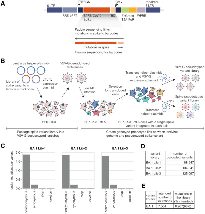
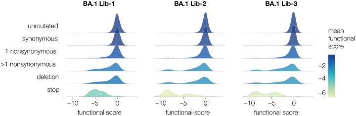
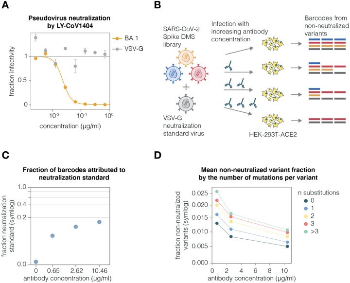
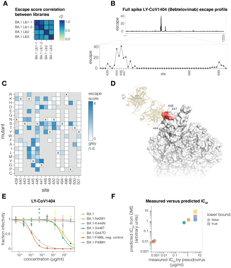
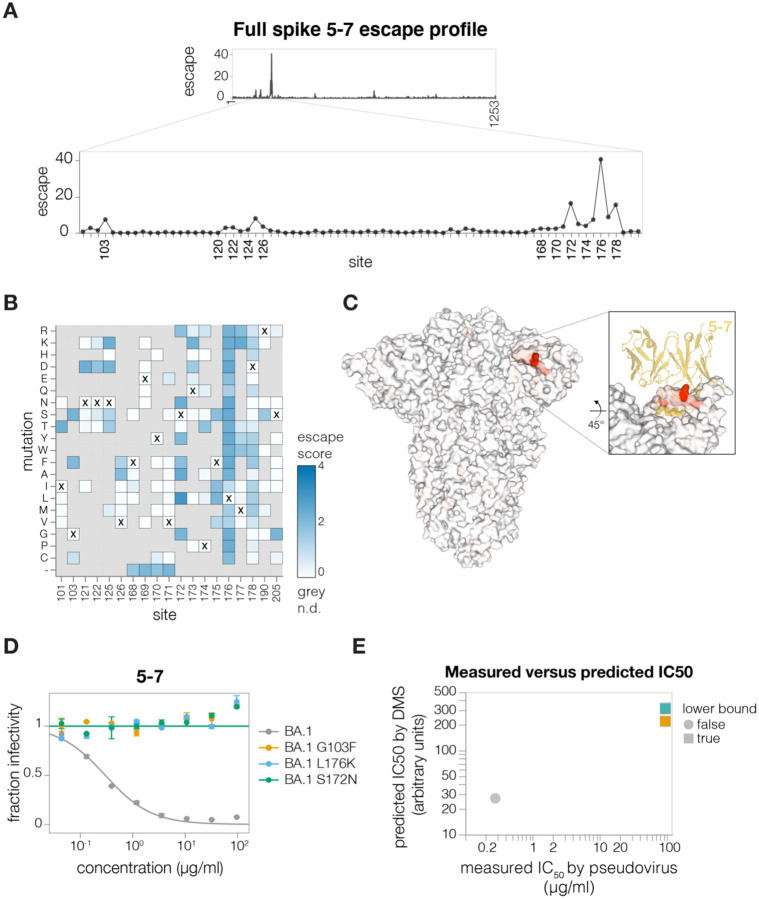
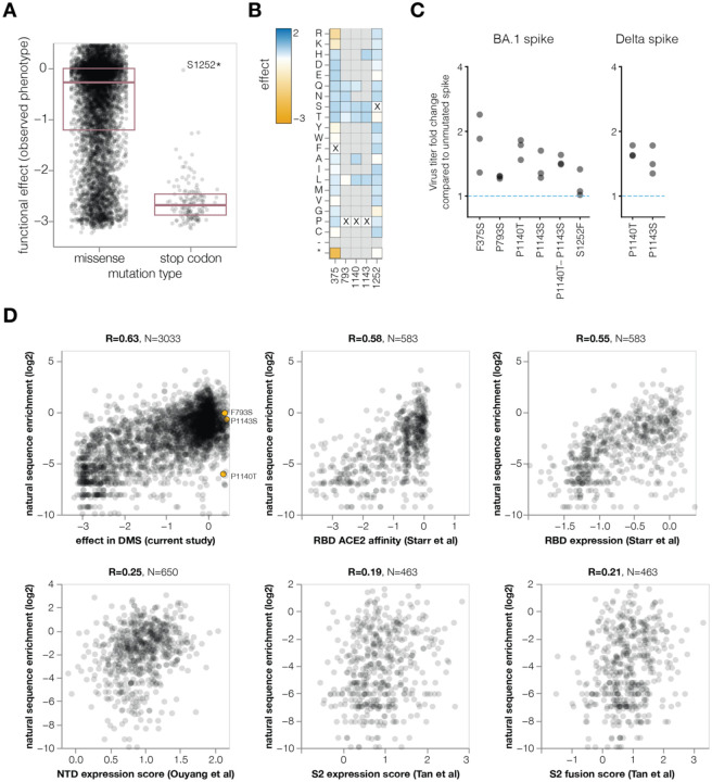
A major challenge in understanding SARS-CoV-2 evolution is interpreting the antigenic and functional effects of emerging mutations in the viral spike protein. Here we describe a new deep mutational scanning platform based on non-replicative pseudotyped lentiviruses that directly quantifies how large numbers of spike mutations impact antibody neutralization and pseudovirus infection. We demonstrate this new platform by making libraries of the Omicron BA.1 and Delta spikes. These libraries each contain ~7000 distinct amino-acid mutations in the context of up to ~135,000 unique mutation combinations. We use these libraries to map escape mutations from neutralizing antibodies targeting the receptor binding domain, N-terminal domain, and S2 subunit of spike. Overall, this work establishes a high-throughput and safe approach to measure how ~10 5 combinations of mutations affect antibody neutralization and spike-mediated infection. Notably, the platform described here can be extended to the entry proteins of many other viruses.


Conflict of interest statement

Competing interests

JDB is on the scientific advisory board of Apriori Bio and Oncorus, and has recently consulted on topics related to viral evolution for Moderna and Merck. JDB, KHDC, and CER receive royalty payments as inventors on Fred Hutch licensed patents related to viral deep mutational scanning. JDB, KHDC, CER and BD are inventors on a pending patent application relating to the viral deep mutational scanning system described in this paper. RB is a consultant for IAVI, Adagio, Adimab, Mabloc, VosBio, Nonigenex, and Radiant. DDH is a co-founder of TaiMed Biologics and RenBio, consultant to WuXi Biologics and Brii Biosciences, and board director for Vicarious Surgical. The other authors declare no competing interests.

Figures

Figure 1.
Figure 1.. Deep mutational scanning platform for spike
(A) Lentivirus backbone used for deep mutational scanning. The backbone contains functional lentiviral 5′ and 3′ long terminal repeat (LTR) regions. The spike gene is under an inducible tet response element 3rd generation (TRE3G) promoter, and there is a 16 nucleotide barcode (BC) downstream of the stop codon. A CMV promoter drives expression of reporter ZsGreen gene that is linked to a puromycin resistance gene (PuR) via a T2A linker. The backbone also contains a woodchuck hepatitis virus posttranscriptional regulatory element (WPRE), Rev response element (RRE), and a central polypurine tract (cPPT). (B) Approach for creating genotype-phenotype linked lentivirus libraries. HEK-293T cells are transfected with spike-carrying lentivirus backbone, VSV-G expression plasmid and lentiviral helper plasmids to generate VSV-G-pseudotyped lentiviruses. These viruses are used to transduce reverse tetracycline-controlled transactivator (rtTA) expressing HEK-293T cells at low multiplicity of infection (MOI), and successfully transduced cells are selected using puromycin. Selected cells can be transfected with helper plasmids and a VSV-G expression plasmid to produce VSV-G-pseudotyped viruses carrying all genomes present in the deep mutational library or selected cells can be induced with doxycycline (dox) to express spike and transfected with only the helper plasmids to generate spike-pseudotyped lentiviruses that have a genotype-phenotype link. (C) Average number of mutations per barcoded spike in BA.1 libraries. (D) Total number of barcoded variants in each BA.1 library. (E) The coverage of intended mutations across all BA.1 libraries.
Figure 2.
Figure 2.. Some types of mutations tend to impair spike-mediated pseudovirus infection
For each barcoded spike variant, we compute a functional score that reflects how well that spike mediates pseudovirus infection relative to the unmutated spike: negative scores indicate impaired infection, positive scores indicate improved infection. The plots show the distribution of functional scores across all variants in each of the three BA.1 libraries for different categories of variants, with each distribution colored by the mean functional score for that variant type.
Figure 3.
Figure 3.. A VSV-G standard enables measurement of absolute neutralization by deep sequencing.
(A) Neutralization assay demonstrating that BA.1-spike-pseudotyped lentivirus is neutralized by antibody LY-CoV1404, but the VSV-G-pseudotyped neutralization standard is not. (B) Use of the VSV-G standard to measure absolute neutralization. Deep mutational scanning libraries are mixed with VSV-G neutralization standard. The virus mixture is incubated with a no-antibody control or increasing antibody concentrations and infected into ACE2-expressing 293T cells. After ~12 hours viral genomes are recovered, barcodes are sequenced, and absolute neutralization of each variant is computed by comparing its barcode counts to those from the VSV-G standard. (C) Fraction of barcodes derived from the VSV-G neutralization standard in infections with increasing LY-CoV1404 concentrations. (D) BA.1 deep mutational scanning library non-neutralized fractions averaged across variants with different numbers of amino-acid mutations at differentLY-CoV1404 concentrations.Note panels C and D use a symlog scale.
Figure 4.
Figure 4.. Antibody LY-CoV1404 escape mapping
(A) Correlation of mutation escape scores between technical replicates (BA1 Lib-1.1 and BA1 Lib-1.2) and biological replicates (BA1 Lib-1, BA1 Lib-2, BA1 Lib-3). (B) Total escape scores at each site in the BA.1 spike, and a zoomed-in plot showing the key escape sites. Sites of mutations chosen for validation experiments are labeled on the x-axis. (C) Heatmap of mutation escape scores at key sites. Residues marked with X are the wild-type amino acids in BA.1. Amino acids not present in our libraries are shown in gray. An interactive heatmap for the entirety of spike is at https://dms-vep.github.io/SARS-CoV-2_Omicron_BA.1_spike_DMS_mAbs/LyCoV-1404_escape_plot.html (D) Surface representation of spike coloured by sum of escape scores at that site. LY-CoV1404 antibody is in yellow. Only the antibody-bound protomer is coloured. PDB IDs 7MMO and 6XM4 were aligned to make this structure. (E) Validation pseudovirus neutralization assays of the indicated BA.1 spike mutants against the LY-CoV1404 antibody. (F) Correlation between predicted IC50 values from deep mutational scanning (DMS) data versus the IC50 values measured in the validation assays in panel (E). The points are colored as in panel (E). Lower bound indicates that the antibody did not neutralize at the highest concentration tested in the validation neutralization assay. Site numbering in all plots is based on the Wuhan-Hu-1 sequence.
Figure 5.
Figure 5.. Antibody 5–7 escape mapping.
(A) Total escape scores for each site in the BA.1 spike and a zoomed-in plot showing the key escape sites. (B) Heatmap of mutation escape scores at key sites. Residues marked with X are the wild-type amino acids in BA.1. Amino acids not present in our libraries are shown in gray. An interactive version of this plot for the entirety of spike is at https://dms-vep.github.io/SARS-CoV-2_Omicron_BA.1_spike_DMS_mAbs/NTD_5-7_escape_plot.html (C) Surface representation of spike coloured by the sum of escape scores at that site. Antibody 5–7 is shown in yellow in the inset. PDB ID: 7RW2. (D) Validation pseudovirus neutralization assays of the indicated BA.1 spike mutants against antibody 5–7. (E) Correlation between predicted IC50 values from deep mutational scanning (DMS) data versus the IC50 values measured in the validation assays in (F). Lower bound indicates that the antibody did not neutralize at the highest concentration tested in the validation neutralization assay. Site numbering in all plots is based on the Wuhan-Hu-1 sequence.
Figure 6.
Figure 6.. Antibody CC9.104 and CC67.105 escape mapping
(A-B) Total escape scores for each site in the BA.1 spike for the CC9.104 (A) and CC67.105 (B) antibodies. (C-D) Escape heatmaps for the S2 stem-helix (sites 1146–1163) for CC9.104 (C) and CC67.105 (D) antibodies. Residues marked with X are the wild-type amino acids in BA.1 sequence. Amino acids that are not present in our libraries are shown in gray. Interactive heatmaps for the entirety of spike are at https://dms-vep.github.io/SARS-CoV-2_Omicron_BA.1_spike_DMS_mAbs/CC67.105_escape_plot.html and https://dms-vep.github.io/SARS-CoV-2_Omicron_BA.1_spike_DMS_mAbs/CC9.104_escape_plot.html (E) Surface representation of spike coloured by the sum of escape scores at that site for CC9.104 (left) and CC67.105 (right) antibodies. Site 1163 is not resolved in the structure. PDB ID: 6XR8. (F) Alignment of SARS-CoV-2 and MERS-CoV spikes at sites 1146–1163. (G) Validation pseudovirus neutralization assay for CC9.104 (left) and CC67.105 (right) antibodies with BA.1 spike carrying the indicated mutations. (H) Correlation between predicted IC50 values from deep mutational scanning (DMS) data versus the IC50 values measured in the validation assays in (G). Lower bound indicates that the antibody did not neutralize at the highest concentration tested in the validation neutralization assay. Site numbering in all plots is based on the Wuhan-Hu-1 sequence.
Figure 7.
Figure 7.. Functional effects of mutations on spike-mediated pseudovirus infection
(A) Distribution of functional effects of mutations in BA.1 deep mutational scanning libraries. Negative values indicate mutations are deleterious for viral entry. The stop codon mutation with a neutral functional effect of ~0 is at the last codon of the spike used in our experiments. (B) Heatmap showing functional effects at sites of mutations with beneficial functional effects that were chosen for validation assays in C. An interactive version of this heatmap for the entire spike is at https://dms-vep.github.io/SARS-CoV-2_Omicron_BA.1_spike_DMS_mAbs/muteffects_observed_heatmap.html (C) Fold change in virus entry titer for spike mutants relative to unmutated spike. There are three points for each mutant, reflecting triplicate measurements. (D) Correlation between enrichment of mutations during actual evolution of human SARS-CoV-2 and functional effects from our lentivirus-based deep mutational scanning or previous RBD expression or ACE2 affinity for yeast-based deep mutational scanning (Starr et al., 2022), and S2 (Tan et al., 2022) or NTD (Ouyang et al., 2022) expression for mammalian display-based deep mutational scanning. Interactive plots that enable mouseovers and show correlations among experiments are at https://dms-vep.github.io/SARS-CoV-2_Omicron_BA.1_spike_DMS_mAbs/natural_enrichment_vs_dms.html

References

    1. Ballal A., Malliaris C.D., Morozov A.V., 2020. Molecular Phenotypes as Key Intermediates in Mapping Genotypes to Fitness, in: Pontarotti P. (Ed.), Evolutionary Biology—A Transdisciplinary Approach. Springer International Publishing, Cham, pp. 15–40. 10.1007/978-3-030-57246-4_2 - DOI
    1. Baum A., Fulton B.O., Wloga E., Copin R., Pascal K.E., Russo V., Giordano S., Lanza K., Negron N., Ni M., Wei Y., Atwal G.S., Murphy A.J., Stahl N., Yancopoulos G.D., Kyratsous C.A., 2020. Antibody cocktail to SARS-CoV-2 spike protein prevents rapid mutational escape seen with individual antibodies. Science eabd0831. 10.1126/science.abd0831 - DOI - PMC - PubMed
    1. Bellusci L., Grubbs G., Zahra F.T., Forgacs D., Golding H., Ross T.M., Khurana S., 2022. Antibody affinity and cross-variant neutralization of SARS-CoV-2 Omicron BA.1, BA.2 and BA.3 following third mRNA vaccination. Nat. Commun. 13, 4617. 10.1038/s41467-022-32298-w - DOI - PMC - PubMed
    1. Benton D.J., Wrobel A.G., Roustan C., Borg A., Xu P., Martin S.R., Rosenthal P.B., Skehel J.J., Gamblin S.J., 2021. The effect of the D614G substitution on the structure of the spike glycoprotein of SARS-CoV-2. Proc. Natl. Acad. Sci. 118, e2022586118. 10.1073/pnas.2022586118 - DOI - PMC - PubMed
    1. Bloom J.D., 2014. An Experimentally Determined Evolutionary Model Dramatically Improves Phylogenetic Fit. Mol. Biol. Evol. 31, 1956–1978. 10.1093/molbev/msu173 - DOI - PMC - PubMed

Publication types