. 2022 Oct 12:22:312-324.
doi: 10.1016/j.bioactmat.2022.10.010. eCollection 2023 Apr.

Enhancing cartilage repair with optimized supramolecular hydrogel-based scaffold and pulsed electromagnetic field

Affiliations

Enhancing cartilage repair with optimized supramolecular hydrogel-based scaffold and pulsed electromagnetic field

Yucong Li et al. Bioact Mater. .

Abstract

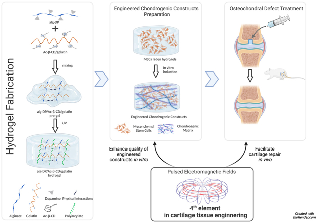
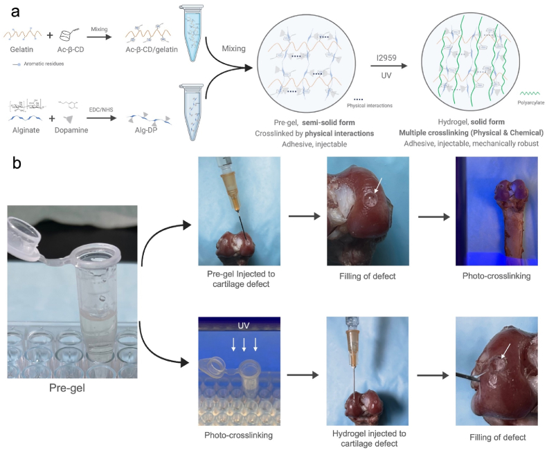
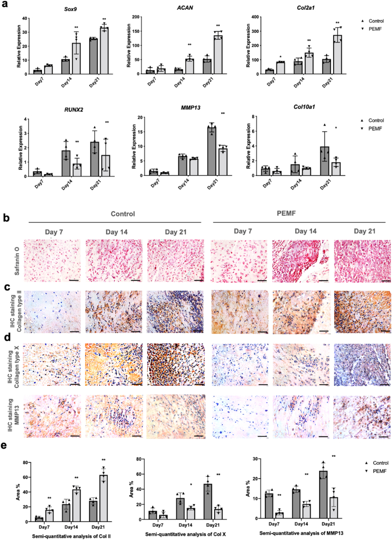
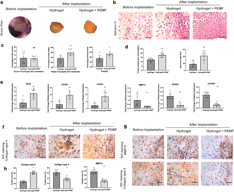
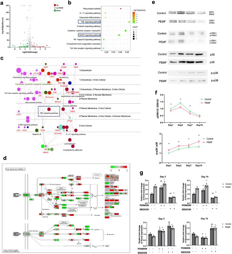
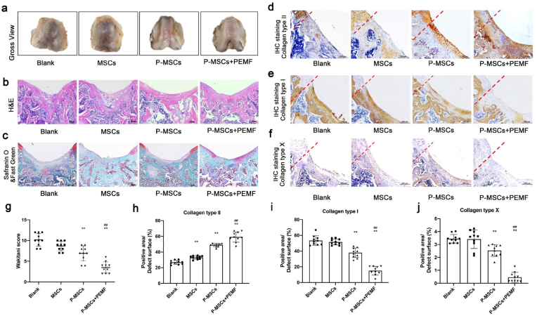
Functional tissue engineering strategies provide innovative approach for the repair and regeneration of damaged cartilage. Hydrogel is widely used because it could provide rapid defect filling and proper structure support, and is biocompatible for cell aggregation and matrix deposition. Efforts have been made to seek suitable scaffolds for cartilage tissue engineering. Here Alg-DA/Ac-β-CD/gelatin hydrogel was designed with the features of physical and chemical multiple crosslinking and self-healing properties. Gelation time, swelling ratio, biodegradability and biocompatibility of the hydrogels were systematically characterized, and the injectable self-healing adhesive hydrogel were demonstrated to exhibit ideal properties for cartilage repair. Furthermore, the new hydrogel design introduces a pre-gel state before photo-crosslinking, where increased viscosity and decreased fluidity allow the gel to remain in a semi-solid condition. This granted multiple administration routes to the hydrogels, which brings hydrogels the ability to adapt to complex clinical situations. Pulsed electromagnetic fields (PEMF) have been recognized as a promising solution to various health problems owing to their noninvasive properties and therapeutic potentials. PEMF treatment offers a better clinical outcome with fewer, if any, side effects, and wildly used in musculoskeletal tissue repair. Thereby we propose PEMF as an effective biophysical stimulation to be 4th key element in cartilage tissue engineering. In this study, the as-prepared Alg-DA/Ac-β-CD/gelatin hydrogels were utilized in the rat osteochondral defect model, and the potential application of PEMF in cartilage tissue engineering were investigated. PEMF treatment were proven to enhance the quality of engineered chondrogenic constructs in vitro, and facilitate chondrogenesis and cartilage repair in vivo. All of the results suggested that with the injectable self-healing adhesive hydrogel and PEMF treatment, this newly proposed tissue engineering strategy revealed superior clinical potential for cartilage defect treatment.

Keywords: Cartilage tissue engineering; Chondrogenesis; Mesenchymal stem cells; Pulsed electromagnetic field; Supramolecular hydrogels.


Conflict of interest statement

The authors have no conflicts of interest to disclose in relation to this article. NZ, EIW and IV are employees of and own stock in Orthofix Medical Inc., USA.

Figures

Image 1
Graphical abstract
Fig. 1
Fig. 1
a. Schematic illustration of the hydrogel design (Created withBioRender.com). b. Under pre-gel state, the mixed solution can be firstly injected to cartilage defect site, and then in situ crosslinked under UV exposure. Per-gel can also be photo-crosslinked first and injected to the defect site, the tissue-adhesive property allows the hydrogel to be tightly attached. Either way, smooth cartilage surface can be restored.
Fig. 2
Fig. 2
Physical properties of the Alg/Ac-β-CD/gelatin and Alg-DA/Ac-β-CD/gelatin hydrogels a Swelling behavior of Alg/Ac-β-CD/gelatin hydrogel and Alg-DA/Ac-β-CD/gelatin hydrogels with different dopamine grafting ratios incubated in PBS solution (n = 4/group). b and c. In vitro degradation properties of Alg/Ac-β-CD/gelatin hydrogel and Alg-DA/Ac-β-CD/gelatin hydrogels with different dopamine grafting ratios incubated in PBS solution (b) and Type-I collagenase/PBS (0.1 mg/mL) (c) (n = 4/group). d. Adhesion properties of Alg/Ac-β-CD/gelatin hydrogel and Alg-DA/Ac-β-CD/gelatin hydrogels with different dopamine grafting ratios through lap-shear adhesion test (n = 5/group). e. Young's compression modulus of Alg/Ac-β-CD/gelatin hydrogel and Alg-DA/Ac-β-CD/gelatin hydrogels with different dopamine grafting ratios (n = 6/group). *p < 0.05.
Fig. 3
Fig. 3
PEMF affected RNA expression and matrix formation of rBMSCs during chondrogenic differentiation in hydrogels in vitro. a. qPCR analysis results showed that compared to control group, PEMF significantly upregulated the expressions of chondrogenic mRNA, and downregulated the expressions of hypertrophic mRNA. Staining of Safranin O (b) and immunohistochemistry of Col II (c, e) demonstrated that after PEMF treatment, significantly larger amounts of aggrecan and Col II were deposited in hydrogels. Immunohistochemistry of Col X and MMP13 (d, e) demonstrated that PEMF treatment could significantly inhibit the formation of hypertrophic proteins, revealing its chondro-protective potential. Scale bar: 50 μm *p < 0.05 **p < 0.01.
Fig. 4
Fig. 4
PEMF promoted rBMSCs chondrogenesis in vivo. a Gross view of hydrogels before and after implantation. b afranin O staining of hydrogels after implantation. c Volume, weight, and density of hydrogels after implantation. d Mechanical property of hydrogels after implantation via compression test. e qPCR analysis of chondrogenic marker genes (Sox9, ACAN and Col2a1) and hypertrophic marker genes (MMP13, RUNX2 and Col10a1). f IHC staining of Col II. g IHC staining of MMP13 and Col X. h Semi-quantitative analysis of IHC staining. n = 8/group. Scale bar: 50 μm *p < 0.05 **p < 0.01.
Fig. 5
Fig. 5
RNA-seq analysis and validation study results. a Volcano plot of control-vs-PEMF revealing up- and down-regulated DEGs after PEMF treatment. b KEGG analysis of DEGs. c Gene-pathway interactions visualized in Cerebral Layout using ClueGo. d Gene expression changes of cells from three individual donors visualized in KEGG pathview of TNF-α pathway. e Protein expression levels of ERK1/2, p-ERK1/2, p38, and p-p38 were analyzed by Western blot. f Semi-quantitative analysis of each band was analyzed by ImageJ, and band intensity ratio (Ap-ERK ⁄ AERK and Ap-p38 ⁄ Ap38) were calculated (n = 4/group/time point). g Expression of chondrogenic marker (Sox9) and hypertrophic marker (Runx2) in hBMSCs during chondrogenic induction after ERK1 ⁄ 2 and p38 inhibition on day 3 and 14 (n = 4/group/time point). *p < 0.05 **p < 0.01. # vs control group without inhibition, ^ vs PEMF group without inhibition, #, ^ p < 0.05, ##, ^^ p < 0.01.
Fig. 6
Fig. 6
PEMF treatment promoted cartilage defect healing in rat osteochondral defect model. a Macroscopic appearance of the rat osteochondral defect. b H&E staining of the rat osteochondral defect sections. c Safranin O & Fast Green staining of the rat osteochondral defect sections. d Immunohistochemical staining of type II collagen. e Immunohistochemical staining of type I collagen. f Immunohistochemical staining of type X collagen. g Cartilage regeneration evaluated by the Wakitani scoring system at week 8 after surgery. h-j Semi-quantitative analysis of IHC staining of collagen type II, I and X. *p < 0.05 vs. blank group, **p < 0.01 vs. blank group, ##p < 0.01 vs P-MSCs group. n = 10/group/time point.

References

    1. Holland T.A., Tessmar J.K., Tabata Y., Mikos A.G. Transforming growth factor-β1 release from oligo (poly (ethylene glycol) fumarate) hydrogels in conditions that model the cartilage wound healing environment. Journal of controlled release. 2004;94(1):101–114. - PubMed
    1. Man Z., Hu X., Liu Z., Huang H., Meng Q., Zhang X., Dai L., Zhang J., Fu X., Duan X. Transplantation of allogenic chondrocytes with chitosan hydrogel-demineralized bone matrix hybrid scaffold to repair rabbit cartilage injury. Biomaterials. 2016;108:157–167. - PubMed
    1. Albani D., Gloria A., Giordano C., Rodilossi S., Russo T., D'Amora U., Tunesi M., Cigada A., Ambrosio L., Forloni G. Hydrogel-based nanocomposites and mesenchymal stem cells: a promising synergistic strategy for neurodegenerative disorders therapy. Sci. World J. 2013;2013 - PMC - PubMed
    1. Wang J., Zhang F., Tsang W.P., Wan C., Wu C. Fabrication of injectable high strength hydrogel based on 4-arm star PEG for cartilage tissue engineering. Biomaterials. 2017;120:11–21. - PubMed
    1. Graca M.F.P., Miguel S.P., Cabral C.S.D., Correia I.J. Hyaluronic acid-Based wound dressings: a review. Carbohydr. Polym. 2020;241 - PubMed

LinkOut - more resources