. 2023 Jan 4;83(1):130-140.
doi: 10.1158/0008-5472.CAN-22-0945.

N-myc-Mediated Translation Control Is a Therapeutic Vulnerability in Medulloblastoma

Affiliations

N-myc-Mediated Translation Control Is a Therapeutic Vulnerability in Medulloblastoma

Duygu Kuzuoglu-Ozturk et al. Cancer Res. .

Abstract

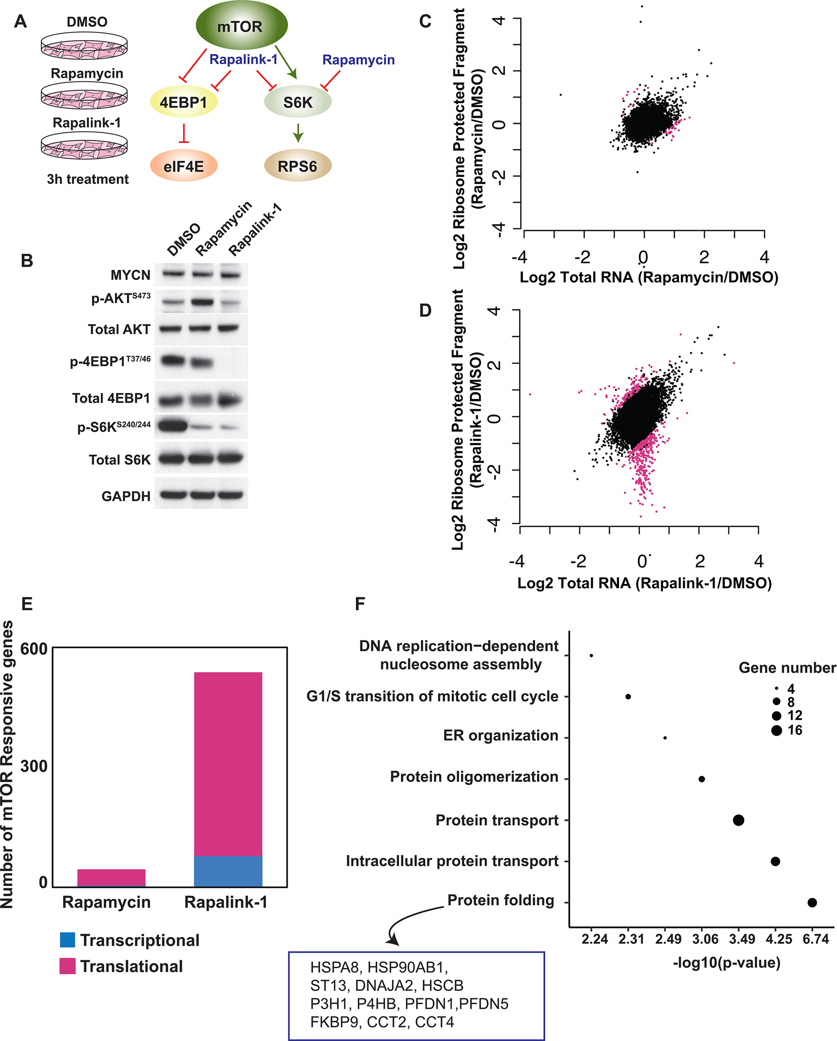
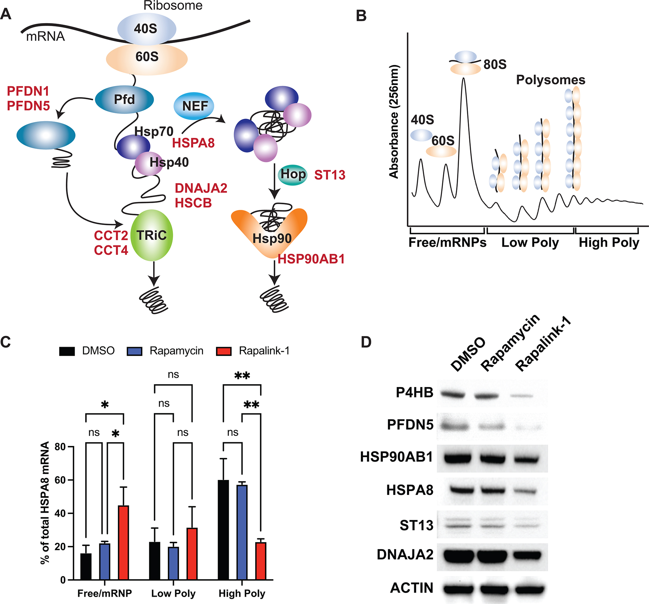
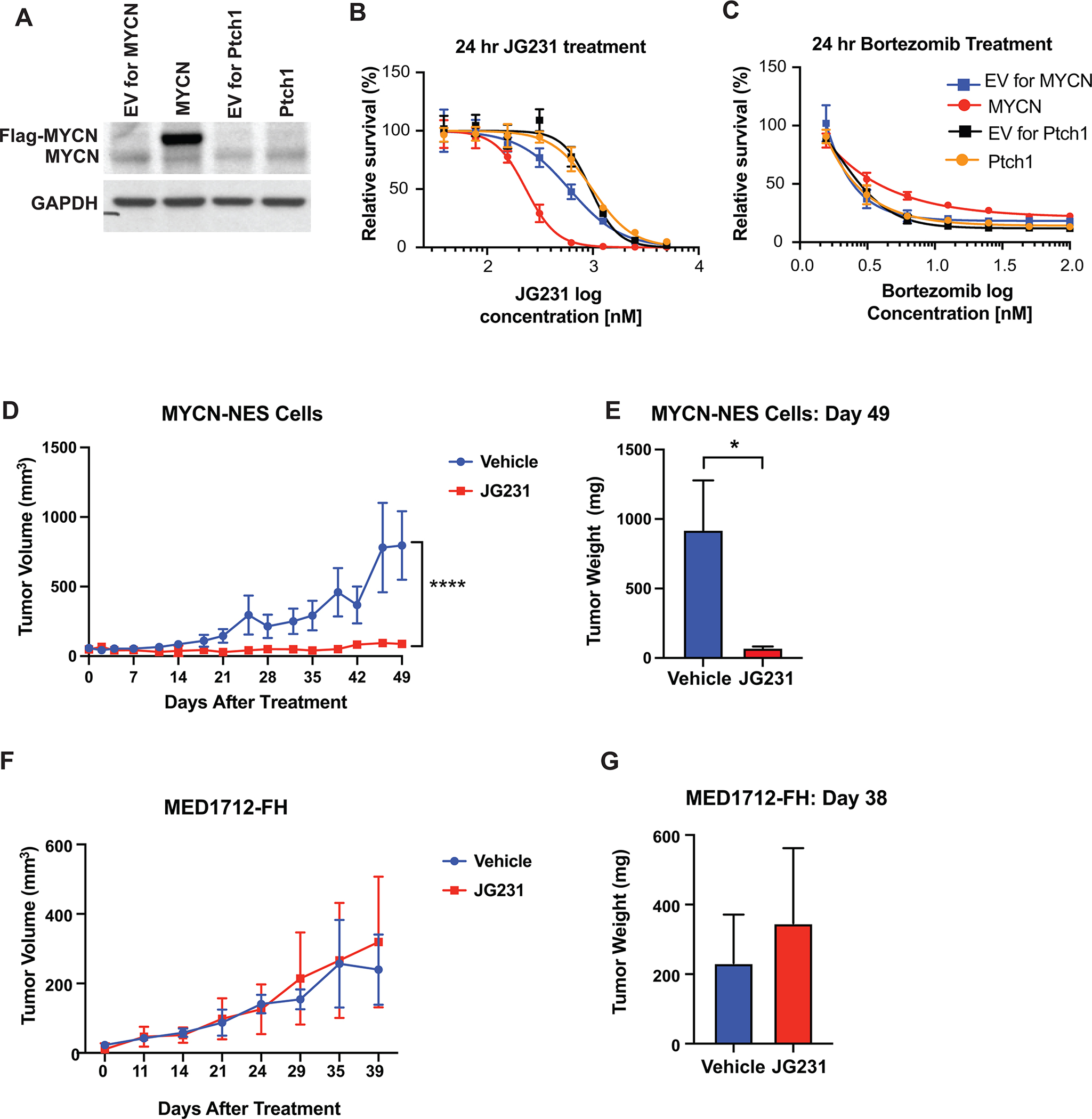
Deregulation of neuroblastoma-derived myc (N-myc) is a leading cause of malignant brain tumors in children. To target N-myc-driven medulloblastoma, most research has focused on identifying genomic alterations or on the analysis of the medulloblastoma transcriptome. Here, we have broadly characterized the translatome of medulloblastoma and shown that N-myc unexpectedly drives selective translation of transcripts that promote protein homeostasis. Cancer cells are constantly exposed to proteotoxic stress associated with alterations in protein production or folding. It remains poorly understood how cancers cope with proteotoxic stress to promote their growth. Here, our data revealed that N-myc regulates the expression of specific components (∼5%) of the protein folding machinery at the translational level through the major cap binding protein, eukaryotic initiation factor eIF4E. Reducing eIF4E levels in mouse models of medulloblastoma blocked tumorigenesis. Importantly, targeting Hsp70, a protein folding chaperone translationally regulated by N-myc, suppressed tumor growth in mouse and human medulloblastoma xenograft models. These findings reveal a previously hidden molecular program that promotes medulloblastoma formation and identify new therapies that may have impact in the clinic.

Significance: Translatome analysis in medulloblastoma shows that N-myc drives selective translation of transcripts that promote protein homeostasis and that represent new therapeutic vulnerabilities.


Figures

Figure 1.
Figure 1.. Ribosome profiling reveals preferentially translated mRNAs in MYCN-driven medulloblastoma.
A, The mechanism of action of two mTOR inhibitors, Rapamycin and Rapalink-1, used in the ribosome profiling. B, Western blot analysis showing the specificity of Rapamycin and Rapalink-1 treatment for 3 hours in GTML cells. C-D, Scatter blots comparing total RNA and ribosome protected fragments of each transcript upon 3 hours Rapamycin (C) and Rapalink-1 (D) treatment compared to DMSO treated samples. Pink dots represent translationally regulated mRNAs. E, Bar graph representing number of mTOR responsive genes regulated at the transcriptional (blue) or translational (pink) level upon treatment. F, GO term analysis of translationally regulated targets uncovering specific gene categories. The diameter of each dot represents number of genes in each category. Genes identified in protein folding cluster are listed.
Figure 2.
Figure 2.. Protein folding machinery is regulated at the translational level by the mTOR pathway
A, Schematic representation of Hsp70 and Hsp90 machineries. B, Cartoon representation of polysome profiling. C, qPCR analysis of HSPA8 mRNA upon Rapalink-1 and Rapamycin treatment, isolated from 10% - 50% sucrose gradient fractions. Pooled fractions corresponding to free ribosomal subunits and messenger ribonucleoprotein (mRNPs) (Free/mRNPs), low-molecular-weight polysomes (Low Poly), and high-molecular-weight polysomes (High Poly) are displayed. A two-way ANOVA test was performed to determine statistical significance (n = 3). D, Western blot analysis of translationally regulated endogenous proteins upon treatment. All values represent the mean + SEM. *p < 0.05, **p < 0.01
Figure 3.
Figure 3.. Inhibition of Hsp70-BAG interactions suppresses tumor growth in vivo
A, Schematic representation of the targets of different compounds used in the study. NEF: Nucleotide Exchange Factor. B-C, Treatment of GTML cell lines derived from two different mice (GTML 5 and GTML 3) and MEFs with JG-231 (B) and Bortezomib (C) for 24 hours. A Cell Titer Glo assay was performed and relative survival for each genotype was plotted normalized to cells treated with DMSO (n > 3). D, Cartoon representing in vivo JG-231 treatment. E, Measurement of tumor volumes over 39 days of JG-231 treatment. A two-way ANOVA test was performed. F, Measurement of tumor weight on Day 39. A multiple t-test was performed. All values represent the mean + SEM. *p < 0.05, ****p < 0.0001
Figure 4.
Figure 4.. eIF4E translationally regulates HSP70 and drives medulloblastoma tumorigenesis
A, qPCR analysis of HSPA8 mRNA in GTML cells expressing Scramble or eIF4E shRNAs, isolated from 10% - 50% sucrose gradient fractions. Fractions are labelled as in Fig. 2c. A two-way ANOVA test was performed to determine statistical significance (n = 3). B, Western blot analysis showing repression in endogenous Hspa8 protein levels.</p>C, Luciferase reporter assay demonstrating that eIF4E is required for 5’ UTR-mediated translation of HSPA8 mRNA. The 5’UTR of HSPA8 mRNA is cloned downstream of Renilla open reading frame and luciferase activity is measured in GTML cells expressing either Scramble or eIF4E shRNAs. Luciferase activity is then normalized to Rluc mRNA levels. A multiple t-test is performed. D-E, Survival plots of GTML;rpS6P−/− (D) and GTML;eIF4E+/− mice (E). n values represent number of animals in each cohort. F, Bioluminescence imaging of GTML and GTML;eIF4E+/− mice at indicated weeks using identical imaging conditions. All values represent the mean + SEM. *p < 0.05, **p < 0.01
Figure 5.
Figure 5.. JG-231 treatment represses tumor growth in human iPSC-derived medulloblastoma
A, Western blot analysis of human iPSC-derived NES cells expressing either empty vector (EV) or Flag-tagged MYCN together with control cells expressing loss of function PTCH1 mutation. B-C, Treatment of NES cells expressing either MYCN or mutant PTCH1 and their corresponding EVs with JG-231 (B) and Bortezomib (C) for 24 hours. A Cell Titer Glo assay was performed and relative survival for each genotype was plotted normalized to cells treated with DMSO (n > 3). D, Measurement of tumor volumes over 49 days of JG-231 treatment. MYCN overexpressing NES cells were orthotopically implanted into immunocompromised mice and treated for 49 days with JG-231. A two-way ANOVA test was performed. E, Measurement of tumor weight on Day 49 of JG-231 treatment, corresponding to (D). A multiple t-test was performed. F, Measurement of tumor volumes over 39 days of JG-231 treatment. MYCN-independent PDX PTCH1 mutant line, MED1712-FH, were grown and treated with JG-231 for 39 days. G, Measurement of tumor weight on Day 49 of JG-231 treatment, corresponding to (F).

References

    1. Hovestadt V, Ayrault O, Swartling FJ, Robinson GW, Pfister SM, Northcott PA. Medulloblastomics revisited: biological and clinical insights from thousands of patients. Nat Rev Cancer. 2020;20:42–56. - PMC - PubMed
    1. Swartling FJ, Grimmer MR, Hackett CS, Northcott PA, Fan Q-W, Goldenberg DD, et al. Pleiotropic role for MYCN in medulloblastoma. Gene Dev. 2010;24:1059–72. - PMC - PubMed
    1. Swartling FJ, Savov V, Persson AI, Chen J, Hackett CS, Northcott PA, et al. Distinct Neural Stem Cell Populations Give Rise to Disparate Brain Tumors in Response to N-MYC. Cancer Cell. 2012;21:601–13. - PMC - PubMed
    1. Knoepfler PS, Cheng PF, Eisenman RN. N-myc is essential during neurogenesis for the rapid expansion of progenitor cell populations and the inhibition of neuronal differentiation. Gene Dev. 2002;16:2699–712. - PMC - PubMed
    1. Hatton BA, Knoepfler PS, Kenney AM, Rowitch DH, Alborán IM de, Olson JM, et al. N-myc Is an Essential Downstream Effector of Shh Signaling during both Normal and Neoplastic Cerebellar Growth. Cancer Res. 2006;66:8655–61. - PubMed

Publication types

Substances