Review
. 2022 Oct 20;13(1):170.
doi: 10.1186/s13244-022-01306-4.

Imaging assessment after pancreaticoduodenectomy: reconstruction techniques-normal findings and complications

Affiliations
Review

Imaging assessment after pancreaticoduodenectomy: reconstruction techniques-normal findings and complications

Lautaro Manuel Florentin et al. Insights Imaging. .

Abstract

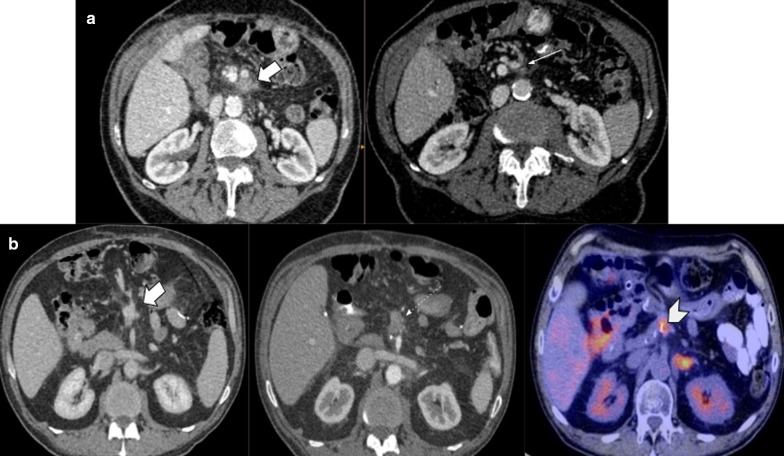
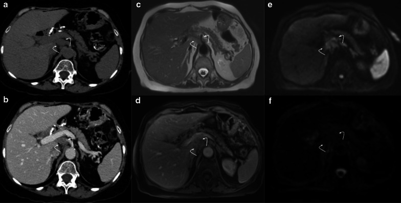
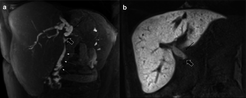
Pancreaticoduodenectomy represents a major surgery for tumors located at the pancreatic head and the ampullary/periampullary region. This complex procedure is associated with a high morbidity rate. Many surgical techniques have been proposed in order to reduce mortality rates, although post-procedure complications represent a current problem. Different imaging findings and complications may appear depending on the surgical technique used. Hence, radiologists should be familiarized with them to distinguish normal findings from real complications. The most challenging scenarios are represented by abdominal fluid collections, and tumor recurrence, that may frequently mimic normal postsurgical changes.

Keywords: Hepaticojejunostomy; Pancreatic cancer; Pancreatic fistula; Pancreaticoduodenectomy; Pancreaticojejunostomy.


Conflict of interest statement

The authors of this study have no competing interests.

Figures

Fig. 1
Fig. 1
Pancreaticoduodenectomy: (a) Classic procedure (Whipple’s) (b) Pylorus-preserving procedure
Fig. 2
Fig. 2
Pancreaticojejunostomy. Main surgical techniques: (a) Invagination (b) Duct-to-mucosa
Fig. 3
Fig. 3
Pancreaticojejunostomy. Duct-to-mucosa. CT-surgical correlation: (a) Surgical photograph. (b) Contrasted Coronal CT scan. References: Duct-to-mucosa pancreaticojejunostomy (discontinuous circle); blind segment of afferent limb (small arrows); liver (asterisk)
Fig. 4
Fig. 4
Hepaticojejunostomy. Radiologic-Surgical correlation: (a) Contrasted Coronal CT scan. (b) Coronal T2WI MRI image. (c) MRI Cholangiography. (d) Surgical photograph References: Partially collapsed afferent limb (white straight arrow). Pneumobilia (white curved arrow). Hepaticojejunostomy (thick arrow). Liver (asterisk)
Fig. 5
Fig. 5
Gastrojejunostomy and duodenojejunostomy: (a, b) Gastrojejunostomy. Axial and sagittal portal venous phase CT scan, respectively. (c, d) Duodenojejunostomy. Coronal and oblique sagittal view portal venous phase CT scan, respectively. Note the importance of using positive oral contrast to denote marked mucosal pattern change in A and B, and to depict the pylorus and latero-terminal anastomosis in duodenojejunostomy. References: White curved arrow: Surgical clips; Thick white arrows: gastric remnant; black arrows: ascended jejunal loop; Thick black arrow: latero-terminal anastomosis; arrowhead: pylorus; curved black arrows: oral contrast reflux to afferent limb. Black arrows: ascended jejunal loop
Fig. 6
Fig. 6
Intestinal reconstruction techniques: (a) Y de Roux intestinal reconstruction. Left: classic type; right: Machado type. (b) Child type reconstruction
Fig. 7
Fig. 7
Normal postoperative findings: (a) Peripancreatic fat stranding; (b) Mild pancreatic duct dilatation; (c) Perivascular cuffing; (d) Intrahepatic biliary duct enhancement and dilatation
Fig. 8
Fig. 8
Lymphadenopathy vs. Tumor recurrence. CT challenge. Two different patients (a) 63-year-old female patient with persisting lymph node along 2-year follow-up. Six months, one- and two-years CT scans, respectively. Note the decreasing size of the node over time (big arrow). Clinical and biological markers level stable. (b) 65-year-old male patient along annual CT scan controls. An increasing lymph node is seen during the follow-up (large arrow). Note the rounded shape appearance and the short axis size greater than 10 mm (dotted circle) after three years from surgery. Tumor recurrence was proven by biopsy in patient B
Fig. 9
Fig. 9
Collapsed loop versus Tumor recurrence. CT challenge. 58-year-old male patient with history of pancreaticoduodenectomy due to periampullary tumor. Control CT scans. (a) Heterogeneous, ill-defined image near pancreaticojejunostomy (big arrow) with peripheral fat stranding and contrast enhancement is seen after three months from surgery. No clinical symptoms or biological marker elevation were present. (b) 18 months CT scan control revealed total fat stranding resolution and demonstrated that the ill-defined enhancing image was due to a collapsed loop, which appears mild distended with normal fluid contain (arrowhead)
Fig. 10
Fig. 10
Collapsed loop vs tumor recurrence. The MRI role. 58-year-old male patient during routine follow-up after pancreaticoduodenectomy due to pancreatic adenocarcinoma. (a, b) Control CT scan. Well-defined enhancing soft tissue image (arrowhead) between the afferent loop (curved arrow) and the pancreaticojejunostomy. Collapsed loop versus tumor recurrence were suggested as possible diagnosis, so MRI was indicated. (c, d, e, f) MRI performed three days later revealed a heterogeneous ill-defined mass located between afferent loop (curved arrow) and pancreaticojejunostomy (straight arrow in d). Note the better tissue differentiation of MRI over CT, ruling out collapsed loop diagnosis. Restriction on DWI (e) and contrast enhancement (f) was also noted (arrowhead in e and f). Local recurrence was confirmed by PET-CT
Fig. 11
Fig. 11
Collapsed loop vs tumor recurrence. The MRI role. 62-year-old female patient during routine follow-up after pancreaticoduodenectomy due to pancreatic adenocarcinoma three months early. (a, c, e) Axial and coronal view contrast-enhanced CT scan. Homogeneous fluid collection (asterisk) in the surgical bed and related to an ill-defined soft tissue enhancing image with peripheral fat stranding associated (dotted arrows). Coronal view denotes a pseudo-mass image (arrowhead in c). Collapsed loop versus inflammatory changes were suggested as possible diagnosis, so strict control was indicated. (b, d, f) MRI performed two months later. Prior collection complete resolution. The ill-defined image related to the hepatic hilum is now better defined as the collapsed loop due to their fluid content and intestinal folds (dotted arrow). Coronal view denotes the blind portion of the afferent loop as the previously described pseudo-mass image (arrowhead in f)
Fig.12
Fig.12
Perivascular cuffing vs. tumor recurrence. CT challenge. Two different patients (a) 81-year-old male patient three weeks after pancreaticoduodenectomy due to ampulloma. (b) 45-year-old male patient with fever 21 days after pancreaticoduodenectomy due to adenocarcinoma. Soft-tissue imaging in the surgical bed (thick white arrows), in both cases strict follow-up was required. In patient A, the focal fat stranding has markedly resolved (thin arrow): while in patient B persists without significant change after a year (dotted arrow). PET/CT shows hypercaptation mass, which confirms tumor recurrence (arrowhead)
Fig. 13
Fig. 13
Tumor recurrence. DWI value. 69-year-old female patient during routine follow-up after pancreaticoduodenectomy due to pancreatic adenocarcinoma. (a, b) Non-contrast and portal venous phase CT scan, respectively. Shows an ill-defined soft tissue enhancing image in the surgical bed (curved arrows). (c, d, e, f) Six month later MRI control. Persistent image surrounding the celiac trunk with no significant progression (curved arrows in c and d). DWI (e) and ADC map (f) shows restriction, raising local recurrence diagnosis (curved arrows in e and f). The patient evolved unfavorably with disease progression
Fig.14
Fig.14
Benign duct dilatation vs. tumor recurrence. (a) 60-year-old female patient during acute abdominal pain. MRI showed pancreatic duct dilatation with filling defect (white straight arrow) just proximal to pancreaticojejunostomy. Acute stump pancreatitis was the final diagnosis. Endoscopic ultrasound confirmed mucoid plug related to pancreaticojejunostomy. Previous MRI (upper) denoted normal wirsung caliber. (b) MRI scan control in a 68-year-old male patient with pancreaticoduodenectomy due to pancreatic cancer. MRI performed three years after surgery reveals increased pancreatic duct dilatation related to an ill-defined hypovascular focal image in the pancreas body (curved arrow in magnified image). Lower: prior CT scan control denotes normal wirsung caliber. Endoscopy ultrasound guided biopsy confirmed adenocarcinoma recurrence
Fig. 15
Fig. 15
Pancreatic fistula, (a, b) Axial and coronal portal venous phase CT scan, respectively. 72-year-old male patient with pancreaticoduodenectomy due to neuroendocrine tumor, during second-month control CT scan. Homogeneous collection (big arrow) in surgical bed. That collection impresses to be in contiguity with the PJ (straight arrow). Normal pancreatic stump (curved arrow) and afferent loop (arrowhead) are also noted
Fig. 16
Fig. 16
Abdominal abscess (a) Axial non-contrast CT scan. (b) Axial portal venous phase CT scan. 52-year-old female patient with fever after 20 days of pancreaticoduodenectomy due to mucinous cystic tumor. CT scan showed heterogeneous fluid collection with air content (big arrow). Drainage (dotted arrow) analysis disclosed purulent material
Fig. 17
Fig. 17
Pancreatic fistula vs. Abscess. Two different patients. Axial portal venous phase CT scans: (a) 73-year-old male patient with abdominal pain 25 days after pancreaticoduodenectomy due to pancreatic neuroendocrine tumor. (b) 45-year-old male patient with fever 21 days after pancreaticoduodenectomy due to ampulloma. Both present an homogeneous fluid collection near the PJ (big arrow). In patient B impresses to exist a thin wall during portal phase (dotted arrow). Both of them were percutaneous drainage with elevated amylase concentration in A and purulent fluid in B.
Fig. 18
Fig. 18
Pancreatojejunostomy stenosis and recurrent pancreatitis (a) 72-year-old male patient with history of pancreaticoduodenectomy due to chronic pancreatitis in the setting of acute stump pancreatitis (big arrow). Endoscopic ultrasound revealed pancreaticojejunostomy with decreased caliber. (b) Control CT scan performed three months later denote normal wirsung
Fig. 19
Fig. 19
Biliary fistula 67-year-old female patient with increased fluid drainage after 60 days of pancreaticoduodenectomy due to ampuloma. (a, b, c) Axial and oblique sagittal portal venous phase CT scan. The intrahepatic biliary duct shows dilatation and fluid level within (white arrows in a and c). A collection in the surgical bed (empty arrow in b and c) and biliary connection (curved arrow in c) is also shown. Drainage analysis disclosed high bilirubin levels, which confirms the diagnosis of biliary fistula
Fig. 20
Fig. 20
Biliary vs pancreatic fistula. Two different patients (a) 65-year-old male patient during abdominal pain three weeks after pancreaticoduodenectomy due to adenocarcinoma. (b) 50-year-old male patient with fever 21 days after pancreaticoduodenectomy due to ampulloma. Both patients presented fluid collection near the surgical bed, the both were homogeneous and no enhancement was present (big arrows). Fluid drainage analysis confirmed biliary fistula in a and pancreatic fistula in b. Note the similar imaging appearance, being indistinguishable one fistula from each other
Fig. 21
Fig. 21
Hepaticojejunostomy stenosis. Two different patients (a) 67-year-old male patient with pancreaticoduodenectomy due to cholangiocarcinoma. All images below to normal postoperative control MRI. (a, b) one month after surgery (c, d) 7 months control. Note the caliber reduction in hepaticojejunostomy due to thickness of hepatic bile duct (curved arrows). Intraoperative cholangiography confirmed total hepaticojejunostomy stenosis (not shown) (b) 59-year-old male patient with abdominal discomfort and unexplained weight loss two years after pancreaticoduodenectomy owing to adenocarcinoma. CT scan denotes abrupt intrahepatic biliary stenosis (dotted arrow) due to ill-defined mass (big arrow). PET/CT fusion confirms local tumor recurrence as causal of stenosis
Fig. 22
Fig. 22
Hepatobiliary-specific contrast agent in hepaticojejunostomy assessment, Hepaticojejunostomy Permeability. Two different patients who underwent hepato-biliary surgeries with hepaticojejunostomy confection. MRI with hepatobiliary-specific contrast agent Gd-EOB-DTPA (gadoxetic acid) was performed due to postoperative biliary complication suspicion. (a, b) Hepaticojejunostomy (thick black arrows) permeability evaluation: MR Cholangiographic image shows afferent limb opacification (dotted arrows in a) that confirmed permeability, while stop in hepaticojejunostomy during hepatobiliary late phase (i.e., 5 h after IV agent injection) depict anastomosis stenosis. Common bile duct (thin black arrow)
Fig. 23
Fig. 23
Delayed gastric emptying. 57-year-old female patient inability to feed orally with abdominal pain and distension 3 weeks after pylorus-preserving pancreaticoduodenectomy. (a, b) CT scans during acute event revealed marked stomach distention (big arrows). (c, d) Normal volume regression after medical treatment (big arrows). Duodenojejunostomy is also noted (dotted arrows in b and d)
Fig. 24
Fig. 24
Gastrojejunostomy dehiscence. 87-year-old male patient with complicated early postoperative duodenopancreatectomy with Y de Roux and gastrojejunostomy due to pancreatic adenocarcinoma. Axial and sagittal non-contrast (ac) and portal venous phase (bd) CT scan. Shows a blood collection (big arrows in a and b) associated with pneumoperitoneum (thin arrows in c and d) adjacent to the gastroenteric anastomosis (dotted arrow in d). Urgent laparoscopy confirmed gastroenteric anastomosis dehiscence
Fig. 25
Fig. 25
Afferent limb syndrome. 55-year-old male patient with Y de Roux reconstruction during abdominal pain and jaundice. Multimodality imaging assessment revealed marked distension of afferent limb (big arrow). Hepaticojejunostomy was permeable (curved arrow in b and d), and intrahepatic biliary dilatation is also noted (dotted arrows in a, b and d). Surgery confirmed afferent limb syndrome caused by limb stricture near entero-enteric anastomosis
Fig. 26
Fig. 26
Postoperative hemorrhage. 67-year-old patient during early complicated postoperative. ac Axial non-contrast, arterial and portal venous phase, respectively. Shows blood collection in the surgical bed (thick white arrow in (a), no active bleeding was seen. Remnant pancreas was preserved (dotted arrow in b and c). (d) CT scan performed three days later due to decreasing hematocrit shows known hematic collection increased in size (curved arrows). (e, f) Digital angiography shows no signs of active bleeding, but there is diffuse spasm of the celiac trunk (dotted black arrows in e) Portal-mesenteric veins were preserved. Exploratory laparotomy confirms hemoperitoneum with retroperitoneal branch-dependent bleeding
Fig. 27
Fig. 27
Right hepatic pseudoaneurysm. 78-year-old male patient with a history of pancreaticoduodenectomy with right hepatic artery reconstruction for pancreatic adenocarcinoma under study due to hematocrit decrease. (a, b, c, d) Multiphase CT shows a focus of focal enhancement in arterial phase in the surgical bed (arrow in b), which persists in post-arterial phases (big arrows in c and d). (e) 3D reconstruction shows saccular dilatation (arrow) dependence of the right branch of the hepatic artery (dotted arrow). (f) Digital angiography confirms the diagnosis of pseudoaneurysm
Fig. 28
Fig. 28
Hepatic infarct. 78-year-old patient with complicated early postoperative due to pseudoaneurysm. Stent in the hepatic artery was placed. Control CT scan due to increased hematic fluid drainage evidence occluded stent (blank arrow in a) and big subcapsular hypodense areas in liver parenchyma (curved arrows in b and d) consisting with infarct. An active bleeding focus depending on the ligated gastroduodenal branch was also detected (big arrow in c)

References

    1. Simon R. Complications after pancreaticoduodenectomy. Surg Clin North Am. 2021;101:865–874. doi: 10.1016/j.suc.2021.06.011. - DOI - PubMed
    1. Busquets J, Fabregat J, Masuet C, et al. Surgical treatment of pancreatic adenocarcinoma by cephalic duodenopancreatectomy (Part 1). Post-surgical complications in 204 cases in a reference hospital. Cir Esp. 2010;88:299–307. doi: 10.1016/j.ciresp.2010.05.006. - DOI - PubMed
    1. (2018) Whipple procedure. AORN J 108(5):P11–P13 - PubMed
    1. Chincarini M, Zamboni GA, Mucelli RP. Major pancreatic resections: normal postoperative findings and complications. Insights Imaging. 2018;9:173–187. doi: 10.1007/s13244-018-0595-4. - DOI - PMC - PubMed
    1. Malleo G, Pulvirenti A, Bassi C, et al. Diagnosis and management of postoperative pancreatic fistula. Langenbecks Arch Surg. 2014;399:801–810. doi: 10.1007/s00423-014-1242-2. - DOI - PubMed

LinkOut - more resources