. 2023 Jan;145(1):127-143.
doi: 10.1007/s00401-022-02510-8. Epub 2022 Oct 20.

Loss of function variants in DNAJB4 cause a myopathy with early respiratory failure

Affiliations

Loss of function variants in DNAJB4 cause a myopathy with early respiratory failure

Conrad C Weihl et al. Acta Neuropathol. 2023 Jan.

Abstract

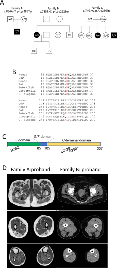
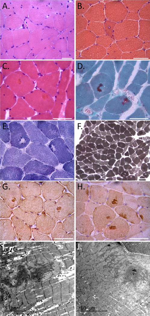
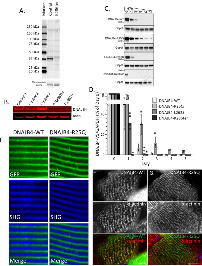
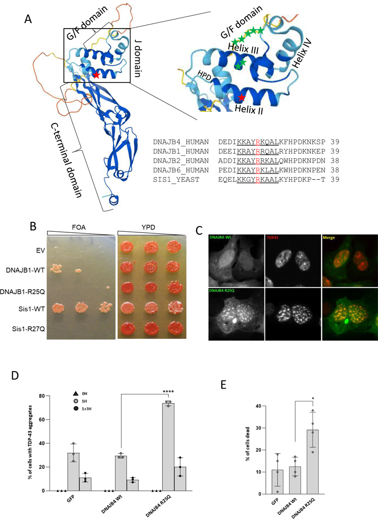
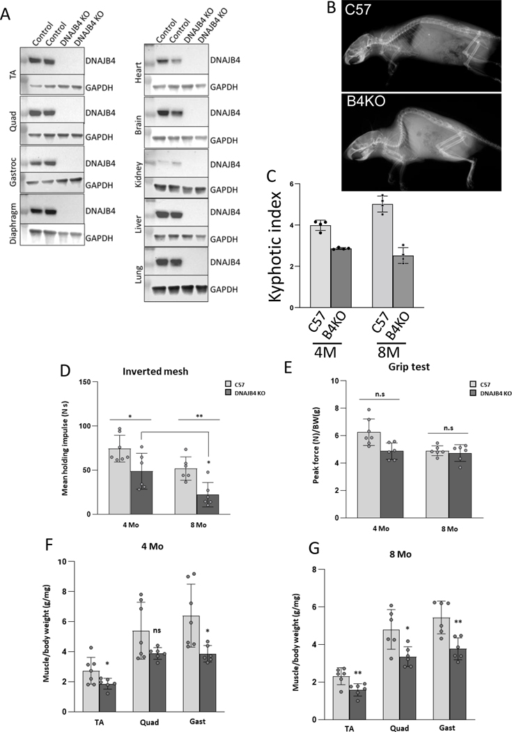
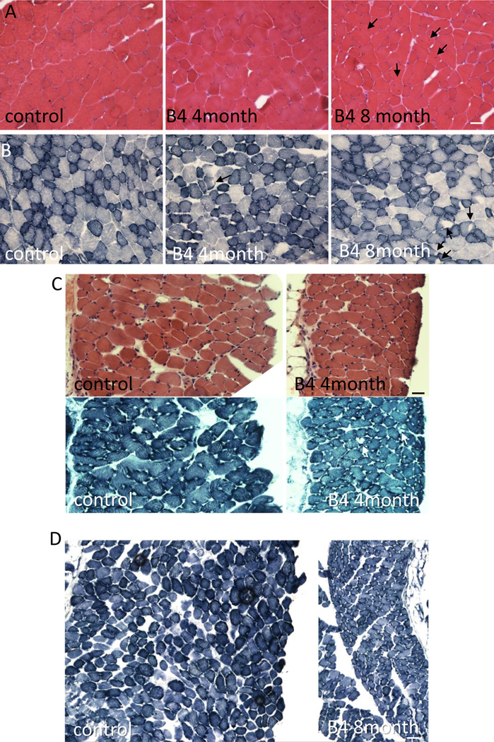
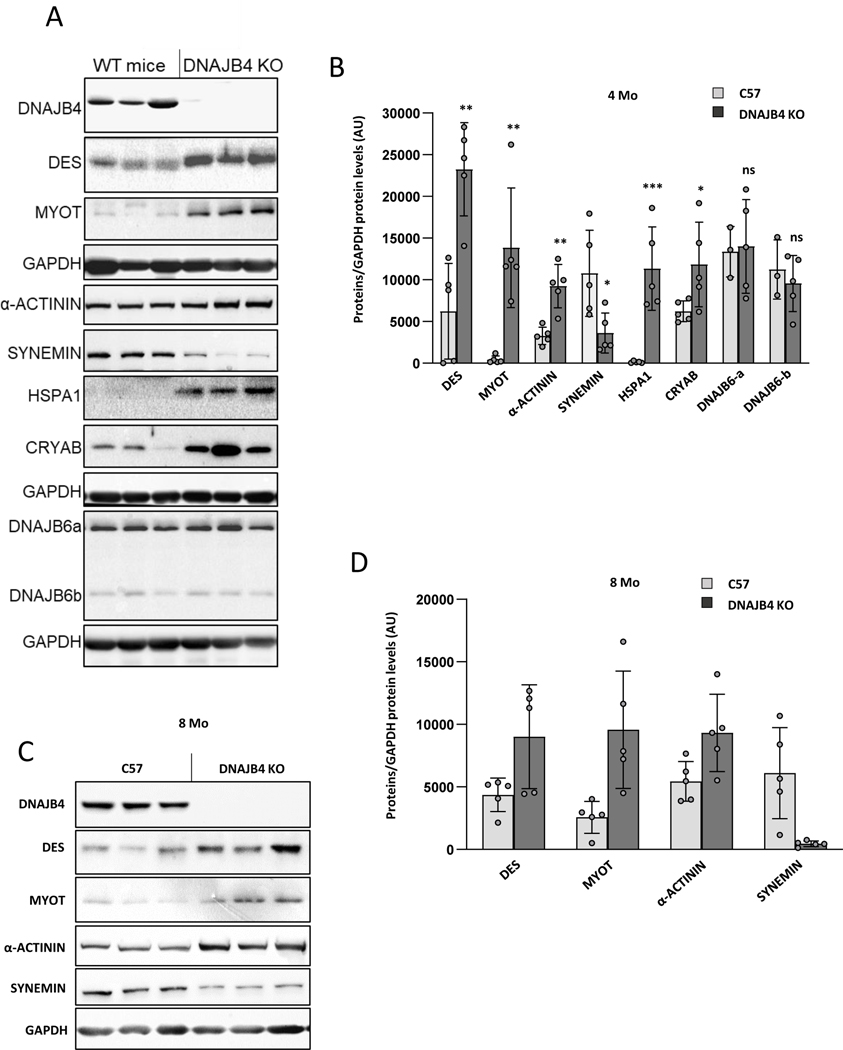
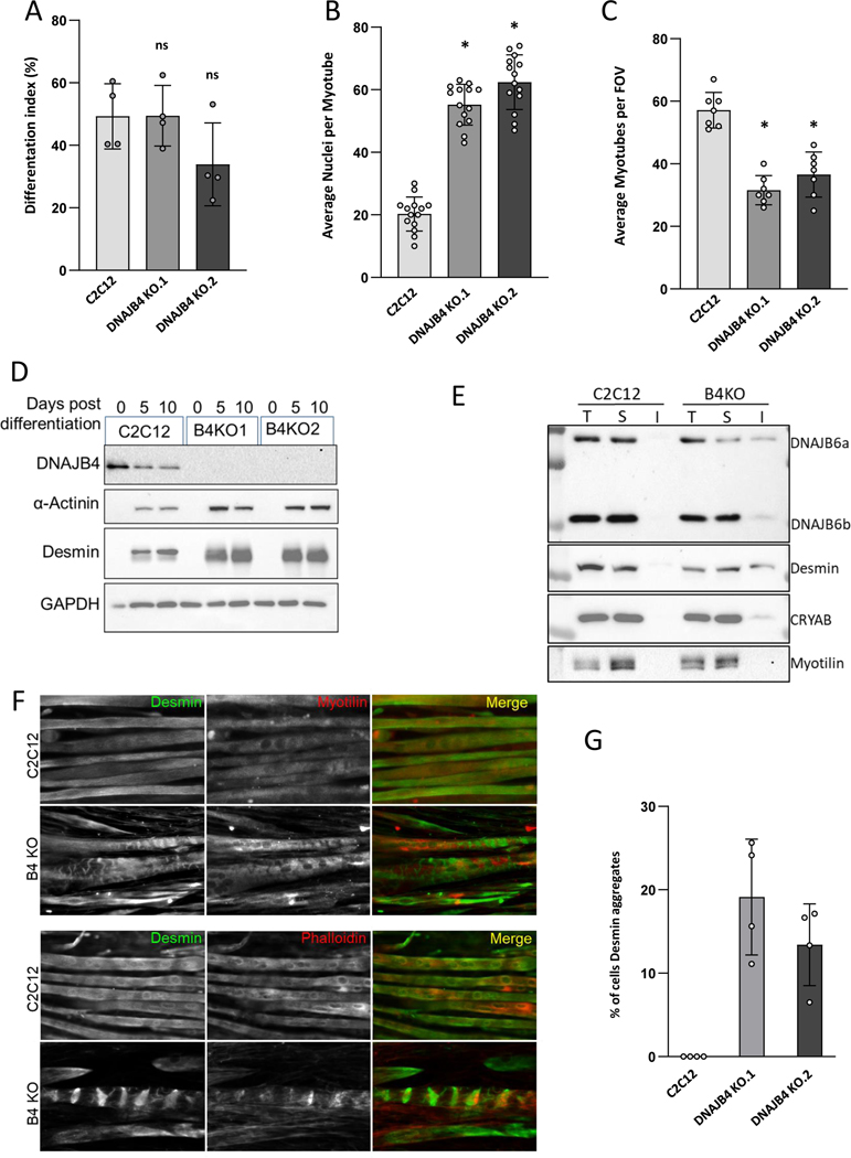
DNAJ/HSP40 co-chaperones are integral to the chaperone network, bind client proteins and recruit them to HSP70 for folding. We performed exome sequencing on patients with a presumed hereditary muscle disease and no genetic diagnosis. This identified four individuals from three unrelated families carrying an unreported homozygous stop gain (c.856A > T; p.Lys286Ter), or homozygous missense variants (c.74G > A; p.Arg25Gln and c.785 T > C; p.Leu262Ser) in DNAJB4. Affected patients presented with axial rigidity and early respiratory failure requiring ventilator support between the 1st and 4th decade of life. Selective involvement of the semitendinosus and biceps femoris muscles was seen on MRI scans of the thigh. On biopsy, muscle was myopathic with angular fibers, protein inclusions and occasional rimmed vacuoles. DNAJB4 normally localizes to the Z-disc and was absent from muscle and fibroblasts of affected patients supporting a loss of function. Functional studies confirmed that the p.Lys286Ter and p.Leu262Ser mutant proteins are rapidly degraded in cells. In contrast, the p.Arg25Gln mutant protein is stable but failed to complement for DNAJB function in yeast, disaggregate client proteins or protect from heat shock-induced cell death consistent with its loss of function. DNAJB4 knockout mice had muscle weakness and fiber atrophy with prominent diaphragm involvement and kyphosis. DNAJB4 knockout muscle and myotubes had myofibrillar disorganization and accumulated Z-disc proteins and protein chaperones. These data demonstrate a novel chaperonopathy associated with DNAJB4 causing a myopathy with early respiratory failure. DNAJB4 loss of function variants may lead to the accumulation of DNAJB4 client proteins resulting in muscle dysfunction and degeneration.

Keywords: Chaperone; Congenital myopathy; Myofibrillar myopathy; Myopathy; Protein aggregation.


Figures

Figure 1:
Figure 1:
A) Pedigrees of three families with homozygous DNAJB4 variants. Affected patients are in black. The genotype of the patient is represented within the pedigree. B) ClustalW alignment of DNAJB4 amino acid sequence from multiple species demonstrating conservation at R25 and L262. C) Schematic of the DNAJB4 denoting the J, G/F rich and C-terminal domains and the location of the identified variants. D) Lower extremity imaging shows involvement of the semitendinosus, biceps femoris and soleus muscles of the proband from family A (MRI) and the involvement of the gluteus maximus and semitendinosus muscles of the proband from family B (CT scan).
Figure 2:
Figure 2:
A) H&E staining of left biceps from patient B:I. B-C) H&E demonstrating a rimmed vacuole (B) and eosinophilic inclusions (C) from a deltoid biopsy of patient A:I. D) Modified Gomori trichrome staining of patient A:I demonstrating dark central inclusions. E) Many fiber regions have diminished oxidative enzyme activity (NADH) that sometimes is accentuated around the larger inclusions. F) These regions have also diminished had reduced ATPase staining, especially in type 1 fibers (ATPase 4,6). Scale bars are 50μM. G-H) Immunohistochemical staining for (G) desmin and (H) myotilin of muscle from patient A:I. I-J) Electron microscopy of muscle from patient A:I. Scale bars are 5 and 10μM respectively.
Figure 3:
Figure 3:
A) Immunoblot of skeletal muscle tissue lysates from control patient muscle or patient A:I carrying homozygous p.Lys286Ter variants in DNAJB4 with an anti-DNAJB4 antibody. B) Immunoblot from lysates of control patient fibroblasts or primary fibroblasts from patient A:I (p.Lys286Ter) and B:I (p.Leu262Ser) with anti-DNAJB4 or anti-actin antibodies. C) Isogenic 293T cells stably expressing V5 tagged DNAJB4-WT, DNAJB4-R25Q, DNAJB4-L262S or DNAJB4-K286Ter under a tetracycline inducible promoter were treated with tetracycline for 48 hours and then lysates were harvested for five days following tetracycline removal. Lysates were then immunoblotted for V5 (DNAJB4) and GAPDH. D) Quantitation of percent change in the ratio of DNAJB4-V5:GAPDH levels from lysates in (C). N=3/condition. * indicate a p value of < 0.01. E) Two-photon confocal microscopy of mouse footpad muscle electroporated with GFP tagged DNAJB4-WT or DNAJB4-R25Q (green) with associated second harmonic generation imaging (SHG). DNAJB4 is at the Z-disc as SHG marks the A-band. F-G) Immunofluorescent images of a single myofiber from mouse tibialis anterior muscle electroporated with a GFP tagged DNAJB4-WT (green) (F) or DNAJB4-R25Q (green) (G) and then immunostained with an antibody to α-actinin (red). Scale bar is 10μM.
Figure 4:
Figure 4:
A) Rendering of DNAJB4 structure denoting the C-terminal, G/F and J domains with and enlargement of the J-domain showing Helix I-IV. Red star marks the R25 residue, green stars denote J domain residues (A50 and E54) and G/F domain residues (F89/F91/F93/N95/P96/D98/F100) mutated in DNAJB6 associated myopathy. Alignment of Helix II (underlined) from DNAJB4, DNAJB1, DNAJB2, DNAJB6 and Sis1. The R25 (DNAJB4/DNAJB1) and the R27 (Sis1) residue is in red. B) Yeast spottings from colonies that delete Sis1 and then complement with empty vector (EV), DNAJB-WT, DNAJB1-R25Q, Sis1-WT or Sis1-R27Q when plated on FOA media (left spottings). Spottings on full media (YPD) are on the right. C) Representative images of Hela cells transfected with GFP-DNAJB4-WT (green), or GFP-DNAJB4-R25Q (green) and mCherry-TDP-43 (red) one hour post heat shock. D) HeLa cells were co-transfected with GFP, GFP-DNAJB4-WT, or GFP-DNAJB4-R25Q and mCherry-TDP-43. The percentage of cells with TDP-43 nuclear inclusions at baseline (0H), post-heat shock (1H) or following heat shock recovery (1+3H) are represented graphically. E) HeLa cells were co-transfected with GFP, GFP-DNAJB4-WT, or GFP-DNAJB4-R25Q and mCherry-TDP-43 transfected with the indicated constructs, subjected to heat shock at 42°C for 1 hour, and the percentage of ethidium homodimer-1–positive cells was quantitated. Data are presented as the percentage of cells found positive/dead.
Figure 5:
Figure 5:
A) Lysates from skeletal muscle and tissues indicated from DNAJB4 homozygous knockout mice or control littermates immunoblotted with an antibody to DNAJB4 or GAPDH. B) X-ray image highlighting the skeleton of 8 month old DNAJB4 knockout (B4KO) or C57 control. C) Quantitation of the kyphotic index from 4 and 8 month old control or DNAJB4 knockout (B4KO) mice. D) Mean holding impulse on an inverted screen for 4 and 8 month old C57 control and DNAJB4 KO mice. E) Peak force forelimb grip strength testing for 4- and 8-month-old C57 control and DNAJB4 KO mice. F-G) Weight of indicated isolated muscles (tibialis anterior (TA), gastrocnemius (Gast) and quadriceps (Quad)) normalized to total body weight for 4- and 8-monthold C57 control and DNAJB4 KO mice.
Figure 6:
Figure 6:
A) H&E staining of quadriceps muscle from 8-month-old control or 4- and 8-month old DNAJB4 KO mouse muscle. Fibers with internal nuclei are denoted with black arrows. B) NADH staining of tibialis anterior muscle from 8-month-old control or 4- and 8-month-old DNAJB4 KO mouse muscle. Fibers with central clearings are denoted with black arrows. C) H&E and NADH staining of diaphragm muscle from 4-month-old control or 4-month-old DNAJB4KO mice. Note myofiber atrophy and decreased diaphragm thickness. White arrows denote central clearings on NADH. D) NADH staining of diaphragm muscle from 8-month-old control or 8-month-old DNAJB4KO mice. Note myofiber atrophy and decreased diaphragm thickness. Scale bars are 50μM.
Figure 7:
Figure 7:
A) Immunoblots of lysates from 4-month-old mouse tibialis anterior of control or DNAJB4 knockout mice with antibodies to DNAJB4, desmin (DES), myotilin (MYOT), α-actinin, synemin, HSPA1, CRYAB, DNAJB6 or GAPDH. B) Quantitation of immunoblotted proteins from TA muscle at 4 months. N=5/condition. *, **, *** indicate p values 0.01, 0.001 and 0.0001. C) Immunoblots of lysates from 8-month-old mouse tibialis anterior of control or DNAJB4 knockout mice with antibodies to DNAJB4, desmin (DES), myotilin (MYOT), α-actinin, synemin or GAPDH. D) Quantitation of immunoblotted proteins from TA muscle at 8 months. N=5/condition. *, **, *** indicate p values 0.01, 0.001 and 0.0001.
Figure 8:
Figure 8:
A-C) Control C2C12 or one of two different DNAJB4 knockout (B4KO) lines were placed in differentiation media for 5 days and then the differentiation index, number of nuclei/myotube and total number of myotubes was quantitated. D) Immunoblots of lysates from undifferentiated (0) or 5- and 10-days post differentiation of control or DNAJB4 knockout C2C12 cells (B4KO1 or B4KO2) with antibodies to DNAJB4, desmin, α-actinin or GAPDH. E) Immunoblots of lysates from differentiated (5 days) C2C12 or B4KO myotubes following detergent lysis and ultracentrifugation into a total (T) lysate, soluble (S) fraction and insoluble (I) fraction with antibodies to DNAJB6, desmin, CRYAB and myotilin. F) Immunofluorescent images of 5 day differentiated C2C12 or B4 KO myotubes using an antibody to desmin (green) and myotilin (red upper panels) or phalloidin (red lower panels). G) Graph of the percentage of fibers with desmin inclusions in control or DNAJB4 KO myotubes.

References

    1. Topf A. et al. Sequential targeted exome sequencing of 1001 patients affected by unexplained limb-girdle weakness. Genet Med 22, 1478–1488, doi:10.1038/s41436-020-0840-3 (2020). - DOI - PMC - PubMed
    1. Sarparanta J, Jonson PH, Kawan S. & Udd B. Neuromuscular Diseases Due to Chaperone Mutations: A Review and Some New Results. Int J Mol Sci 21, doi:10.3390/ijms21041409 (2020). - DOI - PMC - PubMed
    1. Hohfeld J. et al. Maintaining proteostasis under mechanical stress. EMBO Rep 22, e52507, doi:10.15252/embr.202152507 (2021). - DOI - PMC - PubMed
    1. Sarparanta J. et al. Mutations affecting the cytoplasmic functions of the co-chaperone DNAJB6 cause limb-girdle muscular dystrophy. Nat Genet 44, 450–455, S451–452, doi:10.1038/ng.1103 (2012). - DOI - PMC - PubMed
    1. Harms MB et al. Exome sequencing reveals DNAJB6 mutations in dominantly-inherited myopathy. Ann Neurol 71, 407–416, doi:10.1002/ana.22683 (2012). - DOI - PMC - PubMed

Publication types

MeSH terms

Substances