

Methods used for the study of endophytic fungi: a review on methodologies and challenges, and associated tips
- PMID: 36264513
- PMCID: PMC9584250
- DOI: 10.1007/s00203-022-03283-0
Methods used for the study of endophytic fungi: a review on methodologies and challenges, and associated tips
Abstract
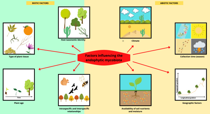
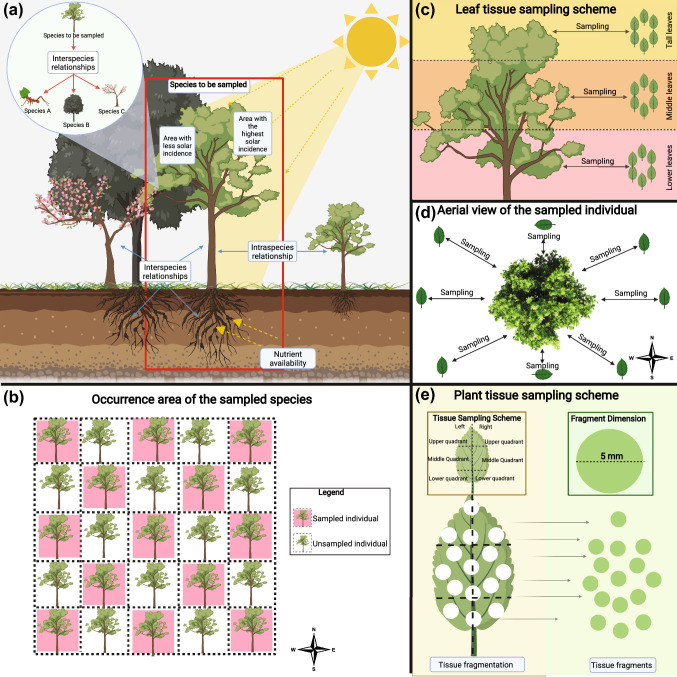
Endophytic fungi are microorganisms that colonize the interior of plant tissues (e.g. leaves, seeds, stem, trunk, roots, fruits, flowers) in intracellular and/or extracellular spaces without causing symptoms of disease in host plants. These microorganisms have been isolated from plant species in a wide variety of habitats worldwide, and it is estimated that all terrestrial plants are colonized by one or more species of endophytic fungus. In addition, these microorganisms have been drawing the attention of researchers because of their ability to synthesize a wide range of bioactive molecules with potential for applications in agriculture, medicine and biotechnology. However, several obstacles come up when studying the diversity and chemical potential of endophytic fungi. For example, the usage of an inappropriate surface disinfection method for plant tissue may not eliminate the epiphytic microbiota or may end up interfering with the endophytic mycobiota, which consequently generates erroneous results. Moreover, the composition of the culture medium and the culture conditions can favor the growth of certain species and inhibit others, which generates underestimated results. Other inconsistencies can arise from the fungus misidentification and consequent exploration of its chemical potential. Based on the methodological biases that may occur at all stages of studies dealing with endophytic fungi, the objective of this review is to discuss the main methods employed in these studies as well as highlight the challenges derived from the different approaches. We also report associated tips to help future studies on endophytic fungi as a contribution.
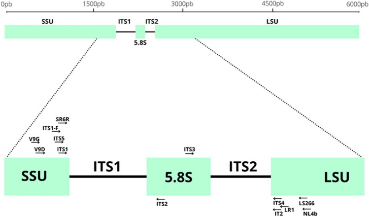
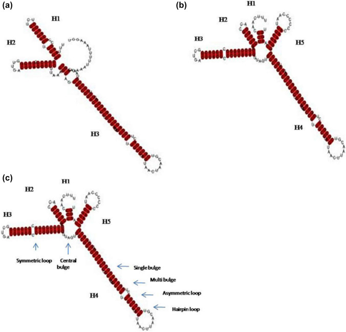
Keywords: Diversity; Endophyte; ITS; Molecular identification; Mycobiome; Symbiosis.
© 2022. The Author(s), under exclusive licence to Springer-Verlag GmbH Germany, part of Springer Nature.
Conflict of interest statement
The authors declare that the work was conducted in the absence of any commercial or financial relationship that could be construed as a potential conflict of interest.
Figures

References
-
- Abarenkov K, Tedersoo L, Nilsson RK, Vellak K, Saar I, Veldre V, et al. PlutoF—a web based workbench for ecological and taxonomic research, with an online implementation for fungal its sequences. Evol Bioinf. 2010;6:189–196. doi: 10.4137/EBO.S6271. - DOI
-
- Agbessenou A, Akutse KS, Yusuf AA, Ekesi S, Subramanian S, Khamis FM. Endophytic fungi protect tomato and nightshade plants against Tuta absoluta (Lepidoptera: Gelechiidae) through a hidden friendship and cryptic battle. Sci Rep. 2020;10(1):22195. doi: 10.1038/s41598-020-78898-8. - DOI - PMC - PubMed
-
- Aoki T, O'Donnell K, Homma Y, Lattanzi AR. Sudden-death syndrome of soybean is caused by two morphologically and phylogenetically distinct species within the fusarium solani species complex— F. virguliforme in North America and F. tucumaniae in South America. Mycologia. 2003;95:660–684. doi: 10.1080/15572536.2004.11833070. - DOI - PubMed
Publication types
MeSH terms
LinkOut - more resources
Full Text Sources
Medical
