

The electrical heart axis in fetuses with congenital heart disease, measured with non-invasive fetal electrocardiography
- PMID: 36264863
- PMCID: PMC9584524
- DOI: 10.1371/journal.pone.0275802
The electrical heart axis in fetuses with congenital heart disease, measured with non-invasive fetal electrocardiography
Abstract
Objectives: To determine if the electrical heart axis in different types of congenital heart defects (CHD) differs from that of a healthy cohort at mid-gestation.
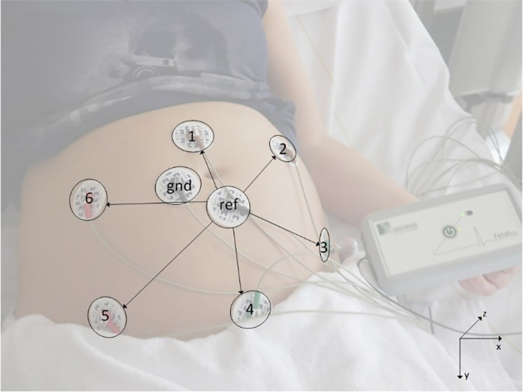
Methods: Non-invasive fetal electrocardiography (NI-fECG) was performed in singleton pregnancies with suspected CHD between 16 and 30 weeks of gestation. The mean electrical heart axis (MEHA) was determined from the fetal vectorcardiogram after correction for fetal orientation. Descriptive statistics were used to determine the MEHA with corresponding 95% confidence intervals (CI) in the frontal plane of all fetuses with CHD and the following subgroups: conotruncal anomalies (CTA), atrioventricular septal defects (AVSD) and hypoplastic right heart syndrome (HRHS). The MEHA of the CHD fetuses as well as the subgroups was compared to the healthy control group using a spherically projected multivariate linear regression analysis. Discriminant analysis was applied to calculate the sensitivity and specificity of the electrical heart axis for CHD detection.
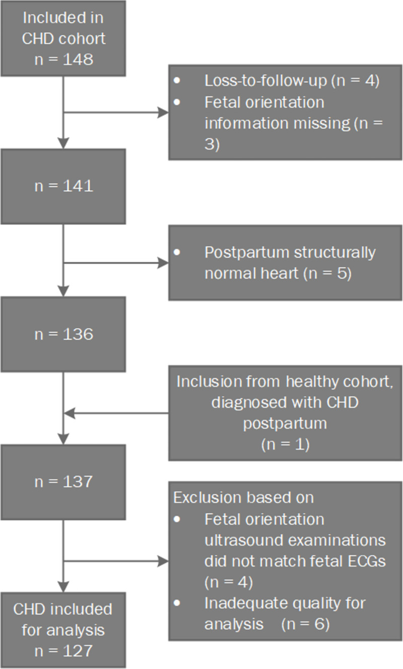
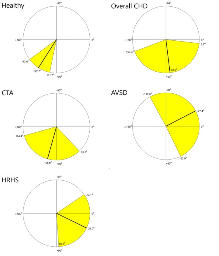
Results: The MEHA was determined in 127 fetuses. The MEHA was 83.0° (95% CI: 6.7°; 159.3°) in the total CHD group, and not significantly different from the control group (122.7° (95% CI: 101.7°; 143.6°). The MEHA was 105.6° (95% CI: 46.8°; 164.4°) in the CTA group (n = 54), -27.4° (95% CI: -118.6°; 63.9°) in the AVSD group (n = 9) and 26.0° (95% CI: -34.1°; 86.1°) in the HRHS group (n = 5). The MEHA of the AVSD and the HRHS subgroups were significantly different from the control group (resp. p = 0.04 and p = 0.02). The sensitivity and specificity of the MEHA for the diagnosis of CHD was 50.6% (95% CI 47.5% - 53.7%) and 60.1% (95% CI 57.1% - 63.1%) respectively.
Conclusion: The MEHA alone does not discriminate between healthy fetuses and fetuses with CHD. However, the left-oriented electrical heart axis in fetuses with AVSD and HRHS was significantly different from the control group suggesting altered cardiac conduction along with the structural defect.
Trial registration: Clinical trial registration number: NL48535.015.14.
Conflict of interest statement
I have read the journal’s policy and the authors of this manuscript have the following competing interests: R. Vullings is shareholder of Nemo Healthcare BV.
Figures
References
MeSH terms
Supplementary concepts
LinkOut - more resources
Full Text Sources
Medical
