

Longitudinal microstructural MRI markers of demyelination and neurodegeneration in early relapsing-remitting multiple sclerosis: Magnetisation transfer, water diffusion and g-ratio
- PMID: 36265199
- PMCID: PMC9668599
- DOI: 10.1016/j.nicl.2022.103228
Longitudinal microstructural MRI markers of demyelination and neurodegeneration in early relapsing-remitting multiple sclerosis: Magnetisation transfer, water diffusion and g-ratio
Abstract
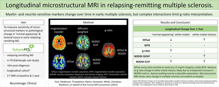
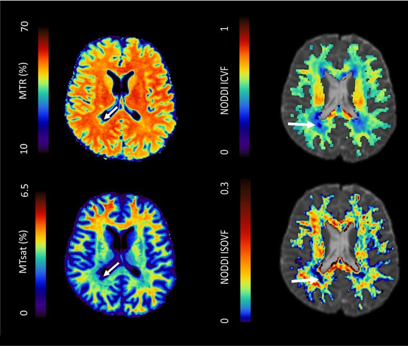
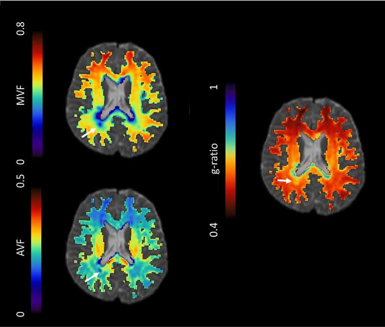
Introduction: Quantitative microstructural MRI, such as myelin-sensitive magnetisation transfer ratio (MTR) or saturation (MTsat), axon-sensitive water diffusion Neurite Orientation Dispersion and Density Imaging (NODDI), and the aggregate g-ratio, may provide more specific markers of white matter integrity than conventional MRI for early patient stratification in relapsing-remitting multiple sclerosis (RRMS). The aim of this study was to determine the sensitivity of such markers to longitudinal pathological change within cerebral white matter lesions (WML) and normal-appearing white matter (NAWM) in recently diagnosed RRMS.
Methods: Seventy-nine people with recently diagnosed RRMS, from the FutureMS longitudinal cohort, were recruited to an extended MRI protocol at baseline and one year later. Twelve healthy volunteers received the same MRI protocol, repeated within two weeks. Ethics approval and written informed consent were obtained. 3T MRI included magnetisation transfer, and multi-shell diffusion-weighted imaging. NAWM and whole brain were segmented from 3D T1-weighted MPRAGE, and WML from T2-weighted FLAIR. MTR, MTsat, NODDI isotropic (ISOVF) and intracellular (ICVF) volume fractions, and g-ratio (calculated from MTsat and NODDI data) were measured within WML and NAWM. Brain parenchymal fraction (BPF) was also calculated. Longitudinal change in BPF and microstructural metrics was assessed with paired t-tests (α = 0.05) and linear mixed models, adjusted for confounding factors with False Discovery Rate (FDR) correction for multiple comparisons. Longitudinal changes were compared with test-retest Bland-Altman limits of agreement from healthy control white matter. The influence of longitudinal change on g-ratio was explored through post-hoc analysis in silico by computing g-ratio with realistic simulated MTsat and NODDI values.
Results: In NAWM, g-ratio and ICVF increased, and MTsat decreased over one year (adjusted mean difference = 0.007, 0.005, and -0.057 respectively, all FDR-corrected p < 0.05). There was no significant change in MTR, ISOVF, or BPF. In WML, MTsat, NODDI ICVF and ISOVF increased over time (adjusted mean difference = 0.083, 0.024 and 0.016, respectively, all FDR-corrected p < 0.05). Group-level longitudinal changes exceeded test-retest limits of agreement for NODDI ISOVF and ICVF in WML only. In silico analysis showed g-ratio may increase due to a decrease in MTsat or ISOVF, or an increase in ICVF.
Discussion: G-ratio and MTsat changes in NAWM over one year may indicate subtle myelin loss in early RRMS, which were not apparent with BPF or NAWM MTR. Increases in NAWM and WML NODDI ICVF were not anticipated, and raise the possibility of axonal swelling or morphological change. Increases in WML MTsat may reflect myelin repair. Changes in NODDI ISOVF are more likely to reflect alterations in water content. Competing MTsat and ICVF changes may account for the absence of g-ratio change in WML. Longitudinal changes in microstructural measures are significant at a group level, however detection in individual patients in early RRMS is limited by technique reproducibility.
Conclusion: MTsat and g-ratio are more sensitive than MTR to early pathological changes in RRMS, but complex dependence of g-ratio on NODDI parameters limit the interpretation of aggregate measures in isolation. Improvements in technique reproducibility and validation of MRI biophysical models across a range of pathological tissue states are needed.
Keywords: Diffusion-weighted imaging; G-ratio; MTsat; Magnetization transfer imaging; Multiple sclerosis; NODDI.
Copyright © 2022 The Authors. Published by Elsevier Inc. All rights reserved.
Conflict of interest statement
Declaration of Competing Interest The authors declare the following financial interests/personal relationships which may be considered as potential competing interests: FutureMS, hosted by Precision Medicine Scotland Innovation Centre (PMS-IC) reports financial support was provided by Biogen UK Ltd.
Figures
References
-
- Alotaibi A., Podlasek A., AlTokhis A., Aldhebaib A., Dineen R.A., Constantinescu C.S. Investigating Microstructural Changes in White Matter in Multiple Sclerosis: A Systematic Review and Meta-Analysis of Neurite Orientation Dispersion and Density Imaging. Brain Sci. 2021;11(9) doi: 10.3390/brainsci11091151. - DOI - PMC - PubMed
-
- Al-Radaideh A., Mougin O.E., Lim S.Y., Chou I.J., Constantinescu C.S., Gowland P. Histogram analysis of quantitative T1 and MT maps from ultrahigh field MRI in clinically isolated syndrome and relapsing-remitting multiple sclerosis. NMR Biomed. 2015;28(11):1374–1382. doi: 10.1002/nbm.3385. - DOI - PubMed
-
- Bates D., Mächler M., Bolker B., Walker S. Fitting Linear Mixed-Effects Models Usinglme4. J Stat Software. 2015;67(1) doi: 10.18637/jss.v067.i01. - DOI
MeSH terms
Substances
Grants and funding
LinkOut - more resources
Full Text Sources
Medical
