. 2022 Nov 3;82(21):3985-4000.e4.
doi: 10.1016/j.molcel.2022.09.026. Epub 2022 Oct 19.

TERRA and RAD51AP1 promote alternative lengthening of telomeres through an R- to D-loop switch

Affiliations

TERRA and RAD51AP1 promote alternative lengthening of telomeres through an R- to D-loop switch

Tribhuwan Yadav et al. Mol Cell. .

Abstract

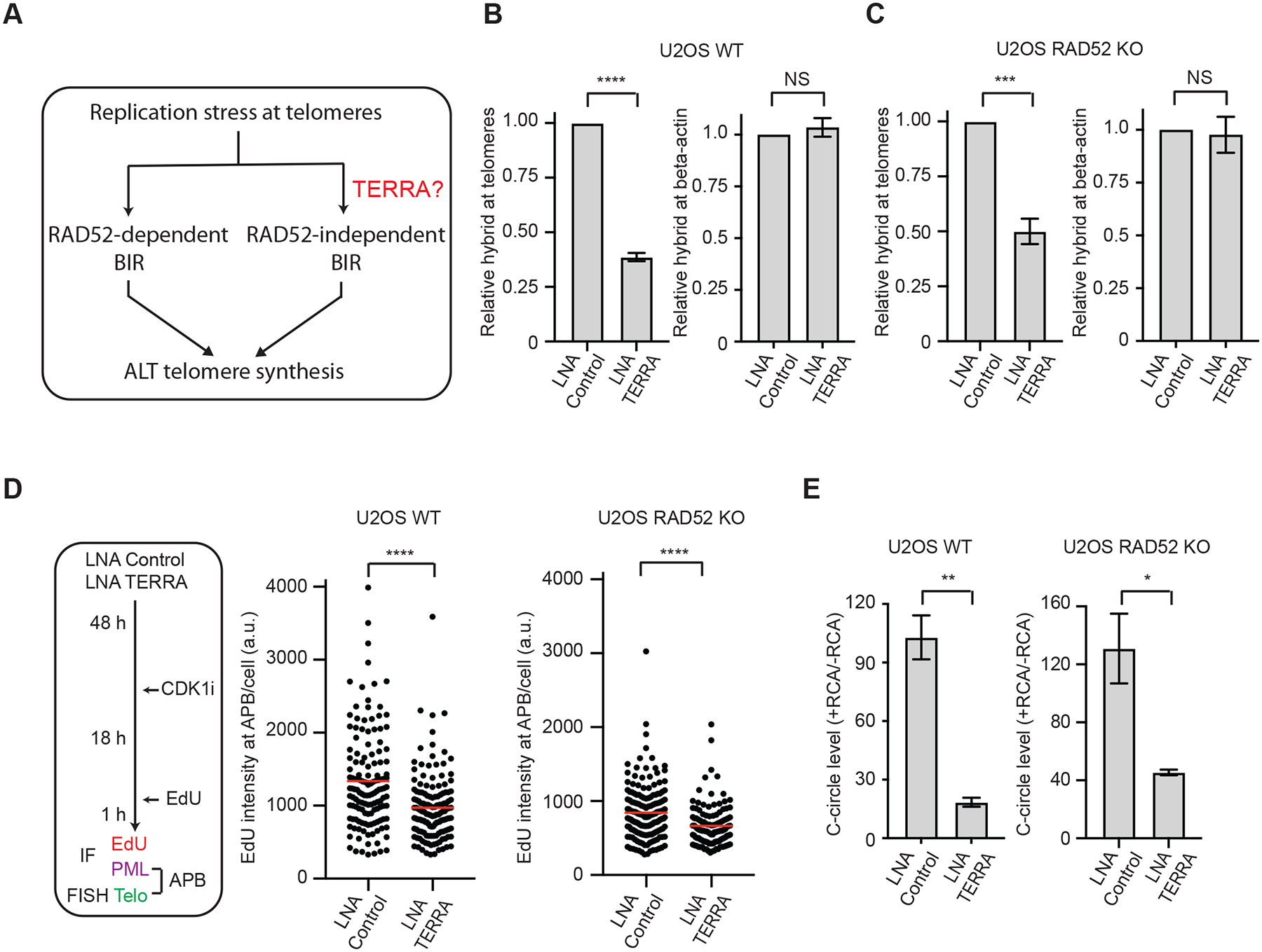
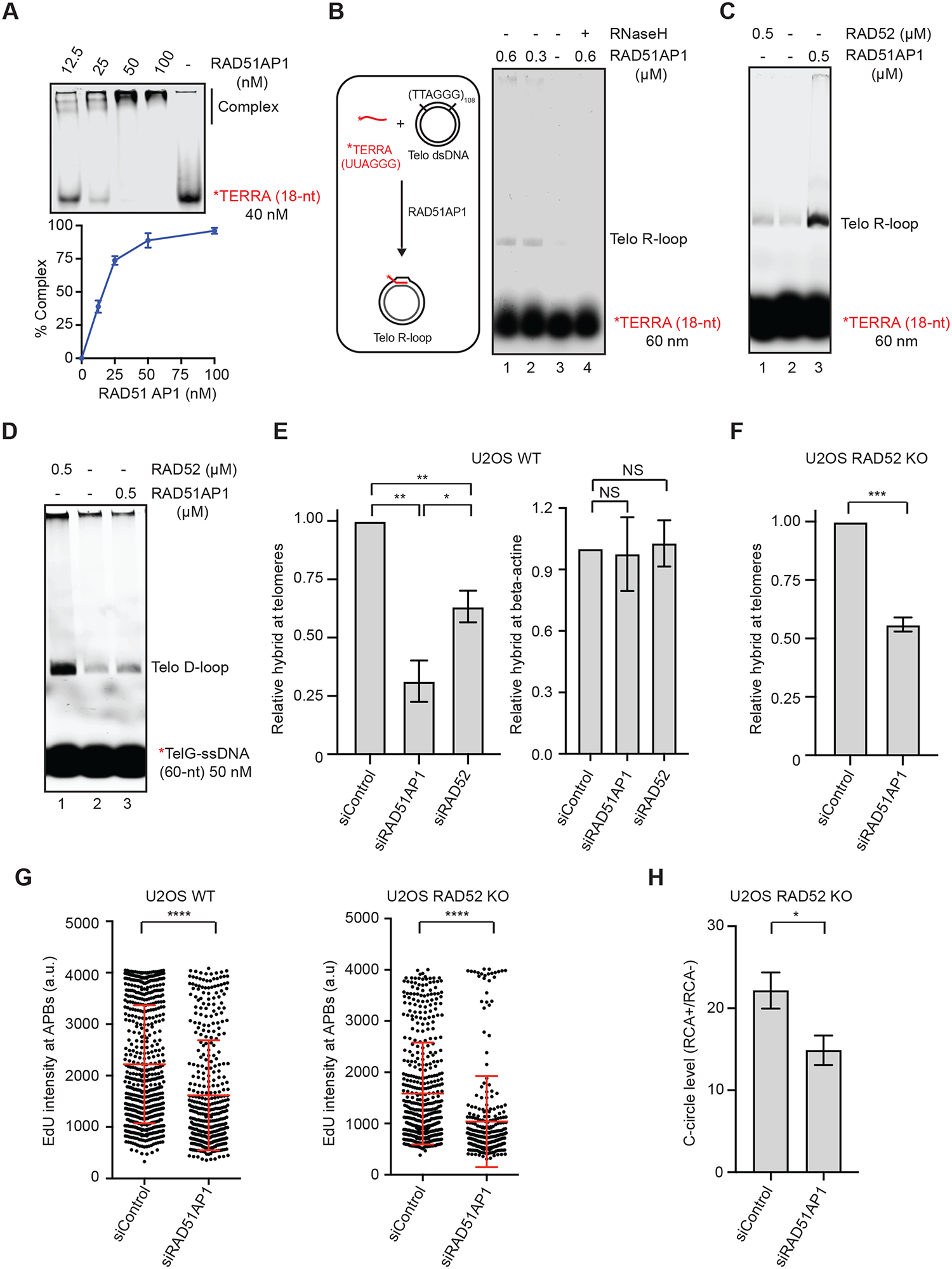
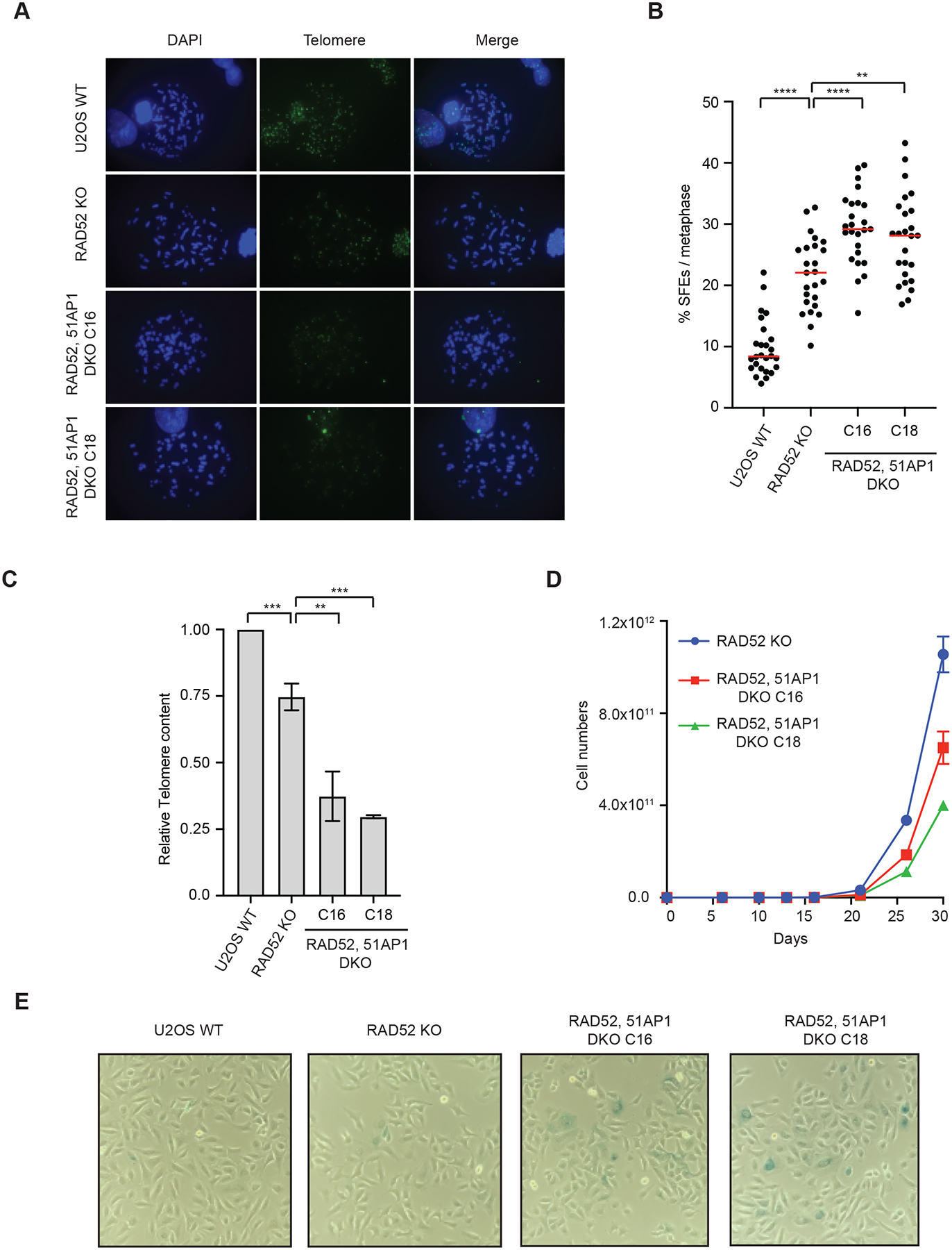
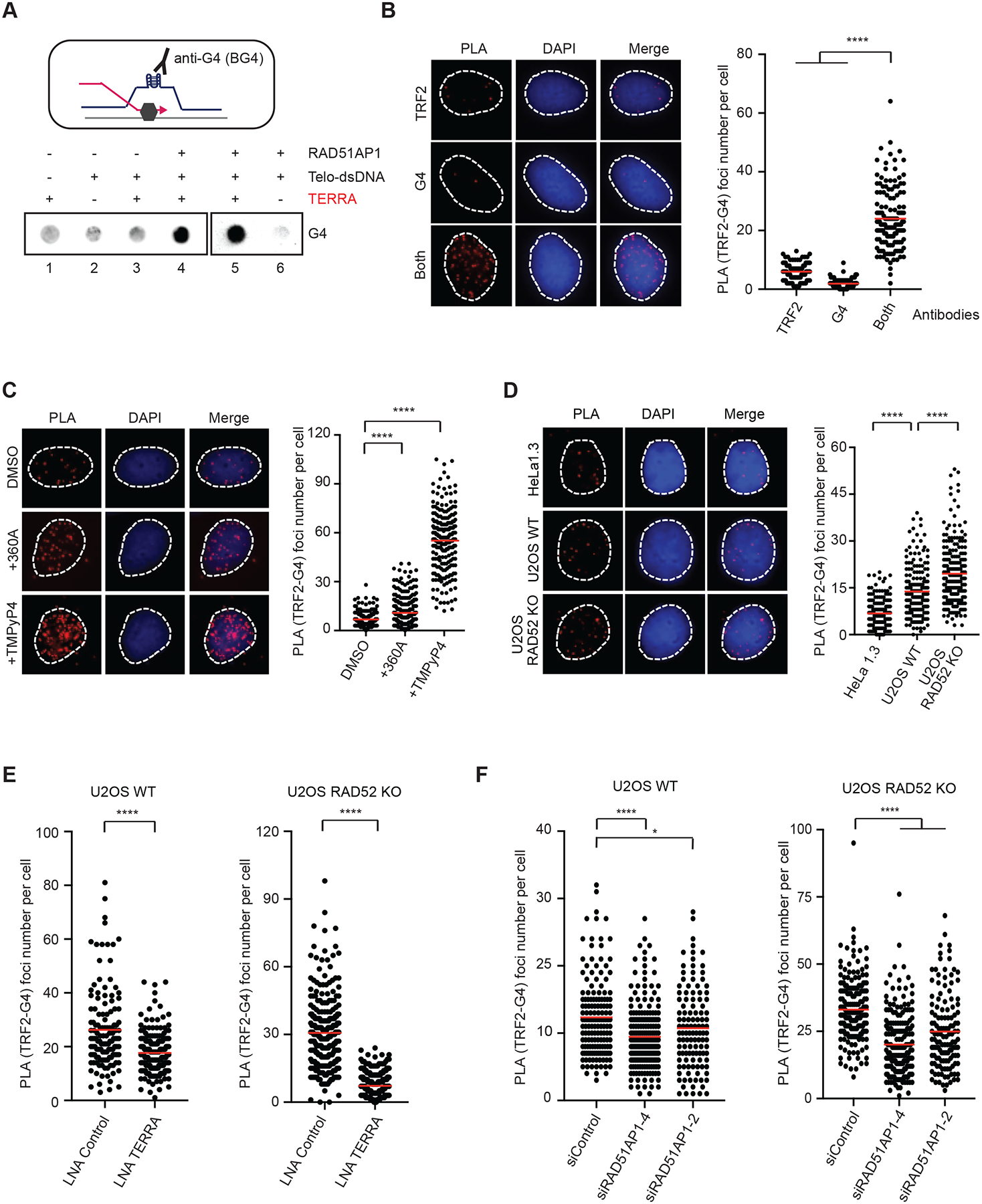
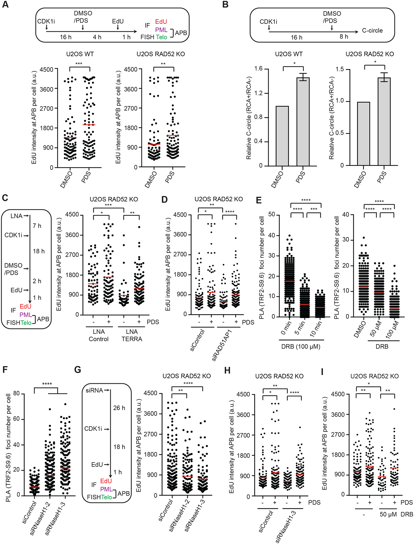
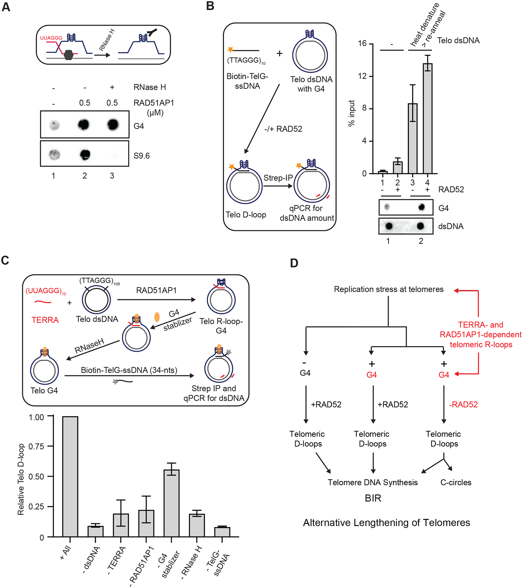
Alternative lengthening of telomeres (ALT), a telomerase-independent process maintaining telomeres, is mediated by break-induced replication (BIR). RAD52 promotes ALT by facilitating D-loop formation, but ALT also occurs through a RAD52-independent BIR pathway. Here, we show that the telomere non-coding RNA TERRA forms dynamic telomeric R-loops and contributes to ALT activity in RAD52 knockout cells. TERRA forms R-loops in vitro and at telomeres in a RAD51AP1-dependent manner. The formation of R-loops by TERRA increases G-quadruplexes (G4s) at telomeres. G4 stabilization enhances ALT even when TERRA is depleted, suggesting that G4s act downstream of R-loops to promote BIR. In vitro, the telomeric R-loops assembled by TERRA and RAD51AP1 generate G4s, which persist after R-loop resolution and allow formation of telomeric D-loops without RAD52. Thus, the dynamic telomeric R-loops formed by TERRA and RAD51AP1 enable the RAD52-independent ALT pathway, and G4s orchestrate an R- to D-loop switch at telomeres to stimulate BIR.

Keywords: ALT; BIR; D-loop; DNA-RNA hybrids; G-quadruplexes; G4; R-loop; RAD51AP1; RAD52; RNaseH1; TERRA; alternative lengthening of telomeres; break-induced replication; telomere.


Conflict of interest statement

Declaration of interests L.Z. is a member of the advisory board of Molecular Cell.

Figures

Figure 1.
Figure 1.. TERRA contributes to the RAD52-independent ALT pathway
(A) A schematic introducing the bifurcated ALT pathway and the question on the role of TERRA in RAD52-independent ALT. (B-C) U2OS WT (B) and RAD52 KO (C) cells transfected with Control or TERRA LNA were analyzed by DRIP coupled with telomere-specific or β-actinspecific qPCR. Error bars: SEM, n=3 (experimental triplicates); ****: P value <0.0001; ***: P value 0.0001; NS: not significant. (D) The experimental scheme to visualize telomere DNA synthesis at APBs in G2 cells (left). U2OS WT and RAD52 KO cells were transfected with Control or TERRA LNA and synchronized in G2. The EdU signals at APBs were quantified in individual cells (n>120) and normalized by nuclear areas. Red lines: mean values. ****: P value <0.0001. (E) U2OS WT and RAD52 KO cells transfected with Control or TERRA LNA were analyzed by the C-circle assay. The RCA+/RCA-ratios of samples reflect the relative levels of C-circle amplification. Error bars: SEM, n=3 (experimental triplicates); **: P value 0.002, *: P value 0.02.
Figure 2.
Figure 2.. RAD51AP1 promotes formation of telomeric R-loops and ALT activity
(A) Binding of RAD51AP1 to TERRA. Increasing concentrations of RAD51AP1 were incubated with TERRA-IR800 (18-nt). Reaction products were separated on a native acrylamide gel. The binding of RAD51AP1 to TERRA was quantified by measuring the reduction of free TERRA in the gel. Mean values of two experiments (n=2) are shown. (B) A schematic of the telomeric R-loop formation assay (left); RAD51AP1 was incubated with TERRA-IR800 and dsDNA containing telomeric sequences for 20 min. In lane 4, the sample was treated with RNaseH (0.5 unit) for 5 min after R-loop formation. R-loops were separated from free TERRA on an agarose gel. (C) RAD51AP1 and RAD52 were compared in the R-loop formation assay shown in (B). (D) RAD51AP1 and RAD52 were compared in the telomeric D-loop formation assay. TelG-ssDNAIR800 was incubated with RAD51AP1 or RAD52 and dsDNA containing telomeric sequences for 20 min. Reaction products were separated on an agarose gel. (E) U2OS WT cells transfected with siControl, siRAD51AP1, or siRAD52 were analyzed by DRIP coupled with telomere-specific or β-actin-specific qPCR. Error bars: SEM, n=3 (experimental triplicates); P value 0.001 (siControl vs siRAD51AP1); 0.005 (siControl vs siRAD52); 0.04 (siRAD51AP1 vs siRAD52); NS: not significant. (F) RAD52 KO cells transfected with siControl or siRAD51AP1 were analyzed by DRIP coupled with telomere-specific qPCR. Error bars: SEM, n=3 (experimental triplicates); ***: P value 0.0001. (G) U2OS WT and RAD52 KO cells were transfected with siControl or siRAD51AP1, synchronized in G2, and analyzed for EdU intensity in APBs. The EdU signals at APBs were quantified in individual cells (n>300) and normalized by nuclear areas. Error bars: SD, n=3 (experimental triplicates); ****: P value <0.0001. (H) U2OS RAD52 KO cells transfected with siControl or siRAD51AP1 were analyzed by the C-circle assay. The RCA+/RCA− ratios of samples reflect the relative levels of C-circle amplification. Error bars: SEM, n=3 (experimental triplicates); *: P value 0.01.
Figure 3.
Figure 3.. RAD51AP1 promotes telomere maintenance and proliferation in RAD52 KO cells
(A) U2OS WT, RAD52 KO, and RAD52, 51AP1 DKO (clones C16 and C18) cells were synchronized in metaphase and telomeric FISH was performed using TelC-488 probe. (B) Signal free ends (SFEs) were quantified from (A). >25 metaphases were quantified from each cell line. Red lines: mean values. ****: P value <0.0001, **: P value 0.003. (C) Telomeres DNA content was quantified in U2OS WT, RAD52 KO, and RAD52, 51AP1 DKO (clones C16 and C18) cells after 1 month of passages. Error bars: SD, n=3 (experimental triplicates); ***: P value 0.001; **: P value 0.003 (D) Cell growth analysis of U2OS WT, RAD52 KO, and RAD52, 51AP1 DKO (clones C16 and C18) cells. (E) Representative images of β -galactosidase staining of U2OS WT, RAD52 KO, and RAD52, 51AP1 DKO (clones C16 and C18) cell populations. All cell lines were passaged in parallel for ~1 month.
Figure 4.
Figure 4.. Telomeric R-loops increases G4s
(A) A schematic of G4 formation at an R-loop (upper panel). Telomeric R-loops were generated by incubating TERRA (60-nt, 60 nM), RAD51AP1 (0.5 μM) and dsDNA containing telomeric sequences for 20 min. Reaction products were analyzed for G4s by dot blot using the BG4 antibody. (B) Detection of telomeric G4s by PLA. U2OS cells were analyzed by PLA using anti-G4 (1H6) and anti-TRF2 antibodies, either alone or in combination. Representative images of PLA foci are shown (left) and quantified in individual cells (right) (n>140). Red lines: mean values. ****: P value <0.0001. (C) U2OS cells were treated with 360A (10 μM) or TMPYP4 (4 μM) for 6 hr, and telomeric G4s were analyzed as in (B) (n>220). Red lines: mean values. ****: P value <0.0001. (D) HeLa1.3, U2OS WT and U2OS RAD52 KO cells were analyzed as in (B) (n>190). Red lines: mean values. ****: P value <0.0001. (E) U2OS WT and RAD52 KO cells transfected with Control or TERRA LNA were analyzed as in (B) (n>240). Red lines: mean values. ****: P value <0.0001. (F) U2OS WT and RAD52 KO cells transfected with siControl or two different siRAD51AP1 were analyzed as in (B) (n>130). Red lines: mean values. ****: P value <0.0001, *: P value 0.03. To specifically detect G4s in DNA, all PLA experiments were performed with cells treated with RNaseA (10 μg/ml) at 37 °C for 1 hr.
Figure 5.
Figure 5.. G4s act downstream of transient R-loops to promote ALT
(A) U2OS WT and RAD52 KO cells were synchronized in G2, treated with DMSO or 2.5 μM PDS, and analyzed for EdU intensity at APBs. The EdU signals at APBs were quantified in individual cells (n>200) and normalized by nuclear areas. Red lines: mean intensities. ***: P value 0.0001, **: P value 0.008. (B) U2OS WT and RAD52 KO cells were treated with 5 μM PDS and analyzed by the C-circle assay. The RCA+/RCA-ratios of samples reflect the relative levels of C-circle amplification. Error bars: SEM, n=3 (experimental triplicates); P values 0.01 (left panel) and 0.03 (right panel). (C) U2OS RAD52 KO cells transfected with Control or TERRA LNA were synchronized in G2 and then treated with DMSO or 2.5 μM PDS as indicated. The EdU signals at APBs were quantified in individual cells (n>230) and normalized by nuclear areas. Red lines: mean intensities. *: P value 0.02, **: P value 0.004, ***: P value 0.0001. (D) U2OS RAD52 KO cells transfected with siControl or siRAD51AP1 were synchronized in G2 and then treated with DMSO or 3 μM PDS for 3 hrs. The EdU signals at APBs were quantified in individual cells (n>100) and normalized by nuclear areas. Red lines: mean intensities. *: P value 0.015, **: P value 0.005, ****: P value <0.0001. (E) U2OS RAD52 KO cells were treated with 100 μM DRB for 0, 5, and 10 min (left), or with 0, 50, and 100 μM DRB for 30 min (right). Telomeric DNA:RNA hybrids were quantified by PLA using S9.6 and TRF2 antibodies. PLA foci were quantified in individual cells (n>220). Red lines: means. ***: P value 0.0001, ****: P value <0.0001. (F) U2OS RAD52 KO cells treated with Control or RNaseH1 siRNA were analyzed for telomeric DNA:RNA hybrids by PLA using S9.6 and TRF2 antibodies. PLA foci were quantified in individual cells (n>170). Red lines: means. ****: P value <0.0001. (G) U2OS RAD52 KO cells treated with Control or RNaseH1 siRNA were synchronized in G2 and analyzed for EdU intensity at APBs. The EdU signals at APBs were quantified in individual cells (n≥150) and normalized by nuclear areas. Red lines: mean intensities. **: P value 0.003, ****: P value <0.0001. (H) U2OS RAD52 KO cells transfected with siControl or siRNaseH1 were synchronized in G2 and then treated with DMSO or 3 μM PDS for 3 hrs. The EdU signals at APBs were quantified in individual cells (n>130) and normalized by nuclear areas. Red lines: mean intensities. *: P value 0.019, **: P value 0.007, ****: P value <0.0001. (I) U2OS RAD52 KO cells were synchronized in G2 and then treated with DMSO, 3 μM PDS, and 50 μM DRB for 3 hr. The EdU signals at APBs were quantified in individual cells (n>125) and normalized by nuclear areas. Red lines: mean intensities. *: P value 0.011, **: P value 0.003.
Figure 6.
Figure 6.. Dynamic telomeric R-loops promote D-loop formation in the absence of RAD52
(A) A schematic for testing the presence of G4s in R-loops after removal of DNA:RNA hybrids (upper panel). Telomeric R-loops were generated by incubating TERRA (60-nt, 60 nM), RAD51AP1 (0.5 μM) and dsDNA containing telomeric sequences for 20 min. In lane 3, the sample was treated with RNaseH (0.5 unit) for 5 min after R-loop formation. Reaction products were analyzed for DNA:RNA hybrids and G4s by dot blot using S9.6 and BG4 antibodies, respectively. (B) The experimental scheme to test whether telomeric G4s promote D-loop formation (left). dsDNA containing telomeric sequences was heat denatured and reannealed slowly to increase G4s. The presence of high or low levels of G4s in dsDNA (1 μg) was confirmed by dot blot (bottom panel). The dsDNA harboring high or low levels of G4s was incubated with biotin-TelG-ssDNA (30 nM) in the presence or absence of RAD52 (0.3 μM) for 20 min. Telomeric D-loops were captured with streptavidin beads (10 uL) and quantified by qPCR using primers specific to the dsDNA. Error bars: SEM, n=2. (C) The experimental scheme to reconstitute the TERRA-and RAD51AP1-mediated formation of telomeric D-loops (upper panel). Telomeric R-loops were generated by incubating TERRA (60-nt, 60 nM), RAD51AP1 (0.3 μM) and dsDNA containing telomeric sequences. PDS (1 mM) was added to stabilize G4s, and the DNA:RNA hybrids in R-loops were removed by RNaseH (0.5 unit, 5 min). Subsequently, biotin-TelG-ssDNA was added, and the resulting D-loops were captured with streptavidin beads (10 μL) and quantified by qPCR. Error bars: SEM: n=3. The effects of omitting individual components of the reaction were tested. (D) A model in which the dynamic telomeric R-loops assembled by TERRA and RAD51AP1 promote G4 accumulation and D-loop formation, enabling the RAD52-independent ALT pathway.

Comment in

References

    1. Amato R, Valenzuela M, Berardinelli F, Salvati E, Maresca C, Leone S, Antoccia A, and Sgura A (2020). G-quadruplex Stabilization Fuels the ALT Pathway in ALT-positive Osteosarcoma Cells. Genes (Basel) 11. - PMC - PubMed
    1. Arora R, Lee Y, Wischnewski H, Brun CM, Schwarz T, and Azzalin CM (2014). RNaseH1 regulates TERRA-telomeric DNA hybrids and telomere maintenance in ALT tumour cells. Nat Commun 5, 5220. - PMC - PubMed
    1. Azzalin CM, Reichenbach P, Khoriauli L, Giulotto E, and Lingner J (2007). Telomeric repeat containing RNA and RNA surveillance factors at mammalian chromosome ends. Science 318, 798–801. - PubMed
    1. Barroso-Gonzalez J, Garcia-Exposito L, Hoang SM, Lynskey ML, Roncaioli JL, Ghosh A, Wallace CT, de Vitis M, Modesti M, Bernstein KA, et al. (2019). RAD51AP1 Is an Essential Mediator of Alternative Lengthening of Telomeres. Mol Cell 76, 11–26 e17. - PMC - PubMed
    1. Bhowmick R, Minocherhomji S, and Hickson ID (2016). RAD52 Facilitates Mitotic DNA Synthesis Following Replication Stress. Mol Cell 64, 1117–1126. - PubMed

Publication types