

Augmented Reality in Spine Surgery: Current State of the Art
- PMID: 36266050
- PMCID: PMC9808789
- DOI: 10.14444/8273
Augmented Reality in Spine Surgery: Current State of the Art
Abstract
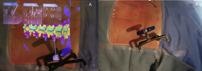
Augmented reality (AR) is the superimposition of a virtual environment on the real world. The use of AR in spine surgery continues to grow, with multiple companies and products becoming available. The proposed benefits of AR include decreased attention shift, decreased line-of-site interruption, opportunity for more minimally invasive approaches, decreased radiation exposure to the operative team, and improved pedicle screw accuracy. In this review, we examine our institutional experiences with utilization and implementation of some of the current AR products.
Keywords: 3D overlays; augmented reality; future technologies; heads-up navigation.
This manuscript is generously published free of charge by ISASS, the International Society for the Advancement of Spine Surgery. Copyright © 2022 ISASS. To see more or order reprints or permissions, see http://ijssurgery.com.
Conflict of interest statement
Declaration of Conflicting Interests: Frank Phillips has stock options from a company involved in the manufacturing of a device examined in this study.
Figures
References
-
- Costa F, Cardia A, Ortolina A, Fabio G, Zerbi A, Fornari M. Spinal navigation: standard preoperative versus intraoperative computed tomography data set acquisition for computer-guidance system: radiological and clinical study in 100 consecutive patients. Spine (Phila Pa 1976). 2011;36(24):2094–2098. 10.1097/BRS.0b013e318201129d - DOI - PubMed
-
- Molina CA, Phillips FM, Colman MW, et al. A cadaveric precision and accuracy analysis of augmented reality–mediated percutaneous pedicle implant insertion: presented at the 2020 AANS/CNS joint section on disorders of the spine and peripheral nerves. J Neurosurg Spine. 2020;1:1–9. - PubMed
LinkOut - more resources
Full Text Sources
Research Materials