. 2022 Oct 20;7(1):63.
doi: 10.1038/s41536-022-00259-y.

Myogenic tissue nanotransfection improves muscle torque recovery following volumetric muscle loss

Affiliations

Myogenic tissue nanotransfection improves muscle torque recovery following volumetric muscle loss

Andrew Clark et al. NPJ Regen Med. .

Abstract

This work rests on our non-viral tissue nanotransfection (TNT) platform to deliver MyoD (TNTMyoD) to injured tissue in vivo. TNTMyoD was performed on skin and successfully induced expression of myogenic factors. TNTMyoD was then used as a therapy 7 days following volumetric muscle loss (VML) of rat tibialis anterior and rescued muscle function. TNTMyoD is promising as VML intervention.


Conflict of interest statement

The authors declare no competing interests.

Figures
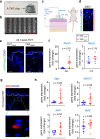
Fig. 1
Fig. 1. TNTMyoD transfects deep into the dermis and causes myogenic reprogramming.
a Image of TNT2.0 device. b SEM images of TNT device showing needle array projections. Scale, 400 µm. c Schematic diagram showing TNT set up for skin. d Distribution of FAM-labeled plasmids immediately after TNT using various voltages on mouse dorsal skin. Scale bar = 40 µm. e Visualization of eGFP signal 24 h after TNT with the eGFP-MyoD plasmid, showing expression of eGFP in epidermis and dermis. No eGFP fluorescence was detected in skin transfected with mock plasmids. The white dashed line indicates the dermal-epidermal junction. Scale, 50 µm. f MyoD expression in skin 24 h and day 10 post-TNT (n = 7,6). g Immunofluorescence staining of MF20 (myosin heavy chain) at 10 days post-TNT in the dermis. The white dashed line indicates the dermal-epidermal junction. The sections were co-stained with DAPI. Scale bar = 20 µm, 5 µm (h) transcript abundance of myogenic genes compared to mock-transfected skin (n = 9). All data are expressed as mean ± SD. Data analyzed by Student’s t-test. Figure c was created with BioRender.com.
Fig. 2
Fig. 2. TNTMyoD improves torque recovery in volumetric muscle loss.
a Image of TNT being performed on tibialis anterior muscle 1 week after VML injury. b Gene expression of MyoD in VML injured muscle 24 h post-TNT (n = 4). c Image of rat undergoing muscle functional testing. d Maximum dorsiflexion torque produced by VML affected limb. e Dorsiflexion torque produced at different frequencies of direct tibialis anterior stimulation 4 weeks after VML(n = 7). f At 4 weeks after VML injury, dorsiflexion torque is produced after repeated electrical stimulation of the tibialis anterior muscle at 70 Hz. Stimulations occurred every 3 s (n = 7). Regression line equations for data between 60–120 stimulations: 7 = −0.0056*x + 2.387 (TNTmock) and y = −0.0086*x + 3.2345 (TNTMyoD). g Images of VML affected tibilais anterior and synergist extensor digitorium longus and their contralateral counterparts 4 weeks post-injury (3 weeks after TNT). h Quantification of muscle weights 4 weeks after VML (n = 7). i Immunohistochemistry of eMyHC and quantification per area of regenerating myofiber at the injury site post-TNT with either mock or MyoD plasmid. Scale, 500 µm (n = 5). j Immunohistochemistry of laminin and quantification per area of regenerating myofiber at the injury site post-TNT with either mock of MyoD. Scale, 100 µm (n = 7,6). Data are expressed as mean ± SD in b, h, and i. *p < 0.05. Data expressed as individual data points with mean trend lines in d and e. Data in b, i, and j were analyzed by Student’s t-test. Data in d, e, f, and h were analyzed by ANOVA.

References

    1. Hinds S, Bian W, Dennis RG, Bursac N. The role of extracellular matrix composition in structure and function of bioengineered skeletal muscle. Biomaterials. 2011;32:3575–3583. doi: 10.1016/j.biomaterials.2011.01.062. - DOI - PMC - PubMed
    1. Dessauge F, Schleder C, Perruchot M-H, Rouger K. 3D in vitro models of skeletal muscle: myopshere, myobundle and bioprinted muscle construct. Vet. Res. 2021;52:72. doi: 10.1186/s13567-021-00942-w. - DOI - PMC - PubMed
    1. Isner JM, et al. Treatment of thromboangiitis obliterans (Buerger’s disease) by intramuscular gene transfer of vascular endothelial growth factor: preliminary clinical results. J. Vasc. Surg. 1998;28:964–973. doi: 10.1016/S0741-5214(98)70022-9. - DOI - PubMed
    1. Vale PR, et al. Left ventricular electromechanical mapping to assess efficacy of phVEGF(165) gene transfer for therapeutic angiogenesis in chronic myocardial ischemia. Circulation. 2000;102:965–974. doi: 10.1161/01.CIR.102.9.965. - DOI - PubMed
    1. Manthorpe M, et al. Gene therapy by intramuscular injection of plasmid DNA: studies on firefly luciferase gene expression in mice. Hum. Gene Ther. 1993;4:419–431. doi: 10.1089/hum.1993.4.4-419. - DOI - PubMed