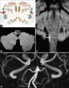
Isolated axial lateropulsion caused by an acute lateral medullary infarction involving the dorsal spinocerebellar tract: A case report
- PMID: 36267436
- PMCID: PMC9578312
- DOI: 10.4103/bc.bc_39_22
Isolated axial lateropulsion caused by an acute lateral medullary infarction involving the dorsal spinocerebellar tract: A case report
Abstract
Lateral medullary syndrome encompasses a broad spectrum of symptoms and signs depending on the bulbar localization of the lesion. Body lateropulsion (BL) can occur without vestibular and cerebellar symptoms, as a unique manifestation of a lateral medullary infarction. However, it is relatively rare and challenging to diagnose. We report a case of a 72-year-old woman who presented with a tendency to fall to the right. She denied having vertigo, cerebellar signs, sensory loss, or motor weakness. No signs of vestibular dysfunction were found on the ENT examination. Neurological evaluation was unremarkable, except for mild ataxia of the right limbs along with BL to the right side when standing and walking. Brain magnetic resonance (MR) imaging showed an acute small infarct in the right lateral aspect of the medulla extending from the rostral to the caudal level. MR angiography found no stenosis or vascular occlusions. We believe that ipsilateral axial lateropulsion shown by our patient may be related to a selective ischemic lesion of the dorsal spinocerebellar tract in its medullary course. A lateral medullary infarction should be seriously considered in patients who present with isolated BL without further signs of bulbar involvement.
Keywords: Isolated body lateropulsion; medulla oblongata; posterior spinocerebellar tract.
Copyright: © 2022 Brain Circulation.
Conflict of interest statement
There are no conflicts of interest.
Figures

References
-
- Wallenberg A. Acute bulbar affection (Embolie der art. cerebellar post. inf. sinistra?) Arch Psychiatr Nervenkr. 1895;27:504–40.
-
- Kim JS. Pure lateral medullary infarction: Clinical-radiological correlation of 130 acute, consecutive patients. Brain. 2003;126:1864–72. - PubMed
-
- Ramaswamy S, Rosso M, Levine SR. Body lateropulsion in stroke: Case report and systematic review of stroke topography and outcome. J Stroke Cerebrovasc Dis. 2021;30:105680. - PubMed
-
- Lee H, Sohn CH. Axial lateropulsion as a sole manifestation of lateral medullary infarction: A clinical variant related to rostral-dorsolateral lesion. Neurol Res. 2002;24:773–4. - PubMed
-
- Kim SH, Cho J, Cho JH, Han SW, Kim SM, Park SC, et al. Isolated lateropulsion by a lesion of the dorsal spinocerebellar tract. Cerebrovasc Dis. 2004;18:344–5. - PubMed
Publication types
LinkOut - more resources
Full Text Sources
Research Materials
Miscellaneous
