. 2021:1:100020.
doi: 10.1016/j.bbadva.2021.100020. Epub 2021 Jul 30.

Fragments of Locusta migratoria apoLp-III provide insight into lipid binding

Affiliations

Fragments of Locusta migratoria apoLp-III provide insight into lipid binding

Blair A Russell et al. BBA Adv. 2021.

Abstract

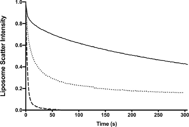
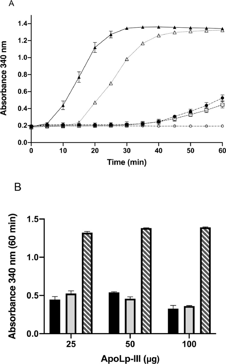
Apolipophorin III (apoLp-III) from Locusta migratoria is an exchangeable apolipoprotein with a critical role in lipid transport in insects. The protein is composed of a bundle of five amphipathic α-helices which undergo a large conformational change upon lipid binding. To better understand the apoLp-III lipid binding interaction, the protein was cleaved by cyanogen bromide upon introduction of a S92M mutation, generating an N-terminal fragment corresponding to the first three helices (NTH1-3) and a C-terminal fragment of the last two helices (CTH4-5). MALDI-TOF analysis of the HPLC purified fragments provided masses of 9863.8 Da for NTH1-3 and 7497.0 Da for CTH4-5 demonstrating that the intended fragments were obtained. Circular dichroism spectra revealed a decrease in helical content from 82% for the intact protein to 57% for NTH1-3 and 41% for CTH4-5. The fragments adopted considerably higher α-helical structure in the presence of trifluoroethanol or phospholipids. Equimolar mixing of the two fragments did not result in changes in helical content or tryptophan fluorescence, indicating recombination into the native protein fold did not occur. The rate of protein induced dimyristoylphosphatidylcholine vesicle solubilization increased 15-fold for NTH1-3 and 100-fold for CTH4-5 compared to the intact protein. Despite the high activity in phospholipid vesicle interaction, CTH4-5 did not protect phospholipase-treated low-density lipoprotein from aggregation. In contrast, NTH1-3 provided protection to lipoprotein aggregation similar to the intact protein, indicating that specific amino acid residues in this part of apoLp-III are essential for lipoprotein binding interaction.

Keywords: Apolipophorin; Apolipoprotein; Diacylglycerol; Phospholipid.


Conflict of interest statement

The authors declare that they have no known competing financial interests or personal relationships that could have appeared to influence the work reported in this paper.

Figures

Image, graphical abstract
Graphical abstract
Fig 1
Fig. 1
ApoLp-III-S92M and fragments generated by CNBr cleavage. The NMR solution structure of apoLp-IIILM is shown in panel A on the left with the three NT helices (grey) and the two CT helices (green). The position of the S92M mutation is shown in yellow, and CNBr digestion produced the NTH1-3 (rotated 180°) and CTH4-5 fragments. Surface contour in Panel B indicates the degree of hydrophobicity with a color gradient of blue (hydrophilic) to red (hydrophobic), showing the increased exposed hydrophobic surface of two fragments (For interpretation of the references to color in this figure legend, the reader is referred to the web version of this article.).
Fig 2
Fig. 2
SDS-PAGE of CNBr digest of apoLp-III-S92M Lane M: Marker protein with molecular masses indicated on the left; lane 1: apoLp-III-S92M; lane 2: CNBr digest of apoLp-III-S92M; lane 3 and 4 show the two fragments purified by reversed-phase HPLC: NTH1-3 (lane 3) and CTH4-5 (lane 4).
Fig 3
Fig. 3
Far UV CD analysis of the apoLp-III fragments. Shown are CD scans of apoLp-III and the two fragments in buffer, at a protein concentration of 0.2 mg/mL. The CD scans are the average of three separate measurements while the corresponding bar graph shows the % α-helical content of each protein using CDSSTR (± SD, n = 3). Panel A shows intact apoLp-III (solid line), and in the presence of 50% TFE (dotted) or DMPC (dash-dotted). Panel B shows intact apoLp-III (solid line), and apoLp-III-NTH1-3 in the absence (dotted) or presence of 50% TFE (dashed) or DMPC (dash-dotted). Panel C shows intact apoLp-III (solid line), and apoLp-III-CTH4-5 in the presence of 50% TFE (dashed) or DMPC (dash-dotted). Panel D shows intact apoLp-III (solid line), NTH1-3 (dotted), CTH4-5 (dash-dotted), and the mixture of NTH1-3 and CTH4-5 (dashed).
Fig 4
Fig. 4
Tryptophan Fluorescence. ApoLp-III samples (30 μg/mL) were excited at 280 nm and the emission intensity was measured between 290 and 460 nm. Shown are NTH1-3 (dotted line), CTH4-5 (dashed line), the NTH1-3/CTH4-5 mixture (dash-dotted line), and intact apoLp-III (solid line). The emission profiles are the average of three independent scans for each protein.
Fig 5
Fig. 5
ANS Fluorescence. Protein samples (5 μM) were excited at 395 nm and the emission intensity was measured between 400 and 650 nm. Shown are intact apoLp-III (solid line), NTH1-3 (dashed line), CTH4-5 (dash-dotted line), and ANS in the absence of protein (dotted line). The emission profiles are the average of three independent scans for each protein.
Fig 6
Fig. 6
SDS-PAGE of DMS protein crosslinking. Protein samples (0.75 mg/mL) were incubated with DMS and crosslinked proteins were analyzed by SDS-PAGE. Shown are apoLp-III-NTH1-3 in the presence (lane 1) or absence of DMS (lane 2), apoLp-III-CTH4-5 in the presence (lane 3) or absence of DMS (lane 4), intact apoLp-III in the presence (lane 5) or absence of DMS (lane 6). Lane M contains marker proteins with molecular masses indicated on the left.
Fig 7
Fig. 7
DMPC vesicle solubilization. LUVs made of DMPC were incubated in the presence of protein (0.125 mg/mL) at a 2:1 weight ratio of lipid to protein at 24.1 °C. Vesicle solubilization was monitored by light scatter intensity at 325 nm. Shown are intact apoLp-III (solid line), NTH1-3 (dotted), and CTH4-5 (dashed) fragments.
Fig 8
Fig. 8
PL-C induced LDL aggregation. Panel A: LDL and 160 mU of PL-C were mixed incubated at 37 °C, and the absorbance at 340 nm measured at 5 min intervals. Shown are LDL (open circles), LDL with PLC in the absence of protein (closed triangles), and in the presence of 25 μg (0.125 mg/mL) of intact apoLp-III (open squares), NTH1-3 (closed circles), and CTH4-5 fragments (open triangles). Panel B shows the absorbance at the 60 min timepoint in the presence of 25, 50, and 100 μg intact apoLp-III (black), NTH1-3 (grey), or CTH4-5 (diagonal hatched pattern).

References

    1. Weers P.M., Ryan R.O. Apolipophorin III: role model apolipoprotein. Insect Biochem. Mol. Biol. 2006;36:231–240. - PubMed
    1. van der Horst D.J., van Hoof D., van Marrewijk W.J.A., Rodenburg K.W. Alternative lipid mobilization: the insect shuttle system. Mol. Cell. Biochem. 2002;239:113–119. - PubMed
    1. Kawooya J.K., Meredith S.C., Wells M.A., Kézdy F.J., Law J.H. Physical and surface properties of insect apolipophorin III. J. Biol. Chem. 1986;261:13588–13591. - PubMed
    1. Ryan R.O., Oikawa K., Kay C.M. Conformational, thermodynamic, and stability properties of Manduca sexta apolipophorin III. J. Biol. Chem. 1993;268:1525–1530. - PubMed
    1. Weers P.M., Kay C.M., Oikawa K., Wientzek M., Van der Horst D.J., Ryan R.O. Factors affecting the stability and conformation of Locusta migratoria apolipophorin III. Biochemistry. 1994;33:3617–3624. - PubMed