CT-based lung motion differences in patients with usual interstitial pneumonia and nonspecific interstitial pneumonia
- PMID: 36267579
- PMCID: PMC9577177
- DOI: 10.3389/fphys.2022.867473
CT-based lung motion differences in patients with usual interstitial pneumonia and nonspecific interstitial pneumonia
Abstract
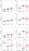
We applied quantitative CT image matching to assess the degree of motion in the idiopathic ILD such as usual interstitial pneumonia (UIP) and nonspecific interstitial pneumonia (NSIP). Twenty-one normal subjects and 42 idiopathic ILD (31 UIP and 11 NSIP) patients were retrospectively included. Inspiratory and expiratory CT images, reviewed by two experienced radiologists, were used to compute displacement vectors at local lung regions matched by image registration. Normalized three-dimensional and two-dimensional (dorsal-basal) displacements were computed at a sub-acinar scale. Displacements, volume changes, and tissue fractions in the whole lung and the lobes were compared between normal, UIP, and NSIP subjects. The dorsal-basal displacement in lower lobes was smaller in UIP patients than in NSIP or normal subjects (p = 0.03, p = 0.04). UIP and NSIP were not differentiated by volume changes in the whole lung or upper and lower lobes (p = 0.53, p = 0.12, p = 0.97), whereas the lower lobe air volume change was smaller in both UIP and NSIP than normal subjects (p = 0.02, p = 0.001). Regional expiratory tissue fractions and displacements showed positive correlations in normal and UIP subjects but not in NSIP subjects. In summary, lung motionography quantified by image registration-based lower lobe dorsal-basal displacement may be used to assess the degree of motion, reflecting limited motion due to fibrosis in the ILD such as UIP and NSIP.
Keywords: computational biomechanics; computed tomography; idiopathic pulmonary fibrosis; image registration; interstitial lung disease; lung motionography; quantitative computed tomography image matching; usual interstitial pneumonia.
Copyright © 2022 Choi, Chae, Jin, Lin, Laroia, Hoffman and Lee.
Conflict of interest statement
EAH is a founder and shareholder of VIDA (Coralville, IA, United States). The remaining authors declare that the research was conducted in the absence of any commercial or financial relationships that could be construed as a potential conflict of interest.
Figures


References
Grants and funding
LinkOut - more resources
Full Text Sources

