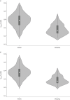
Infant tidal flow-volume parameters and arousal state
- PMID: 36267897
- PMCID: PMC9574559
- DOI: 10.1183/23120541.00163-2022
Infant tidal flow-volume parameters and arousal state
Abstract
Background: Infant lung function can be assessed with tidal flow-volume (TFV) loops. While TFV loops can be measured in both awake and sleeping infants, the influence of arousal state in early infancy is not established. The aim of the present study was to determine whether TFV loop parameters in healthy infants differed while awake compared to the sleeping state at 3 months of age.
Methods: From the population-based Scandinavian Preventing Atopic Dermatitis and ALLergies in children (PreventADALL) birth cohort, 91 infants had reproducible TFV loops measured with Exhalyzer® D in both the awake and sleeping state at 3 months of age. The TFV loops were manually selected according to a standardised procedure. The ratio of time to peak tidal expiratory flow (t PTEF) to expiratory time (t E) and the corresponding volume ratio (V PTEF/V E), as well as tidal volume (V T) and respiratory rate were compared using nonparametric tests.
Results: The mean (95% CI) t PTEF/t E was significantly higher while awake compared to the sleeping state: 0.39 (0.37-0.41) versus 0.28 (0.27-0.29); with the corresponding V PTEF/V E of 0.38 (0.36-0.40) versus 0.29 (0.28-0.30). The V T was similar, while the respiratory rate was higher while awake compared to the sleeping state: 53 (51-56) breaths·min-1 versus 38 (36-40) breaths·min-1.
Conclusion: Higher t PTEF/t E, V PTEF/V E and respiratory rate, but similar V T while awake compared to the sleeping state suggests that separate normative TFV loop values according to arousal state may be required in early infancy.
Copyright ©The authors 2022.
Conflict of interest statement
Conflicts of interest: The authors have no conflicts of interest to disclose. The authors have no conflict of interest relating to or relationship with Exhalyzer® D or its manufacturer.
Figures


References
LinkOut - more resources
Full Text Sources
Miscellaneous
