Review
. 2023 Jan;21(1):3-27.
doi: 10.1111/gbi.12527. Epub 2022 Oct 21.

A review of microbial-environmental interactions recorded in Proterozoic carbonate-hosted chert

Affiliations
Review

A review of microbial-environmental interactions recorded in Proterozoic carbonate-hosted chert

Kelsey R Moore et al. Geobiology. 2023 Jan.

Abstract

The record of life during the Proterozoic is preserved by several different lithologies, but two in particular are linked both spatially and temporally: chert and carbonate. These lithologies capture a snapshot of dominantly peritidal environments during the Proterozoic. Early diagenetic chert preserves some of the most exceptional Proterozoic biosignatures in the form of microbial body fossils and mat textures. This fossiliferous and kerogenous chert formed in shallow marine environments, where chert nodules, layers, and lenses are often surrounded by and encased within carbonate deposits that themselves often contain kerogen and evidence of former microbial mats. Here, we review the record of biosignatures preserved in peritidal Proterozoic chert and chert-hosting carbonate and discuss this record in the context of experimental and environmental studies that have begun to shed light on the roles that microbes and organic compounds may have played in the formation of these deposits. Insights gained from these studies suggest temporal trends in microbial-environmental interactions and place new constraints on past environmental conditions, such as the concentration of silica in Proterozoic seawater, interactions among organic compounds and cations in seawater, and the influence of microbial physiology and biochemistry on selective preservation by silicification.

Keywords: biosignature; carbonate; chert; fossil; proterozoic.


Figures

FIGURE 1
FIGURE 1
Side‐by‐side comparisons of some of the most diagnostic Proterozoic microbial fossils and their modern analogs. (a) Eoentophysalis (Butterfield, 2015), (b) Modern Entophysalis (see Moore et al.,  for culturing conditions), (c) Obruchevella (Butterfield, 2015), (d) Modern Spirulina (see Moore et al.,  for culturing conditions), (e) Bangiomorpha (Butterfield, 2000), (f) Modern red algae (Sheath & Vis, 2015), (g) Polybessurus (Butterfield, 2001), (h) modern stalk forming cyanobacteria (Demoulin et al., 2019), (i) Eohyella (Butterfield, 2015), (j) modern endolithic cyanobacteria (Demoulin et al., 2019). [Correction added on 26 October 2022, after first online publication: the figure caption related to part figure (f) was corrected in this version.]
FIGURE 2
FIGURE 2
Pustular supratidal microbial mats in Shark Bay, Western Australia. A photomicrograph (inset) shows the coccoidal cyanobacteria that form pustular mats and produce concentric envelopes of EPS around cells and cell clusters (see Moore et al., , and Skoog et al., , for culturing conditions).
FIGURE 3
FIGURE 3
Subtidal to intertidal stromatolites in Shark Bay, Western Australia with photomicrograph of Spirulina (inset), one of the cyanobacteria enriched from these stromatolites (see Moore et al.,  for culturing conditions).
FIGURE 4
FIGURE 4
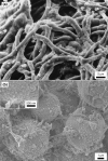
SEM images showing modern examples of microbial silicification. (a) Filamentous cyanobacteria coated in silica that were experimentally silicified under supersaturated conditions (see Phoenix et al., , for experimental conditions). (b) Coccoidal cyanobacteria that were experimentally silicified in artificial seawater with 1.5 mM silica (see Moore et al., , for experimental conditions). The inset shows nanoscopic colloidal silica coating cell surfaces.
FIGURE 5
FIGURE 5
Experimentally silicified filamentous cyanobacteria. SEM image shows the silicified microbial mat (left). The corresponding EDS spectra (right) show elevated magnesium, calcium, and silica in these mats (see Moore et al., ; Morgenstein, for experimental conditions).
FIGURE 6
FIGURE 6
Pustular microbial mats built by coccoidal cyanobacteria in supratidal environments. (a) Modern, Shark Bay, Australia. (b) Proterozoic, Belcher Island Group, Canada (Butterfield, 2015).
FIGURE 7
FIGURE 7
Examples of black, fossiliferous chert nodules, lenses, and layers within carbonate strata from the Proterozoic Angmaat Formation (Manning‐Berg et al., 2018).
FIGURE 8
FIGURE 8
Photomicrographs of coccoidal cyanobacteria from Shark Bay, Western Australia grown at different conditions. (a) Cyanobacteria grown under the exposure to LED UV lights (400–420 nm wavelength) and a fluorescent light. (b). Cyanobacteria grown in liquid cultures under a fluorescent light. The same cyanobacteria produce thicker EPS envelopes in response to UV radiation stress (see Moore et al.,  for culturing conditions).
FIGURE 9
FIGURE 9
Examples of microbial‐associated dolomite. (a) An environmental sample from the sabkhas of Abu Dhabi (Bontognali et al., 2010). (b) Experimentally fossilized cells encased in dolomite that nucleated on the surfaces of cells (see Daye et al.,  for experimental conditions).
FIGURE 10
FIGURE 10
Kerogen‐rich microbial lamination in dolomite‐hosted chert in the Balbirini Dolomite. The chert preserves microbial body fossils, but dolomite does not.
FIGURE 11
FIGURE 11
Schematics of possible formation mechanisms for biosignature‐preserving chert and dolomite. Scenario shown in the top panel shows contemporaneous precipitation of silica and dolomite within a microbial mat. Dolomite forms in anoxic portions of the mat, whereas silica precipitates in surface portions of the mat around actively photosynthesizing cyanobacteria. Bottom panel depicts an alternate scenario in which dolomite precipitates in deeper portions of the mat where heterotrophic processes continuously degrade cyanobacterial cells and surface organic matter. The evaporation during the dry season concentrates silica, magnesium, and other cations and promotes the precipitation of silica in surface portions of the mats dominated by actively photosynthesizing cyanobacteria.

References

    1. Aitken, J. D. (1988). Giant “algal” reefs, Middle/Upper Proterozoic Little Dal Group (>770, >1200 Ma), Mackenzie Mountains, N.W.T., Canada. Reef: Canada and Adjacent Areas Memoir, 13, 13–23.
    1. Aitken, J. D. , & Narbonne, G. M. (1989). Two occurrences of Precambrian thrombolites from the Mackenzie Mountains, Northwestern Canada. PALAIOS, 4, 384–388. 10.2307/3514563 - DOI
    1. Allen, M. A. , Goh, F. , Burns, B. P. , & Neilan, B. A. (2009). Bacterial, archaeal and eukaryotic diversity of smooth and pustular microbial mat communities in the hypersaline lagoon of Shark Bay. Geobiology, 7, 82–96. 10.1111/j.1472-4669.2008.00187.x - DOI - PubMed
    1. Alleon, J. , Bernard, S. , Le Guillou, C. , Marin‐Carbonne, J. , Pont, S. , Beyssac, O. , McKeegan, K. D. , & Robert, F. (2016). Molecular preservation of 1.88 Ga Gunflint organic microfossils as a function of temperature and mineralogy. Nature Communications, 7, 11977. 10.1038/ncomms11977 - DOI - PMC - PubMed
    1. Amard, B. , & Bertrand‐Sarfati, J. (1997). Microfossils in 2000 Ma old cherty stromatolites of the Franceville Group, Gabon. Precambrian Research, 81, 197–221.

Publication types