Review
. 2022 Oct 31;51(21):9127-9173.
doi: 10.1039/d2cs00618a.

Bio-macromolecular design roadmap towards tough bioadhesives

Affiliations
Review

Bio-macromolecular design roadmap towards tough bioadhesives

Hossein Montazerian et al. Chem Soc Rev. .

Abstract

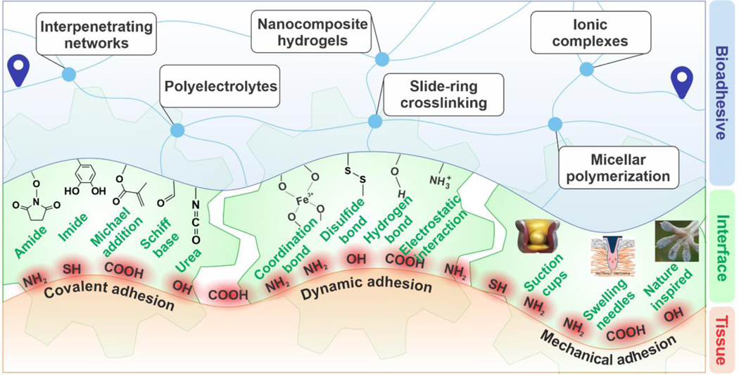
Emerging sutureless wound-closure techniques have led to paradigm shifts in wound management. State-of-the-art biomaterials offer biocompatible and biodegradable platforms enabling high cohesion (toughness) and adhesion for rapid bleeding control as well as robust attachment of implantable devices. Tough bioadhesion stems from the synergistic contributions of cohesive and adhesive interactions. This Review provides a biomacromolecular design roadmap for the development of tough adhesive surgical sealants. We discuss a library of materials and methods to introduce toughness and adhesion to biomaterials. Intrinsically tough and elastic polymers are leveraged primarily by introducing strong but dynamic inter- and intramolecular interactions either through polymer chain design or using crosslink regulating additives. In addition, many efforts have been made to promote underwater adhesion via covalent/noncovalent bonds, or through micro/macro-interlock mechanisms at the tissue interfaces. The materials settings and functional additives for this purpose and the related characterization methods are reviewed. Measurements and reporting needs for fair comparisons of different materials and their properties are discussed. Finally, future directions and further research opportunities for developing tough bioadhesive surgical sealants are highlighted.


Conflict of interest statement

The authors declare no competing financial interest.

Figures

Figure 1.
Figure 1.. An overview of the material design roadmap for tough bioadhesives.
Development of bioadhesive materials involves: (i) design for cohesion where a combination of the polymer backbone and crosslinking strategy is selected to ensure mechanical durability in a tough hydrogel, and (ii) design for bioadhesion where a mixture of covalent and dynamic interactions, as well as mechanical interlocks, are incorporated into the material design.
Figure 2.
Figure 2.. Examples of interpenetrating hydrogel networks (IPN) with improved toughness.
(A) Schematic of components and interactions in an IPN of poly(acrylamide) (PAM)/poly(ethylene oxide) (PEO) hydrogel crosslinked by N,N’-methylene-bis(acrylamide) (MBAA) toughened due to the synergy between hydrogen-bonding and covalent networks. (B) Hydrogel resistance against tensile, compressive, and torsion deformations. The hydrogels were able to stretch by ~8.8×. (C) Hydrogels showed anti-puncturing characteristics. (D) The punctured hydrogels still maintained their stretchability. Reproduced with permission from ref . Copyright 2019, Elsevier. (E) Schematic of processing poly(diallyldimethylammonium chloride) (PDDA)-poly(sodium 4-styrenesulfonate) (PSS)/branched poly(ethylenimine) (PEI)-poly(acrylic acid) (PAA) hydrogels. (F) Chemical structure of PSS, PAA, PEI, and PDDA components. (G) Toughening mechanism of the hydrogels: the effect of electrostatic interactions and hydrogen bonding. Reprinted with permission from ref . Copyright 2019 American Chemical Society. (H) Effect of pH on the conformation of polyelectrolytes (PE). At low pH, chitosan is ionized resulting in polyelectrolyte repulsion. Hyaluronic acid (HA), on the other hand, tends to fold due to intramolecular interactions. As the pH increases (above pKa of chitosan), a random coil conformation is formed by chitosan. Any further increase above pKa of HA leads to its extended conformation. Schematic of molecular reformation of the chains before (I) and after (J) the application of stretching loads. The dissipative process stems from the interchain hydrogen bonding and crosslinking through the polyelectrolyte complex (PEC) aggregates and hydrogen bonding. Reproduced with permission from ref . Copyright 2017, Royal Society of Chemistry.
Figure 3.
Figure 3.. Engineering tough and stretchable ionic hydrogels.
(A) Processing scheme for the ionic hydrogels based on poly(acrylic acid‐co‐acrylamide)/CoCl2 composition. (B) Effect of the Co2+ concentration on the tensile mechanical properties of the hydrogels. (C, D) Self-healing properties of the hydrogels and mechanical properties of the healed hydrogels at different time points. Reprinted with permission from ref . Copyright 2019 American Chemical Societ€(E) Fabrication procedure of the carbon nanotubes (CNT)/poly(vinyl alcohol) (PVA) hydrogels with borax to form the PVA complex through (F) attraction of borax to the hydroxyl groups of the PVA chain. (G) Tensile deformation of the hydrogel by 1000%. Reproduced with permission from ref . Copyright 2017, Wiley-VCH Verlag GmbH & Co. KGaA, Weinheim.
Figure 4.
Figure 4.. Creating tougher hydrogels through functionalization of the crosslinker and polymeric backbone.
(A) Functionalization of polyetheramine (PEA) with tertiary amines allowed the polymeric backbone to act as both the initiator and crosslinker during the radical polymerization in the presence of poly(acrylamide) (PAM) network. (B, C) Tough and stretchable hydrogels were obtained with stretchability of up to 2000%. Reproduced with permission from ref . Copyright 2016, Royal Society of Chemistry. (D) Schematic illustration of the synthesis and deformation mechanism of poly(acrylic acid) (PAA) hydrogels crosslinked with Pluronic F127 (F127DA). Ionogels were fabricated by adding ionic liquid 1-ethyl-3-methylimidazolium dicyanamide ([EMIm][DCA]) through a solvent exchange process. The hydrogel showed high fatigue resistance for strains of up to 850%. Reprinted with permission from ref . Copyright 2019 American Chemical Society. (E) Rapid synthesis procedure of the liquid metal-based hydrogels acrylamide (AAm) and 2-hydroxyethyl acrylate (HEA) formed via a redox catalyzed reaction. (F) Elasticity of the hydrogel and (G) the stress-strain curves showing stretchability at the optimized concentration of liquid metal (up to ~1500%). (H) Cyclic stress-strain curves of the hydrogel for 100 cycles. Reproduced with permission from ref . Copyright 2019, Royal Society of Chemistry.
Figure 5.
Figure 5.. Examples of slide-ring polymers to form hydrogels with improved toughness.
(A) Molecular design of a hydroxypropylated polyrotaxane (HPR) crosslinker (HPR-C) based on α-cyclodextrin (α-CD) and (B) schematic of free radical copolymerization of N-isopropyl(acrylamide) (NIPA) and sodium acrylic acid (AAcNa) using the developed crosslinker. (C) Demonstration of highly stretchable and deformable hydrogels. (D) Stress-strain curves for (i) NIPA–AAcNa–N,N′-methylene-bis(acrylamide) (BIS) (0.65 wt%), (ii) NIPA–AAcNa–BIS (0.065 wt%), (iii) NIPA–AAcNa–HPR-C (2.00 wt%), (iv) NIPA–AAcNa–HPR-C (1.21 wt%) and (v) NIPA–AAcNa–HPR-C (0.65 wt%) shows the hydrogels containing the same amount of crosslinkers but different amounts of HPR-C crosslinker can stretch up to 912%, which is significantly higher than that of BIS crosslinker, (i.e., 29%). Reprinted by permission from ref . Nature Publishing Group, Copyright 2014. (E) Schematic illustration of the gelation via metal coordination through pseudo-polyrotaxanes. (F) The mechanism proposed for the thermal relaxation and shear-induced gelation effects. (G) Chain conformation changes with stretching the hydrogel, and (H) digital photographs of the hydrogel stretched by ~30×. Reprinted by permission from ref . Nature Publishing Group, Copyright 2019.
Figure 6.
Figure 6.. Examples of tough hydrogels created by micellar polymers.
(A) (i) Schematic illustration of the crosslinker based on hydrophobic interactions. (ii) A crosslinker that consists of an acrylic head, a hydrophobic alkyl spacer connected by carbamate, and a 2‐ureido‐4‐pyrimidone (UPy) tail (UPyHCBA) and the micelles loaded with UPyHCBA in an acrylamide solution. (iii, iv) micellar copolymerization of acrylamide using the developed hydrophobic crosslinkers. (B) Demonstration of the stretchability of tough acrylamide hydrogels stretched by 100×. Reproduced with permission. Copyright 2016, Wiley-VCH Verlag GmbH & Co. KGaA, Weinheim. (C) Dual crosslinking mechanism in the hydrophobically crosslinked polyacrylamide (PAM)/GO composite hydrogel. Hydrophobic domains formed by the interactions between hydrophobic sides of stearyl methacrylate (SMA) and sodium dodecyl benzene sulfonate (SDBS), which led to abundant dynamic crosslinking points well dispersed within the polymer network. (D) The stress-strain curves under cyclic loads demonstrating elasticity and strain-recovery of the hydrogels. (E) Digital photographs of the hydrogel under bending, knotting, and stretching conditions. Reproduced with permission from ref . Copyright 2015, Royal Society of Chemistry.
Figure 7.
Figure 7.. Examples of nanocomposite (NC) hydrogels.
(A) Internal components of a casein-reinforced polyacrylamide (PAM) hydrogel, and (B) schematic illustration of the toughening mechanism of casein additives due to energy dissipation through hydrophobic interactions. (C) The tensile mechanical properties, and (D) transmission electron microscopy (TEM) images of casein micelles in the hydrogels (reprinted with permission from ref ). (E) Fabrication process of tough nanocomposites mediated by incorporating calcium hydroxide (Ca(OH)2) nano-spherulites (CNS) in a PAM network. The Ca3SiO5 releases Ca2+ and OH- in a hydration process during which the small-sized CNS particles (<5 nm in size) are crystallized at 0 °C. The persulfate ions from the ammonium persulfate (APS) initiator are attracted electrostatically to CNS and act as crosslinkers. (F) Significant improvement of stretchability in PAM networks with small amounts of CNS. Reprinted by permission from ref . Nature Publishing Group, Copyright 2016. (G) Fabrication steps of montmorillonite (MMT)/PAM composite hydrogels. (H) Digital photographs of bow-tied hydrogel, and tensile stretching of the hydrogels over 12000%. Reprinted with permission from ref . Copyright 2015 American Chemical Society. (I) Preparation of vinyl functionalized hybrid silica nanoparticles (VSNPs)-poly(acrylic acid) (PAA) hydrogels where VSNP nanoparticles act as crosslinking points. (J) The mechanism explaining the improved hydrogel stretchability and molecular mechanism of deformation. (K) Stress-strain characteristics of the hydrogels with different amounts of VSNP nanoparticles, and (L) illustration of manually stretched hydrogels. Reprinted by permission from ref . Nature Publishing Group, Copyright 2015.
Figure 8.
Figure 8.. Methods and procedures for developing synthetic bioadhesives.
(A) Crosslinking mechanisms and the molecular interactions between the substrate leading to adhesion in nucleobase materials. (B) Demonstration of adhesion of polyphosphoesters to glass vials and (C) human skin. Reprinted with permission from ref . Copyright 2019 American Chemical Society. (D) Schematic of the tough hydrogels comprised of a dissipative layer matrix and bridging polymers containing primary amines, which can diffuse into the substrate and the sealant. Propagation of a crack at the tissue interface is inhibited by the energy absorbed through the dynamic ionic bonds between the calcium ions and alginate chains. (E) Illustration of the tough adhesive adhered to the myocardium tissue while peeling off, and (F) under internal pressure. Reprinted with permission from ref . Copyright AAAS. (G) Schematic of the xylose-based polyurethane (PU) sealant and their mechanism of adhesion. Reprinted with permission from ref . Copyright 2016 American Chemical Society. (H) Chemistry of adhesion in the polyethylene glycol (PEG)-lysozyme (LZM) hydrogels formed via the amidation reaction between the egg-derived lysozyme protein and 4-arm-PEG-N-hydroxysuccinimide. (I) Demonstration of the conformation of hydrogels onto the tissue under different deformation scenarios. (J) In vitro analysis of the burst tests on the porcine vessels, and (K) the burst pressure results showing that the strength values are greater than those of the normal arterial blood pressure. Reproduced with permission from ref . Copyright 2019, Elsevier. (L) Schematic of the composite fiber deposition on the wounded tissue using an airbrush acting as a surgical sealant. (M) Burst pressure data for the sealants show enhanced strength with increasing silica particle size in PEG/poly(lactic-co-glycolic acid) (PLGA)-based hydrogel. Reproduced with permission from ref . Copyright 2019, Elsevier. N-hydroxysuccinimide, NHS.
Figure 9.
Figure 9.. Different chemical modification strategies used to create gelatin-based bioadhesives.
(A) Chemical synthesis route for the development of highly adhesive and strong hydrogels based on modified hyaluronic acid (HA) and gelatin methacryloyl (GelMA) and its application in sealing (B) heart- and (C) arterial-related incisions. Reproduced with permission from ref . Copyright 2019, Wiley-VCH Verlag GmbH & Co. KGaA, Weinheim. (D) Ex vivo evaluation of the methacryloyl-substituted tropoelastin (MeTro) sealant for sealing pig lung. (E) The sealing pressure was increased significantly using a MeTro hydrogel at 20 wt.% pre-polymer concentration. (F, G) Application of MeTro for sealing rat artery, and (H) the burst pressure suggesting the MeTro hydrogel is on the same order as a healthy artery. From ref . Reprinted with permission from AAAS. (I) Synthesis of the MeTro and (J) GelMA pre-polymers for application on the wound site. (J) MeTro/GelMA hydrogels in a crosslinked network. Reproduced with permission from ref . Copyright 2017, Elsevier.
Figure 10.
Figure 10.. Mechanisms of introducing covalent and non-covalent interactions in catechol-terminated hydrogels.
(A) The crosslinking scheme of catechol functionalized chitosan (CHI-C) and thiolated Pluronic F127 (Plu-SH), resulting in thermosensitive in situ crosslinking at body temperature. Reprinted with permission from ref . Copyright 2011 American Chemical Society. (B) Schematic of the crosslinking mechanism of catechol conjugated chitosan via vanadium. Rheological characterization of a pre-polymer containing 4 wt.% CHI-C with different compositions of (C) vanadium and (D) Fe3+ ions. Reprinted with permission from . Copyright 2014 American Chemical Society. (E) Schematic of the mussel-inspired silk fibroin (SF)-based hydrogels, and (F) the different mechanisms of hydrogel-tissue adhesion. (G) Lap shear adhesive strength of the SF hydrogel sealant (SFT) is significantly higher than that of commercially available fibrin sealant. Reproduced with permission from ref . Copyright 2019, Royal Society of Chemistry.
Figure 11.
Figure 11.. Mussel foot proteins (mfp)-inspired adhesive biomaterials.
(i) A mussel anchored by plaques and threads to a rock. (ii) Illustration of the mussel adhesive protein distribution within a plaque. (iii) The primary sequence of the mussel foot protein (mfp)-5, and (iv) distribution of the functionalities in mfp-5. (v) A polymer design based on small molecules to mimic the functionalities inside mfp-5, and (vi) micrograph photo of the liquid phase-separated zwitterionic surfactant (scale bar = 200 μm). Reprinted by permission from ref . Nature Publishing Group, Copyright 2015.
Figure 12.
Figure 12.. Examples of copolymerization of catechol-containing monomers, and conjugating catechol groups to the hydrogel polymer backbones for enhancing adhesion.
(A) Schematic of the fit-to-shape photocurable sealants and underlying mechanisms of crosslinking and adhesion to the tissue in wound-closure applications. The hydrogel is composed of PEGDF, maleic-modified chitosan (MCS), dopamine methacrylate (DMA), and poly(ethylene glycol)diacrylate (PEGDA) components. Reproduced with permission from ref . Copyright 2019, Royal Society of Chemistry. (B) Molecular design of statistical co-polymer adhesives consisting of hydrophobic, interfacial bonding region, and crosslinking portions. (C) Application of the hydrogel for sealing incision and defects on the myocardium tissue. Reprinted with permission from ref . Copyright 2019 American Chemical Society. (D) Carbodiimide coupling of the caffeic acid to the gelatin backbone as a wound dressing material. The caffeic-acid-bioconjugated gel (CBG) was crosslinked through Michael addition, biaryl coupling, as well as Schiff base formation mechanisms. Reproduced with permission from ref . Copyright 2015, Royal Society of Chemistry. (E) Chemical conjugation of the dopamine to the gelatin backbone using carbodiimide chemistry. (F) Mechanism of dual ionic and covalent crosslinking of the catechol conjugated gelatin using Fe3+ ions and genipin. (G) The lap shear adhesion test and (H) the molecular mechanisms of adhesion to the tissue substrates. Reproduced with permission from ref . Copyright 2016, Elsevier.
Figure 13.
Figure 13.. Coating nanoparticles with polydopamine (PDA) for nanocomposite (NC) hydrogel development and improvement in adhesion and cohesion.
(A) Design and processing of adhesive nanocomposite hydrogels based on Laponite® nanoclays. Multi-arm poly(ethylene glycol) (PEG) was capped with the catechol groups. (B) Curing occurred via a mixture of dynamic interactions and covalent bonds between the catechol-functionalized PEG and Laponite®. (C) Reversible and loose crosslinking as well as adhesion through catechol groups, and (D) moldability of the cured hydrogels. (E) Application of the adhesive hydrogel to the convex pericardium and collagen tubing and the mechanism of covalent bonding explaining adhesion. Reproduced with permission from ref . Copyright 2017, Wiley-VCH Verlag GmbH & Co. KGaA, Weinheim. (F) Schematic of nanocomposite hydrogels based on PDA-coated carbon nanotubes (CNT). The UV-assisted crosslinking of the hydrogel network and internal strong hydrogen-bonding interactions between the polymer molecules led to gelation in the hydrogels. Reproduced with permission from ref . Copyright 2018, WILEY-VCH Verlag GmbH & Co. KGaA, Weinheim.
Figure 14.
Figure 14.. Illustration of octopus-inspired mechanical bioadhesives.
(A) Anatomical structure of O. vulgaris tentacles. (B) Suction cups on the O. vulgaris tentacles. (C) The fabrication process of the octopus-inspired adhesive tapes. (D) Illustration of the attachment mechanism of adhesive layers with octopus suction-inspired surface architecture. Reprinted by permission from ref . Nature Publishing Group, Copyright 2017.
Figure 15.
Figure 15.. The application of organ-attachable wrinkled octopus-inspired adhesives for sealing incisions.
Demonstration of sealing wounds in (A) heart and (B) liver tissues using protuberance-inspired architectures (PIA) with soft wrinkles (sw). Reprinted with permission from ref . Copyright 2019 American Chemical Society.
Figure 16.
Figure 16.. Swelling-mediated adhesive microneedles.
(A) Illustration of mechanical interlock adhesion enabled by swelling of the needle tip in the microneedle patches. (B) The fabrication process of the microneedle array using a polydimethylsiloxane (PDMS) mold and double-layered deposition of stiff PS core and swellable polystyrene-block-poly(acrylic acid) (PS-b-PAA) coating. (C) The mechanisms showing the swelling effect on the improved interlock with the tissue. (D-H) Magnified images of the fabricated needle tip on the microneedle arrays. Reprinted by permission from ref . Nature Publishing Group, Copyright 2013.

References

    1. US Outpatient Surgical Procedures Market by Surgical Procedure Type, Patient Care Setting - US Forecast to 2023; United States, January 2019; p 59.
    1. Cuddy LC Wound Closure, Tension-Relieving Techniques, and Local Flaps. 2017; Vol. 47, p 1221–35. - PubMed
    1. Wallen TJ; Habertheuer A; Gottret JP; Kramer M; Abbas Z; Siki M; Hobbs R; Vasquez C; Molina M; Kanchwala S; Low D; Acker M; Vallabhajosyula P.Sternal Wound Complications in Patients Undergoing Orthotopic Heart Transplantation. J. Card. Surg 2019, 34, 186–189. - PubMed
    1. Haghniaz R; Rabbani A; Vajhadin F; Khan T; Kousar R; Khan AR; Montazerian H; Iqbal J; Libanori A; Kim H-J; Wahid F.Anti‐Bacterial and Wound Healing‐Promoting Effects of Zinc Ferrite Nanoparticles. J. Nanobiotechnol 2021, 19, 38. - PMC - PubMed
    1. Kazemzadeh-Narbat M; Annabi N; Khademhosseini A.Surgical Sealants and High Strength Adhesives. Mater. Today 2015, 18, 176–177.