. 2022 Dec 1;43(45):4679-4693.
doi: 10.1093/eurheartj/ehac543.

Restrictive cardiomyopathy: definition and diagnosis

Affiliations

Restrictive cardiomyopathy: definition and diagnosis

Claudio Rapezzi et al. Eur Heart J. .

Abstract

Restrictive cardiomyopathy (RCM) is a heterogeneous group of diseases characterized by restrictive left ventricular pathophysiology, i.e. a rapid rise in ventricular pressure with only small increases in filling volume due to increased myocardial stiffness. More precisely, the defining feature of RCM is the coexistence of persistent restrictive pathophysiology, diastolic dysfunction, non-dilated ventricles, and atrial dilatation, regardless of ventricular wall thickness and systolic function. Beyond this shared haemodynamic hallmark, the phenotypic spectrum of RCM is wide. The disorders manifesting as RCM may be classified according to four main disease mechanisms: (i) interstitial fibrosis and intrinsic myocardial dysfunction, (ii) infiltration of extracellular spaces, (iii) accumulation of storage material within cardiomyocytes, or (iv) endomyocardial fibrosis. Many disorders do not show restrictive pathophysiology throughout their natural history, but only at an initial stage (with an evolution towards a hypokinetic and dilated phenotype) or at a terminal stage (often progressing from a hypertrophic phenotype). Furthermore, elements of both hypertrophic and restrictive phenotypes may coexist in some patients, making the classification challenge. Restrictive pathophysiology can be demonstrated by cardiac catheterization or Doppler echocardiography. The specific conditions may usually be diagnosed based on clinical data, 12-lead electrocardiogram, echocardiography, nuclear medicine, or cardiovascular magnetic resonance, but further investigations may be needed, up to endomyocardial biopsy and genetic evaluation. The spectrum of therapies is also wide and heterogeneous, but disease-modifying treatments are available only for cardiac amyloidosis and, partially, for iron overload cardiomyopathy.

Keywords: Amyloidosis; Classification; Myocardial disease; RCM; Restrictive cardiomyopathy.


Conflict of interest statement

Conflict of interest: None declared.

Figures

Graphical Abstract
Graphical Abstract
Proposed classification of restrictive cardiomyopathy according to myocardial histology, the genetic basis and the transient or permanent nature of restriction. APOA, apolipoprotein A; ATTRv, variant amyloid transthyretin amyloidosis; EMF, endomyocardial fibrosis; HES, hypereosinophilic syndrome; PRKAG2, protein kinase AMP-activated non-catalytic subunit gamma 2.
Figure 1
Figure 1
Classification of cardiomyopathies according to the 2008 European Society of Cardiology position statement. ARVC, arrhythmogenic right ventricular cardiomyopathy; DCM, dilated cardiomyopathy; HCM, hypertrophic cardiomyopathy; RCM, restrictive cardiomyopathy. Reprinted with permission from: Elliott et al.
Figure 2
Figure 2
Myocardial tissue in nine different forms of restrictive cardiomyopathy (A) idiopathic restrictive cardiomyopathy; (B) cardiac amyloidosis (with enlargement of the extracellular spaces by amyloid fibres); (C) Danon disease (with intracellular glycogen deposits); (D) endomyocardial fibrosis (with extensive fibrosis in the endocardium and myocardium); (E) hypereosinophilic syndrome (with tissue accumulation of eosinophils); (F) glycogenosis (with tissue accumulation of glycogen); (G) Anderson–Fabry disease (with lipid deposits as seen through electron microscopy); (H) sarcoidosis (with tissue granulomas); (I) end-stage hypertrophic cardiomyopathy (with extensive fibrosis). Courtesy of Dr Ornella Leone, Bologna, Italy.
Figure 3
Figure 3
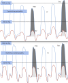
Simultaneous right and left ventricular haemodynamic assessment in constrictive pericarditis and restrictive cardiomyopathy. (Top) Left ventricular (blue) and right ventricular (orange) hemodynamic pressure tracings in constrictive pericarditis. End-diastolic filling pressures are elevated, and a ‘square root’ sign is present on both tracings (*). Enhanced ventricular interdependence is present, demonstrated by visualization of the systolic area index, right ventricular (light grey) and left ventricular (dark grey) areas under the curve for both inspiration (Insp) and expiration (Exp). During inspiration, there is an increase in the area of the right ventricular pressure curve and a decrease in the area of the left ventricular pressure curve. (Bottom) left ventricular and right ventricular pressure tracings in restrictive cardiomyopathy. End-diastolic pressures are elevated and a square root sign (*) is seen; there is no evidence of enhanced ventricular interdependence, with parallel changes in LV and RV pressure curve areas. Reprinted with permission from Geske et al.
Figure 4
Figure 4
Echocardiography in restrictive cardiomyopathies. (A) Small left ventricular cavity size in presence of significantly increased wall thickness and severe left atrial dilatation; (B) biventricular wall thickening in absence of pulmonary hypertension; (C and D) restrictive filling pattern with elevated E/E′ ratio in keeping with increased left ventricular filling pressures; (E) myocardial strain analysis showing an apical sparing pattern in a patient with cardiac amyloidosis.
Figure 5
Figure 5
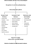
Proposed flowchart for contemporary diagnostic work-up of restrictive cardiomyopathy. CMR, cardiovascular magnetic resonance; DCM, dilated cardiomyopathy; ECG, electrocardiogram; HCM, hypertrophic cardiomyopathy.
Figure 6
Figure 6
Red flag-based diagnostic approach. The main disorders presenting with normal or increased left ventricular wall thickness are listed. CMR, cardiovascular magnetic resonance; EMB, endomyocardial biopsy; EMF, endomyocardial fibrosis; HES, hypereosinophilic syndrome; LV, left ventricle; PRKAG2, protein kinase AMP-activated non-catalytic subunit gamma-2; RCM, restrictive cardiomyopathy.
Figure 7
Figure 7
Cardiovascular magnetic resonance findings in a patient with early stage endomyocardial fibrosis. In a 50-year-old woman, the cardiovascular magnetic resonance examination showed evidence of active inflammation of the subendocardium (as demonstrated by myocardial oedema on T2-weighted images), and multiple intraventricular thrombi (dark images on early enhancement images).
Figure 8
Figure 8
Patterns of late gadolinium enhancement at cardiovascular magnetic resonance across different stages of cardiac amyloidosis. Four-chamber late gadolinium enhancement images acquired 10–15 min after gadolinium administration (with an inversion time set to null the normal ‘healthy’ myocardium) in a healthy subject (left) and in patients with different stages of amyloid light-chain (above) or transthyretin (ATTR; below) cardiac amyloidosis. The circumferential subendocardial late gadolinium enhancement pattern is particularly evident in the patient with intermediate-stage amyloid light-chain amyloidosis, and the diffuse transmural pattern in patients with advanced amyloid light-chain or ATTR amyloidosis.
Figure 9
Figure 9
Mapping analyses by cardiovascular magnetic resonance. Left panel: low native T1 values due to iron accumulation; global myocardial T2* values were very low too (4 ms, r.v. >20 ms) Right panel: high native T1 values due to amyloid accumulation. Segmental T1 values are listed (±SD); Z-score values from a reference population are indicated in brackets.

References

    1. Elliott P, Andersson B, Arbustini E, Bilinska Z, Cecchi F, Charron P, et al. . Classification of the cardiomyopathies: a position statement from the European Society of Cardiology Working Group on Myocardial and Pericardial Diseases. Eur Heart J 2008;29:270–276. - PubMed
    1. Coppini R, Ho CY, Ashley E, Day S, Ferrantini C, Girolami F, et al. . Clinical phenotype and outcome of hypertrophic cardiomyopathy associated with thin-filament gene mutations. J Am Coll Cardiol 2014;64:2589–2600. - PMC - PubMed
    1. Porcari A, Pagura L, Rossi M, Porrazzo M, Dore F, Bussani R, et al. . Light-chain cardiac amyloidosis: a case report of extraordinary sustained pathological response to cyclophosphamide, bortezomib, and dexamethasone combined therapy. Eur Heart J Case Rep 2022;6:ytac130. - PMC - PubMed
    1. Knight DS, Zumbo G, Barcella W, Steeden JA, Muthurangu V, Martinez-Naharro A, et al. . Cardiac structural and functional consequences of amyloid deposition by cardiac magnetic resonance and echocardiography and their prognostic roles. JACC Cardiovasc Imaging 2019;12:823–833. - PubMed
    1. Goldstein JA, Kern MJ. Hemodynamics of constrictive pericarditis and restrictive cardiomyopathy. Catheter Cardiovasc Intervent 2020;95:1240–1248. - PubMed

MeSH terms