. 2022 Nov 15;3(11):100792.
doi: 10.1016/j.xcrm.2022.100792. Epub 2022 Oct 20.

AAV-mediated gene therapy produces fertile offspring in the Lhcgr-deficient mouse model of Leydig cell failure

Affiliations

AAV-mediated gene therapy produces fertile offspring in the Lhcgr-deficient mouse model of Leydig cell failure

Kai Xia et al. Cell Rep Med. .

Abstract

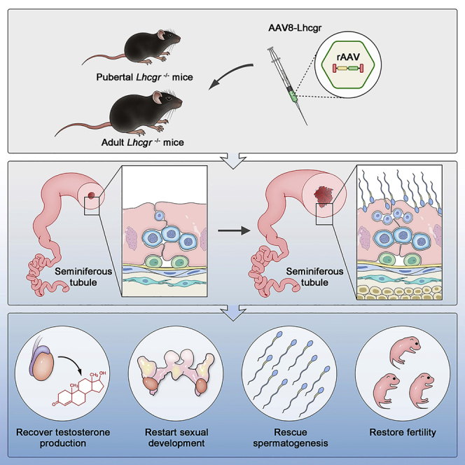
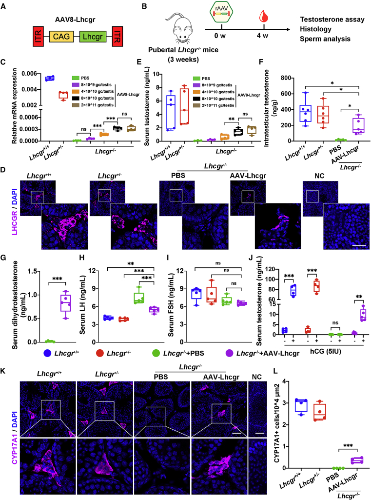
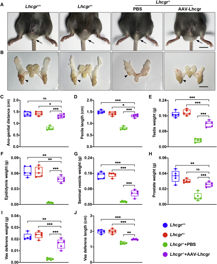
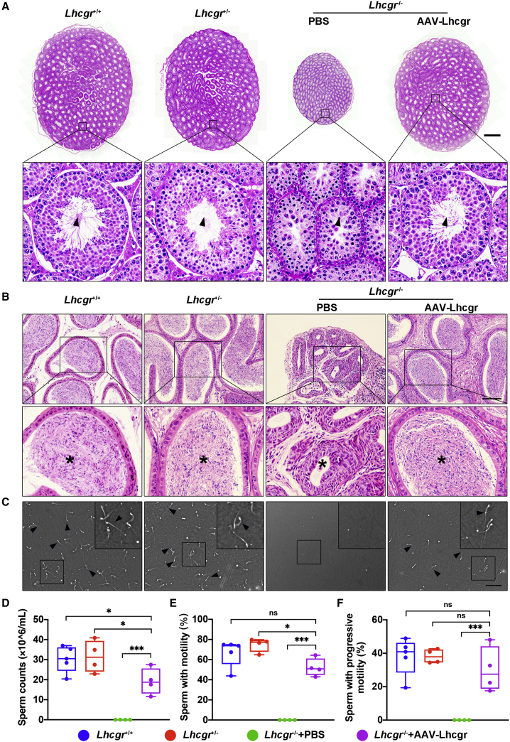
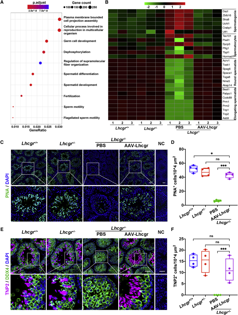
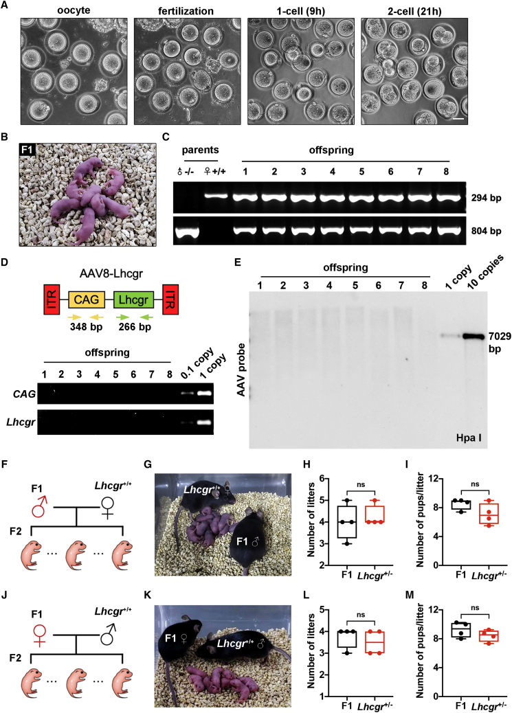
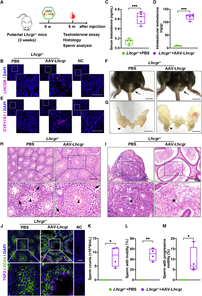
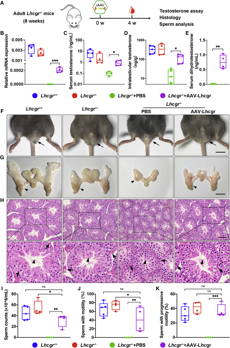
Leydig cell failure (LCF) caused by gene mutation results in testosterone deficiency and infertility. Serum testosterone levels can be recovered via testosterone replacement; however, established therapies have shown limited success in restoring fertility. Here, we use a luteinizing hormone/choriogonadotrophin receptor (Lhcgr)-deficient mouse model of LCF to investigate the feasibility of gene therapy for restoring testosterone production and fertility. We screen several adeno-associated virus (AAV) serotypes and identify AAV8 as an efficient vector to drive exogenous Lhcgr expression in progenitor Leydig cells through interstitial injection. We observe considerable testosterone recovery and Leydig cell maturation after AAV8-Lhcgr treatment in pubertal Lhcgr-/- mice. Of note, this gene therapy partially recovers sexual development, substantially restores spermatogenesis, and effectively produces fertile offspring. Furthermore, these favorable effects can be reproduced in adult Lhcgr-/- mice. Our proof-of-concept experiments in the mouse model demonstrate that AAV-mediated gene therapy may represent a promising therapeutic approach for patients with LCF.

Keywords: AAV; Leydig cell failure; fertility; gene therapy; offspring; sexual development; spermatogenesis; testosterone.


Conflict of interest statement

Declaration of interests The authors declare no competing interests.

Figures

None
Graphical abstract
Figure 1
Figure 1
Testicular injection of AAV8-Lhcgr rescues Leydig cell function and recovers testosterone levels in pubertal Lhcgr−/− mice (A) Schematic of the AAV vector used in the study. (B) Experimental overview of the in vivo studies. (C) Quantitative RT-PCR was used to quantify Lhcgr mRNA transcripts in testicular tissues from Lhcgr+/+ mice, Lhcgr+/− mice, and Lhcgr−/− mice injected with PBS or increasing doses of AAV8-Lhcgr (n = 4). β-actin was used for normalization. (D) Representative images of LHCGR in the testes of Lhcgr+/+ mice, Lhcgr+/− mice, and Lhcgr−/− mice injected with PBS or AAV8-Lhcgr (n = 4). Nuclei were counterstained with DAPI. NC, negative control. Scale bar: 25 μm. (E) The concentrations of serum testosterone were analyzed in Lhcgr+/+ mice (n = 5), Lhcgr+/− mice (n = 5), and Lhcgr−/− mice injected with PBS (n = 3) or increasing doses of AAV8-Lhcgr (8 × 109 gc/testis [n = 4], 4 × 1010 gc/testis [n = 5], 8 × 1010 gc/testis [n = 5], and 2 × 1011 gc/testis [n = 4]). (F) The concentrations of intratesticular testosterone were detected in Lhcgr+/+ mice (n = 6), Lhcgr+/− mice (n = 6), and Lhcgr−/− mice injected with PBS (n = 5) or AAV8-Lhcgr (n = 5). (G) The concentrations of serum dihydrotestosterone testosterone were analyzed in Lhcgr−/− mice injected with PBS or AAV8-Lhcgr (n = 5). (H and I) The concentrations of serum LH (H) and FSH (I) were analyzed in Lhcgr+/+ mice, Lhcgr+/− mice, and Lhcgr−/− mice injected with PBS or AAV8-Lhcgr (n = 5). (J) The concentrations of serum testosterone were analyzed before and 1 h after hCG injection in Lhcgr+/+ mice (n = 4), Lhcgr+/− mice (n = 4), and Lhcgr−/− mice injected with PBS (n = 5) or AAV8-Lhcgr (n = 5). (K) CYP17A1 was evaluated by immunostaining of testes from Lhcgr+/+ mice, Lhcgr+/− mice, and Lhcgr−/− mice injected with PBS or AAV8-Lhcgr (n = 4). NC indicates negative control. Scale bar: 50 μm. (L) CYP17A1+ cells were quantified in the different groups. Data are represented by boxplots, and whiskers show the minimum to maximum values. ∗p < 0.05; ∗∗p < 0.01; ∗∗∗p < 0.001; ns, not significant.
Figure 2
Figure 2
AAV8-Lhcgr restarts sexual development in pubertal Lhcgr−/− mice (A and B) Representative photographs of the external (A) and internal genitalia (B) of Lhcgr+/+ mice, Lhcgr+/− mice, and Lhcgr−/− mice injected with PBS or AAV8-Lhcgr (n = 5). Arrows (A) and arrowheads (B) indicate the testes. Scale bar: 1 cm. (C–J) Ano-genital distance (C), penile length (D), testis weight (E), epididymis weight (F), seminal vesicle weight (G), prostate weight (H), vas deferens weight (I), and vas deferens length (J) of Lhcgr+/+ mice, Lhcgr+/− mice, and Lhcgr−/− mice injected with PBS or AAV8-Lhcgr (n = 5). Data are represented by boxplots, and whiskers show the minimum to maximum values. ∗p < 0.05; ∗∗p < 0.01; ∗∗∗p < 0.001; ns, not significant.
Figure 3
Figure 3
AAV8-Lhcgr rescues spermatogenesis in pubertal Lhcgr−/− mice (A) Representative light micrographs of testis sections from Lhcgr+/+ mice, Lhcgr+/− mice, and Lhcgr−/− mice injected with PBS or AAV8-Lhcgr (n = 3). Scale bar: 500 μm. Arrowheads indicate full spermatogenesis in testes. (B) Histological analysis of cauda epididymis collected from Lhcgr+/+ mice, Lhcgr+/− mice, and Lhcgr−/− mice injected with PBS or AAV8-Lhcgr (n = 3). Stars indicate spermatozoa in the cauda epididymis. Scale bar: 100 μm. (C) Representative light micrographs of sperm obtained from the cauda epididymis of Lhcgr+/+ mice (n = 5), Lhcgr+/− mice (n = 4), and Lhcgr−/− mice injected with PBS (n = 4) or AAV8-Lhcgr (n = 4). Scale bar: 200 μm. (D–F) The sperm counts (D), proportions of sperm with motility (E), and progressive motility (F) were analyzed (Lhcgr+/+ mice [n = 5], Lhcgr+/− mice [n = 4], and Lhcgr−/− mice injected with PBS [n = 4] or AAV8-Lhcgr [n = 4]). Data are represented by boxplots, and whiskers show the minimum to maximum values. ∗p < 0.05; ∗∗∗p < 0.001; ns, not significant.
Figure 4
Figure 4
AAV8-Lhcgr promotes the formation of elongating spermatids (A) Representative GO terms of the top 500 upregulated genes in male Lhcgr−/− mice treated with AAV8-Lhcgr versus PBS (n = 3). (B) Heatmap showing the expression of marker genes for the four major germ cell types in the testicular transcriptional profiles. (C–F) Representative images of testis sections from the four groups (n = 4). Sections were immunostained for PNA (C), and DDX4 and TNP2 (E), and counterstained with DAPI. Quantitative analysis showing the percentage of PNA+ (D) and TNP2+ (F) germ cells in the seminiferous tubules of the testes. NC, negative control. Scale bar: 50 μm. Data are represented by boxplots, and whiskers show the minimum to maximum values. ∗p < 0.05; ∗∗∗p < 0.001; ns, not significant.
Figure 5
Figure 5
AAV8-Lhcgr restores fertility and produces fertile offspring (A) Representative images of oocytes, fertilization, one-cell embryos, and two-cell embryos. Scale bar: 50 μm. (B) Offspring (F1) derived from AAV8-Lhcgr-treated Lhcgr−/− male mice. (C) Genotyping of the offspring derived from AAV8-Lhcgr-treated Lhcgr−/− males and Lhcgr+/+ females. The amplified wild-type (wt) DNA is 294 bp, while the mutant (mt) DNA fragment is 804 bp. (D) PCR analysis of AAV8-Lhcgr integration in the genomes of offspring. CAG promoter- and Lhcgr-specific primers were used. As a control, tail DNA from Lhcgr+/− mice was spiked with viral particles representing 0.1 and 1 copies of the viral genome. (E) Southern blot analysis of DNA samples from offspring mice hybridized with AAV vector. Controls represent viral DNA in amounts equivalent to one and 10 copies of viral DNA per diploid genome. DNA was digested with the indicated restriction enzymes. (F) Mating scheme used to produce the second-generation (F2) mice. (G) Male F1 mice were used to produce F2 by mating with Lhcgr+/+ females. (H and I) Continuous breeding assay starting at 6 weeks of age, showing numbers of litters (H) and pups per litter (I) between F1 males and Lhcgr+/− males within 4 months (n = 4). (J) Mating scheme used to produce F2 mice. (K) Female F1 mice were used to produce F2 pups by mating with Lhcgr+/+ males. (L and M) Continuous breeding assay starting at 6 weeks of age, showing numbers of litters (L) and pups per litter (M) between F1 females and Lhcgr+/− females within 4 months (n = 4). Data are represented by boxplots, and whiskers show the minimum to maximum values. ns, not significant.
Figure 6
Figure 6
AAV-mediated gene therapy may ensure long-term benefits with a single treatment (A) Experimental overview of the in vivo studies. (B) Representative images of LHCGR expression in the testicular interstitium 6 months after treatment in Lhcgr−/− mice injected with PBS or AAV8-Lhcgr (n = 4). Nuclei were counterstained with DAPI. NC, negative control. Scale bar: 50 μm. (C and D) The concentrations of serum (C) and intratesticular (D) testosterone were analyzed 6 months after treatment in 3-week-old Lhcgr−/− mice injected with PBS or AAV8-Lhcgr (n = 5). (E) Representative images of CYP17A1expression in the testicular interstitium in Lhcgr−/− mice injected with PBS or AAV8-Lhcgr (n = 4). Nuclei were counterstained with DAPI. NC, negative control. Scale bar: 50 μm. (F and G) Representative photographs of the external (F) and internal genitalia (G) of Lhcgr−/− mice injected with PBS or AAV8-Lhcgr (n = 4). Arrows (F) and arrowheads (G) indicate the testes. Scale bar: 1 cm. (H and I) Representative light micrographs of testes (H) and epididymis (I) sections from Lhcgr−/− mice injected with PBS or AAV8-Lhcgr (n = 4). Arrows indicate Leydig cells, and arrowheads indicate full spermatogenesis in the testis (H). Stars indicate spermatozoa in the cauda epididymis (I). Scale bars: 100 μm. (J) Representative images of testicular sections from Lhcgr−/− mice injected with PBS or AAV8-Lhcgr (n = 4). Sections were immunostained for DDX4 and TNP2 and counterstained with DAPI. Scale bars: 50 μm. (K–M) The sperm counts (K) and proportions of sperm with motility (L) and progressive motility (M) were analyzed in Lhcgr−/− mice injected with PBS or AAV8-Lhcgr (n = 4). Data are represented by boxplots, and whiskers show the minimum to maximum values. ∗p < 0.05, ∗∗p < 0.01, ∗∗∗p < 0.001.
Figure 7
Figure 7
AAV8-Lhcgr restarts sexual development and rescues spermatogenesis in adult Lhcgr-deficient mice (A) Experimental overview of the in vivo studies. (B) Quantitative RT-PCR was used to quantify Lhcgr mRNA transcripts in testis tissues from Lhcgr+/+, Lhcgr+/−, and Lhcgr−/− mice injected with PBS or AAV8-Lhcgr (n = 4). β-actin was used for normalization. (C) The concentrations of serum testosterone were analyzed in Lhcgr+/+, Lhcgr+/−, and Lhcgr−/− mice injected with PBS or AAV8-Lhcgr (n = 4). (D) The concentrations of intratesticular testosterone were analyzed in Lhcgr+/+ (n = 5), Lhcgr+/− (n = 4), and Lhcgr−/− mice injected with PBS (n = 5) or AAV8-Lhcgr (n = 5). (E) The concentrations of serum dihydrotestosterone testosterone were analyzed in Lhcgr−/− mice injected with PBS or AAV8-Lhcgr (n = 4). (F and G) Representative photographs of the external and internal genitalia of mice from the four groups (n = 4). Arrows (F) and arrowheads (G) indicate the testes. Scale bar: 1 cm. (H) Representative light micrographs of testis sections from four groups (n = 4). Arrows indicate Leydig cells and arrowheads indicate full spermatogenesis in the testis. Scale bars: 100 μm. (I–K) The sperm counts (I), proportions of sperm with motility (J), and progressive motility (K) were analyzed (Lhcgr+/+ [n = 5], Lhcgr+/− [n = 4], and Lhcgr−/− mice injected with PBS [n = 4] or AAV8-Lhcgr [n = 4]). Data are represented by boxplots, and whiskers show the minimum to maximum values. ∗p < 0.05; ∗∗p < 0.01; ∗∗∗p < 0.001; ns, not significant.

References

    1. Salonia A., Rastrelli G., Hackett G., Seminara S.B., Huhtaniemi I.T., Rey R.A., Hellstrom W.J.G., Palmert M.R., Corona G., Dohle G.R., et al. Paediatric and adult-onset male hypogonadism. Nat. Rev. Dis. Primers. 2019;5:38. doi: 10.1038/s41572-019-0087-y. - DOI - PMC - PubMed
    1. Zirkin B.R., Papadopoulos V. Leydig cells: formation, function, and regulation. Biol. Reprod. 2018;99:101–111. doi: 10.1093/biolre/ioy059. - DOI - PMC - PubMed
    1. Teerds K.J., Huhtaniemi I.T. Morphological and functional maturation of Leydig cells: from rodent models to primates. Hum. Reprod. Update. 2015;21:310–328. doi: 10.1093/humupd/dmv008. - DOI - PubMed
    1. Mendonca B.B., Costa E.M.F., Belgorosky A., Rivarola M.A., Domenice S. 46, XY DSD due to impaired androgen production. Best Pract. Res. Clin. Endocrinol. Metab. 2010;24:243–262. doi: 10.1016/j.beem.2009.11.003. - DOI - PubMed
    1. Bhasin S., Brito J.P., Cunningham G.R., Hayes F.J., Hodis H.N., Matsumoto A.M., Snyder P.J., Swerdloff R.S., Wu F.C., Yialamas M.A. Testosterone therapy in men with hypogonadism: an endocrine society clinical practice guideline. J. Clin. Endocrinol. Metab. 2018;103:1715–1744. doi: 10.1210/jc.2018-00229. - DOI - PubMed

Publication types