. 2023 Jan 1:660:114929.
doi: 10.1016/j.ab.2022.114929. Epub 2022 Oct 19.

Protein A-Nanoluciferase fusion protein for generalized, sensitive detection of immunoglobulin G

Affiliations

Protein A-Nanoluciferase fusion protein for generalized, sensitive detection of immunoglobulin G

Suman Nandy et al. Anal Biochem. .

Abstract

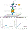
Detection and quantification of antibodies, especially immunoglobulin G (IgG), is a cornerstone of ELISAs, many diagnostics, and the development of antibody-based drugs. Current state-of-the-art immunoassay techniques for antibody detection require species-specific secondary antibodies and carefully-controlled bioconjugations. Poor conjugation efficiency degrades assay performance and increases the risk of clinical false positives due to non-specific binding. We developed a generic, highly-sensitive platform for IgG quantification by fusing the IgG-Fc binding Z domain of Staphylococcal Protein A with the ultrabright bioluminescence reporter Nanoluc-luciferase (Nluc). We demonstrated the application of this fusion protein in a sandwich IgG detection immunoassay using surface-bound antigens to capture target IgG and protein A-Nanoluc fusion as the detector. We optimized the platform's sensitivity by incorporating multiple repeats of the Z domain into the fusion protein constructs. Using rabbit and mouse anti-SARS-CoV-2 Nucleoprotein IgGs as model analytes, we performed ELISAs in two different formats, either with SARS-CoV-2 Nucleoprotein as the capture antigen or with polyclonal chicken IgY as the capture antibody. Using standard laboratory equipment, the platform enabled the quantitation of antibody analytes at concentrations as low as 10 pg/mL (67 fM).

Keywords: Bioluminescence; IgG detection; Immunoassay; Nanoluciferase; Protein A; SARS-CoV-2 Nucleoprotein.


Conflict of interest statement

Conflicts of Interest The authors declare no competing interests.

Figures

Fig. 1.
Fig. 1.
Gene fragment assembly and recombinant protein characterization. (A) Gene schematics. (B and C) Agarose gel electrophoresis of colony PCR amplicons: five bacterial transformants were selected from LB-kanamycin plates, grown at 37 °C for 4 h, centrifuged briefly to collect the cell pellet which was incubated for 5 min at 98 °C to extract plasmids. PCR amplification of the insert gene fragments was performed with the sequencing primer pair (NdeI-N-Z-F, XhoI-Nluc-R). After electrophoresis, amplicons were visualized with SYBR green I dye (B) Z3-Nluc and (C) Z5-Nluc transformants. (D and E) SDS-PAGE analysis of IgG affinity and immobilized metal affinity (IMAC) purification fractions of (D) Z3-Nluc and (E) Z5-Nluc protein.
Fig. 2.
Fig. 2.
The predicted tertiary structures of the Z3-Nluc and Z5-Nluc proteins. The Z domain helices are shown in green, the Nluc in pink, and the His6 tag in red. (A) Z3–Nluc (a single Nluc with three repeats of the Z domain). (B) Z5–Nluc (a single Nluc with five repeats of the Z domain). Loop modeling and sequence alignments were generated using Phyre2 (Protein Homology/analogy Recognition Engine V 2.0, Imperial College, London). The structures were drawn using PyMOL (Schrödinger, Inc, NY, USA).
Fig. 3.
Fig. 3.
MALDI-ToF mass spectra of purified recombinant fusion proteins. (A) His6 tagged Z3-NLuc protein (predicted: 41,870 Da, observed: 41,882 Da, additional prominent peak at 21,030 attributed to the doubly charged ion); (B) His6 tagged Z5-NLuc protein (predicted: 54,983 Da, observed: 54,865 Da, additional prominent peak at 27,372 attributed to the doubly charged ion). Mass spectrometry analyses was performed in Tufts University Core Facility.
Fig. 4.
Fig. 4.
IgG quantification by Z3–Nluc-ELISA using different capture molecules. Quantification of (A) anti-NP mouse antibodies and (B) anti-NP rabbit antibodies using NP (directly adsorbed on the well) as the capture molecule. Quantification of (C) anti-NP mouse antibodies and (D) anti-NP rabbit antibodies using NP captured on anti-NP chicken IgY as the capture molecule. Each analyte concentration was tested in duplicate wells and each well was measured 3 times; error bars represent the standard deviation of six replicates. The LOD was taken as the intercept with the dashed line representing the average luminescence value of the no-antibody control plus three times the standard deviation (σ) of the no-antibody control (n=6; σ was 1–7% of the signal).
Fig. 5.
Fig. 5.
(A) Schematic of the Z5–Nluc-ELISA used in panels (B) and (C). (B) Detection of SARS-CoV-2 NP spiked in 10% human serum using anti-NP chicken IgY as the capture molecule. (C) Detection of anti-NP rabbit IgG spiked in 10% human serum using NP captured on anti-NP chicken IgY as the capture molecule. The LOD was taken as the intercept with the dashed line representing the average luminescence value of the no-antibody control plus three times the standard deviation (σ) of the no-antibody control.

References

    1. Zhu L, Cui X, Wu J, Wang Z, Wang P, Hou Y, Yang M, Fluorescence immunoassay based on carbon dots as labels for the detection of human immunoglobulin G, Anal. Methods 6 (2014) 4430–4436. 10.1039/C4AY00717D. - DOI
    1. Zhao H, Chang Y, Liu M, Gao S, Yu H, Quan X, A universal immunosensing strategy based on regulation of the interaction between graphene and graphene quantum dots, Chem. Commun 49 (2012) 234–236. 10.1039/C2CC35503E. - DOI - PubMed
    1. Song HY, Ngai MH, Song ZY, MacAry PA, Hobley J, Lear MJ, Practical synthesis of maleimides and coumarin-linked probes for protein and antibody labelling via reduction of native disulfides, Org. Biomol. Chem 7 (2009) 3400–3406. 10.1039/B904060A. - DOI - PubMed
    1. Brinkley M, A brief survey of methods for preparing protein conjugates with dyes, haptens and crosslinking reagents, Bioconjug. Chem 3 (1992) 2–13. https://pubs.acs.org/doi/10.1021/bc00013a001. - DOI - PubMed
    1. Nilsson BL, Hondal RJ, Soellner MB, Raines RT, Protein assembly by orthogonal chemical ligation methods, J. Am. Chem. Soc 125 (2003) 5268–5269. https://pubs.acs.org/doi/10.1021/ja029752e. - DOI - PubMed

Publication types