Benign Lobular Inner Nuclear Layer Proliferations of the Retina Associated with Congenital Hypertrophy of the Retinal Pigment Epithelium
- PMID: 36270406
- PMCID: PMC9974858
- DOI: 10.1016/j.ophtha.2022.10.011
Benign Lobular Inner Nuclear Layer Proliferations of the Retina Associated with Congenital Hypertrophy of the Retinal Pigment Epithelium
Abstract
Purpose: To report the clinical and imaging findings of 4 patients with benign intraretinal tumors, 2 of which were associated with retinal pigment epithelium (RPE) hypertrophy. To our knowledge, this condition has not been described previously and should be distinguished from retinoblastoma and other malignant retinal neoplasms.
Design: Retrospective case series.
Participants: Four patients from 3 institutions.
Methods: Four patients with intraretinal tumors of the inner nuclear layer (INL) underwent a combination of ophthalmic examination, fundus photography, fluorescein angiography, OCT, OCT angiography, and whole exome sequencing.
Main outcome measures: Description of multimodal imaging findings and systemic findings from 4 patients with benign intraretinal tumors and whole exome studies from 3 patients.
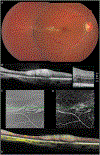
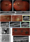
Results: Six eyes of 4 patients 5, 13, 32, and 27 years of age were found to have white intraretinal tumors that remained stable over the follow-up period (range, 9 months-4 years). The tumors were unilateral in 2 patients and bilateral in 2 patients. The tumors were white, centered on the posterior pole, and multifocal, with some consisting of multiple lobules with arching extensions that extended beyond the central tumor mass. OCT demonstrated these lesions to be centered within the INL at the border of the inner plexiform layer. In addition, 2 patients demonstrated congenital hypertrophy of the RPE (CHRPE) lesions. Three of 4 patients underwent whole exome sequencing of the blood that revealed no candidate variants that plausibly could account for the phenotype.
Conclusions: We characterize a novel benign tumor of the INL that, in 2 patients, was associated with separate CHRPE lesions. We propose the term benign lobular inner nuclear layer proliferation to describe these lesions.
Financial disclosure(s): Proprietary or commercial disclosure may be found after the references.
Keywords: Congenital hypertrophy of the retinal pigment epithelium; Inner nuclear layer; Retinal mass; Retinal tumor; Retinoblastoma.
Copyright © 2022 American Academy of Ophthalmology. Published by Elsevier Inc. All rights reserved.
Figures


References
-
- Shields CL, Say EAT, Fuller T, et al. Retinal Astrocytic Hamartoma Arises in Nerve Fiber Layer and Shows “Moth-Eaten” Optically Empty Spaces on Optical Coherence Tomography. Ophthalmology 2016;123(8):1809–16. - PubMed
-
- Pichi F, Massaro D, Serafino M, et al. RETINAL ASTROCYTIC HAMARTOMA: Optical Coherence Tomography Classification and Correlation With Tuberous Sclerosis Complex. Retina 2016;36(6):1199–208. - PubMed
-
- Cao C, Markovitz M, Ferenczy S, Shields CL. Hand-held spectral-domain optical coherence tomography of small macular retinoblastoma in infants before and after chemotherapy. J Pediatr Ophthalmol Strabismus 2014;51(4):230–4. - PubMed
Publication types
MeSH terms
Grants and funding
LinkOut - more resources
Full Text Sources
Medical

