. 2022 Oct 21;13(1):6265.
doi: 10.1038/s41467-022-33759-y.

Long-term whole blood DNA preservation by cost-efficient cryosilicification

Affiliations

Long-term whole blood DNA preservation by cost-efficient cryosilicification

Liang Zhou et al. Nat Commun. .

Abstract

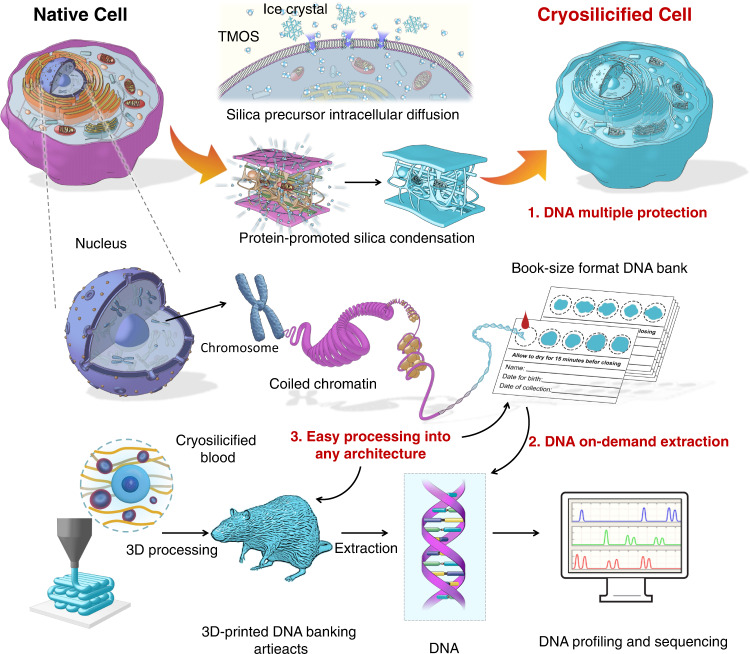
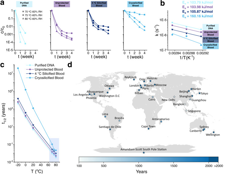
Deoxyribonucleic acid (DNA) is the blueprint of life, and cost-effective methods for its long-term storage could have many potential benefits to society. Here we present the method of in situ cryosilicification of whole blood cells, which allows long-term preservation of DNA. Importantly, our straightforward approach is inexpensive, reliable, and yields cryosilicified samples that fulfill the essential criteria for safe, long-term DNA preservation, namely robustness against external stressors, such as radical oxygen species or ultraviolet radiation, and long-term stability in humid conditions at elevated temperatures. Our approach could enable the room temperature storage of genomic information in book-size format for more than one thousand years (thermally equivalent), costing only 0.5 $/person. Additionally, our demonstration of 3D-printed DNA banking artefacts, could potentially allow 'artificial fossilization'.


Conflict of interest statement

The authors declare no competing interests.

Figures

Fig. 1
Fig. 1. Schematic representation of the cell cryosilicification process and the construction of whole blood DNA banking.
The silica precursor diffuses into the cell and nucleus under dormancy, and subsequently, condenses under the catalysis of cellular protein. This self-limited silica condensation on sub-cellular templates preserves the entire cellular architectures, mimicking the hermetic sealing of DNA in natural fossils. Through simple drop casting or three-dimensional (3D) printing technologies, whole-blood genomic DNA banking could be constructed for cryosilicified blood samples with various substrates for further on-demanded DNA profiling and sequencing.
Fig. 2
Fig. 2. In situ cryosilicification of whole-blood cells.
a Schematic representation of the procedure of cell cryosilicification and desilicification. be SEM images of b native leukocyte, c cryosilicified leukocyte, d native erythrocyte, and e cryosilicified erythrocyte, Scale bars = 1 μm. A representative image of three biological replicates is shown. f FTIR spectroscopy of native blood (purple), silica (gray), and cryosilicified blood (blue). A representative spectrum of three biological replicates is shown. g Silicon content of cryosilicified blood prepared with different silica precursors concentration determined by ICP-OES. Six biological replicates are shown. h Fluorescent microscopy images of native and cryosilicified leukocytes. Cell nucleus was stained by Hoechst 33342 (blue), and the cell membrane was stained by DiD (red), Scale bars = 20 μm. A representative image of three biological replicates is shown. i Cell diameter of leukocyte and erythrocyte after cryosilicification. Box and whiskers represent mean ± 25–75 percentile, center values 7.393, 7.242, 7.871, and 8.016, successively, n = 102 cells of three biological replicates. j 3D confocal microscopy images of cryosilicified leukocyte. Cell nucleus was stained by Hoechst 33342 (blue), and silica was stained by FITC-modified silane (green) during synthesis, Scale bars = 2 μm. A representative image of three biological replicates is shown. k Bright-field images of native and cryosilicified leukocyte under different aspiration pressures, Scale bars = 5 μm. A representative image of three biological replicates is shown. l Aspiration length changes of native and cryosilicified leukocyte under different aspiration pressures. Three biological replicates are shown. m Young’s modules of native and cryosilicified leukocytes. Box and whiskers represent mean ± 25–75 percentile, center values 0.042 and 0.234, n = 33 biological replicates. Statistical significance was calculated with two-tailed Student’s t test. Data are presented as mean values ± SD. Source data are provided as a Source Data file.
Fig. 3
Fig. 3. Whole blood DNA preservation.
a Gel electrophoresis of DNA extracted from fresh blood, cryosilicified blood, 4 °C silicified blood, and formaldehyde-fixed blood samples. A representative image of three biological replicates is shown. b Gel electrophoresis of the amplified target genomic fragments (G1–G5) from fresh blood, cryosilicified blood, 4 °C silicified blood, and formaldehyde-fixed blood. A representative image of three biological replicates is shown. c Circos plot summary of mutations between fresh and cryosilicified blood samples in all human chromosomes (1–22+XY). d SNP mutations between fresh and cryosilicified blood samples. e InDel mutations between fresh and cryosilicified blood samples.
Fig. 4
Fig. 4. Robust and long-term DNA preservation.
a, b Gel electrophoresis of DNA extracted from fresh blood sample and purified DNA, unprotected blood, 4 °C silicified blood, and cryosilicified blood samples after a H2O2 and b UV treatment. A representative image of three biological replicates is shown. c Gel electrophoresis of DNA extracted from a fresh blood sample, and unprotected blood and cryosilicified blood samples aging at 70 °C and 60% RH. A representative image of three biological replicates is shown. d Gel electrophoresis of the amplified target genomic fragments (G1–G5) from fresh blood sample, unprotected blood, and cryosilicified blood samples after aging at 70 °C and 60% RH. A representative image of three biological replicates is shown. e STR analysis of DNA from fresh blood samples and cryosilicified blood samples after aging 21 days at 70 °C and 60% RH.
Fig. 5
Fig. 5. Degradation kinetics of DNA storage.
a Concentration of target genomic fragments (G6) from purified DNA, unprotected blood, 4 °C silicified blood, and cryosilicified blood samples after aging at 70, 75, 80 °C and 60% RH. b Activation energies of target genomic fragments (G6) from purified DNA, unprotected blood, 4 °C silicified blood, and cryosilicified blood samples after aging at 70, 75, 80 °C, and 60% RH. c Half-lives extrapolated from the experimental decay rate constants. d Estimated half-lives of DNA from cryosilicified blood samples based on the average annual temperature of cities.
Fig. 6
Fig. 6. Construction of whole blood DNA banking with paper-based cards or 3D-printed artifacts.
a Schematic representation of the preparation of paper-based whole blood genomic DNA banking and its application for forensic identification. b STR analysis of DNA extracted from fresh blood samples and paper-based whole blood DNA bank after aging for 28 days at 70 °C and 60% RH. c Schematic representation of the fabrication of whole blood genomic DNA banking with 3D-printed artifacts (middle, scale bars = 2 cm) and on-demanded DNA extraction. d Microscopy images of cryosilicified leukocytes and erythrocytes in 3D-printed whole blood DNA banking scaffold; Cell nucleus was stained by Hoechst 33342 (blue), and cell membrane was stained by DiO (green), Scale bars = 200 μm. e 3D-printed mouse model. f 3D-printed mouse figurine with embedded cryosilicified mouse blood samples. g Gel electrophoresis of DNA extracted from fresh blood, cryosilicified blood, and 3D-printed DNA banking artifacts before and after its aging for 7 days at 70 °C and 60% RH. A representative image of three biological replicates is shown.

References

    1. Cavalli-Sforza LL. The Human Genome Diversity Project: past, present and future. Nat. Rev. Genet. 2005;6:333–340. - PubMed
    1. Gibbs RA. The Human Genome Project changed everything. Nat. Rev. Genet. 2020;21:575–576. - PMC - PubMed
    1. Liggett SB. Pharmacogenetic applications of the Human Genome project. Nat. Med. 2001;7:281–283. - PubMed
    1. National Research Council. DNA Technology in Forensic Science. (The National Academies Press, Washington, DC; 1992). - PubMed
    1. Quillin JM, Bodurtha JN, Smith TJ. Genetics assessment at the end of life: Suggestions for implementation in clinic and future research. J. Palliat. Med. 2008;11:451–458. - PubMed

Publication types