. 2022 Oct 21;12(1):17647.
doi: 10.1038/s41598-022-20542-8.

Mapping periplasmic binding protein oligosaccharide recognition with neutron crystallography

Affiliations

Mapping periplasmic binding protein oligosaccharide recognition with neutron crystallography

Shantanu Shukla et al. Sci Rep. .

Abstract

Numerous studies have shown how periplasmic binding proteins (PBPs) bind substrates with exquisite specificity, even distinguishing between sugar epimers and anomers, or structurally similar ions. Yet, marked substrate promiscuity is also a feature encoded in some PBPs. Except for three sub-Ångström crystal structures, there are no reports of hydrogen atom positions in the remaining (> 1000) PBP structures. The previous X-ray crystal structure of the maltodextrin periplasmic-binding protein from Thermotoga maritima (tmMBP) complexed with oligosaccharide showed a large network of interconnected water molecules stretching from one end of the substrate binding pocket to the other. These water molecules are positioned to form multiple hydrogen bonds, as well as forming interactions between the protein and substrate. Here we present the neutron crystal structure of tmMBP to a resolution of 2.1 Å. This is the first neutron crystal structure from the PBP superfamily and here we unambiguously identify the nature and orientation of the hydrogen bonding and water-mediated interactions involved in stabilizing a tetrasaccharide in the binding site. More broadly, these results demonstrate the conserved intricate mechanisms that underlie substrate-specificity and affinity in PBPs.


Conflict of interest statement

The authors declare no competing interests.

Figures
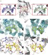
Figure 1
Figure 1
The neutron crystal structure of tmMBP. (a) Center, overall structure of tmMBP bound to maltotetraose (stick representation with yellow carbon atoms). The N- and C-termini are indicated as well as the nomenclature for the maltotetraose rings (R1–R4). Left, 2Fo–Fc electron density for maltotetraose contoured at 1.4 sigma. Right, 2Fo–Fc nuclear density for maltotetraose contoured at 1.1 sigma. (b) Top, close-up view of the 2Fo–Fc nuclear density for the R1/R2 subsite contoured at 1.0 sigma. Bottom, close-up view of the R1/R2 hydrogen bonding network identified from the nuclear density maps. Hydrogen bonds are represented as black dashed lines. (c) Top, close-up view of the 2Fo–Fc nuclear density for the R3/R4 subsite 1.0 sigma. Bottom, close-up view of the R3/R4 hydrogen bonding network identified from the nuclear density maps. Hydrogen bonds are represented as black dashed lines.
Figure 2
Figure 2
Water structure in the tmMBP binding site. (a) tmMBP water subsite 1. (b) tmMBP water subsite 2. (c) tmMBP water subsite 3. (d) ecMBP water subsite 3. Protein sidechains are represented as sticks with grey carbon atoms, maltotetraose is represented as sticks with yellow carbon atoms, and maltose is represented as sticks with green carbon atoms. Hydrogen bonds are indicated as black dashed lines. Nuclear density is show for the tmMBP water subsites and is contoured at 1.0 sigma.

References

    1. Duan X, Hall JA, Nikaido H, Quiocho FA. Crystal structures of the maltodextrin/maltose-binding protein complexed with reduced oligosaccharides: flexibility of tertiary structure and ligand binding. J. Mol. Biol. 2001;306:1115–1126. doi: 10.1006/jmbi.2001.4456. - DOI - PubMed
    1. Cuneo MJ, Beese LS, Hellinga HW. Ligand-induced conformational changes in a thermophilic ribose-binding protein. BMC Struct. Biol. 2008;8:50. doi: 10.1186/1472-6807-8-50. - DOI - PMC - PubMed
    1. Magnusson U, et al. Hinge-bending motion of D-allose-binding protein from Escherichia coli: Three open conformations. J. Biol. Chem. 2002;277:14077–14084. doi: 10.1074/jbc.M200514200. - DOI - PubMed
    1. Sharff AJ, Rodseth LE, Spurlino JC, Quiocho FA. Crystallographic evidence of a large ligand-induced hinge-twist motion between the two domains of the maltodextrin binding protein involved in active transport and chemotaxis. Biochemistry. 1992;31:10657–10663. doi: 10.1021/bi00159a003. - DOI - PubMed
    1. Vergara R, et al. The interplay of protein–ligand and water-mediated interactions shape affinity and selectivity in the LAO binding protein. FEBS J. 2020;287:763–782. doi: 10.1111/febs.15019. - DOI - PubMed

Publication types