

Structure of the pre-mRNA leakage 39-kDa protein reveals a single domain of integrated zf-C3HC and Rsm1 modules
- PMID: 36271106
- PMCID: PMC9586977
- DOI: 10.1038/s41598-022-22183-3
Structure of the pre-mRNA leakage 39-kDa protein reveals a single domain of integrated zf-C3HC and Rsm1 modules
Abstract
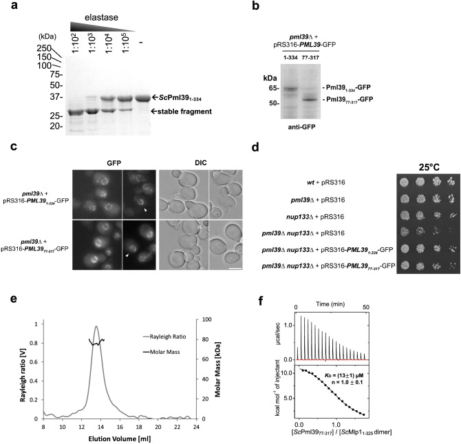
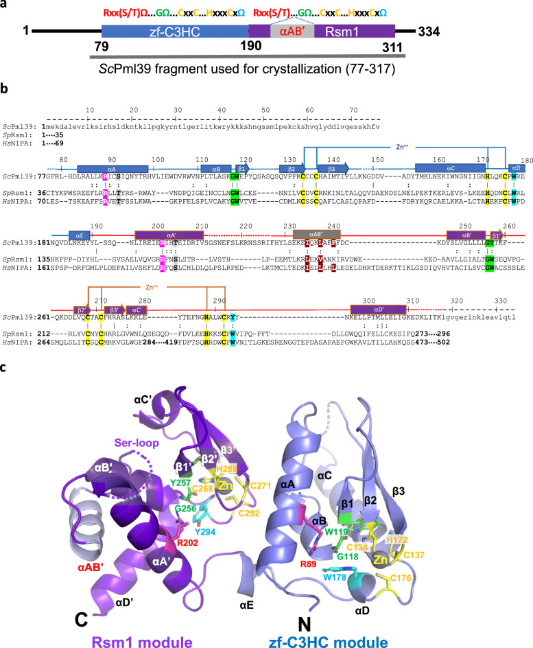
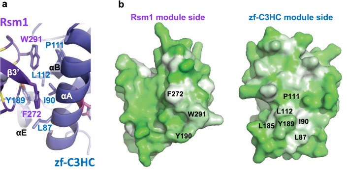
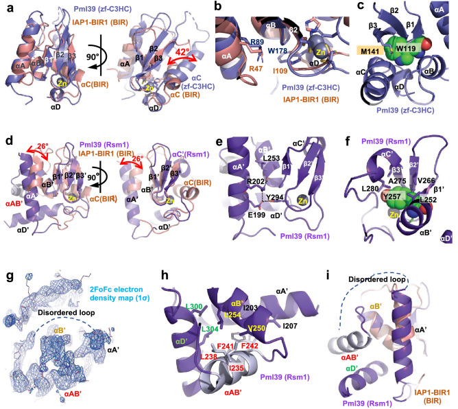
In Saccharomyces cerevisiae, the pre-mRNA leakage 39-kDa protein (ScPml39) was reported to retain unspliced pre-mRNA prior to export through nuclear pore complexes (NPCs). Pml39 homologs outside the Saccharomycetaceae family are currently unknown, and mechanistic insight into Pml39 function is lacking. Here we determined the crystal structure of ScPml39 at 2.5 Å resolution to facilitate the discovery of orthologs beyond Saccharomycetaceae, e.g. in Schizosaccharomyces pombe or human. The crystal structure revealed integrated zf-C3HC and Rsm1 modules, which are tightly associated through a hydrophobic interface to form a single domain. Both zf-C3HC and Rsm1 modules belong to the Zn-containing BIR (Baculovirus IAP repeat)-like super family, with key residues of the canonical BIR domain being conserved. Features unique to the Pml39 modules refer to the spacing between the Zn-coordinating residues, giving rise to a substantially tilted helix αC in the zf-C3HC and Rsm1 modules, and an extra helix αAB' in the Rsm1 module. Conservation of key residues responsible for its distinct features identifies S. pombe Rsm1 and Homo sapiens NIPA/ZC3HC1 as structural orthologs of ScPml39. Based on the recent functional characterization of NIPA/ZC3HC1 as a scaffold protein that stabilizes the nuclear basket of the NPC, our data suggest an analogous function of ScPml39 in S. cerevisiae.
© 2022. The Author(s).
Conflict of interest statement
The authors declare no competing interests.
Figures
Similar articles
-
Proteomics analysis reveals stable multiprotein complexes in both fission and budding yeasts containing Myb-related Cdc5p/Cef1p, novel pre-mRNA splicing factors, and snRNAs.Mol Cell Biol. 2002 Apr;22(7):2011-24. doi: 10.1128/MCB.22.7.2011-2024.2002. Mol Cell Biol. 2002. PMID: 11884590 Free PMC article.
-
Genome-wide analysis of pre-mRNA splicing: intron features govern the requirement for the second-step factor, Prp17 in Saccharomyces cerevisiae and Schizosaccharomyces pombe.J Biol Chem. 2004 Dec 10;279(50):52437-46. doi: 10.1074/jbc.M408815200. Epub 2004 Sep 27. J Biol Chem. 2004. PMID: 15452114
-
Structural and functional analysis of Nro1/Ett1: a protein involved in translation termination in S. cerevisiae and in O2-mediated gene control in S. pombe.RNA. 2011 Jul;17(7):1213-24. doi: 10.1261/rna.2697111. Epub 2011 May 24. RNA. 2011. PMID: 21610214 Free PMC article.
-
Nuclear envelope insertion of spindle pole bodies and nuclear pore complexes.Nucleus. 2012 May-Jun;3(3):226-36. doi: 10.4161/nucl.20148. Epub 2012 May 1. Nucleus. 2012. PMID: 22572959 Free PMC article. Review.
-
Nuclear export of mRNA.FEBS Lett. 2001 Jun 8;498(2-3):150-6. doi: 10.1016/s0014-5793(01)02482-6. FEBS Lett. 2001. PMID: 11412847 Review.
Cited by
-
An evolutionarily conserved bimodular domain anchors ZC3HC1 and its yeast homologue Pml39p to the nuclear basket.Mol Biol Cell. 2023 May 1;34(5):ar40. doi: 10.1091/mbc.E22-09-0402. Epub 2023 Mar 1. Mol Biol Cell. 2023. PMID: 36857168 Free PMC article.
-
Structure, function and assembly of nuclear pore complexes.Nat Rev Mol Cell Biol. 2025 Sep 9. doi: 10.1038/s41580-025-00881-w. Online ahead of print. Nat Rev Mol Cell Biol. 2025. PMID: 40926106 Review.
References
-
- Hoelz A, Debler EW, Blobel G. The structure of the nuclear pore complex. Annu. Rev. Biochem. 2011;80:613–643. - PubMed
Publication types
MeSH terms
Substances
Grants and funding
LinkOut - more resources
Full Text Sources
Molecular Biology Databases
Research Materials
