. 2023 Jan;391(1):173-188.
doi: 10.1007/s00441-022-03698-7. Epub 2022 Oct 22.

Platelet lysate can support the development of a 3D-engineered skin for clinical application

Affiliations

Platelet lysate can support the development of a 3D-engineered skin for clinical application

I Banakh et al. Cell Tissue Res. 2023 Jan.

Abstract

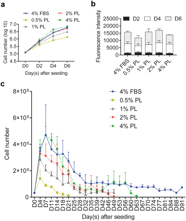
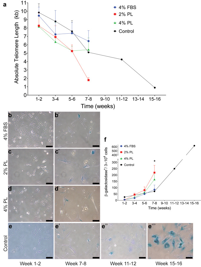
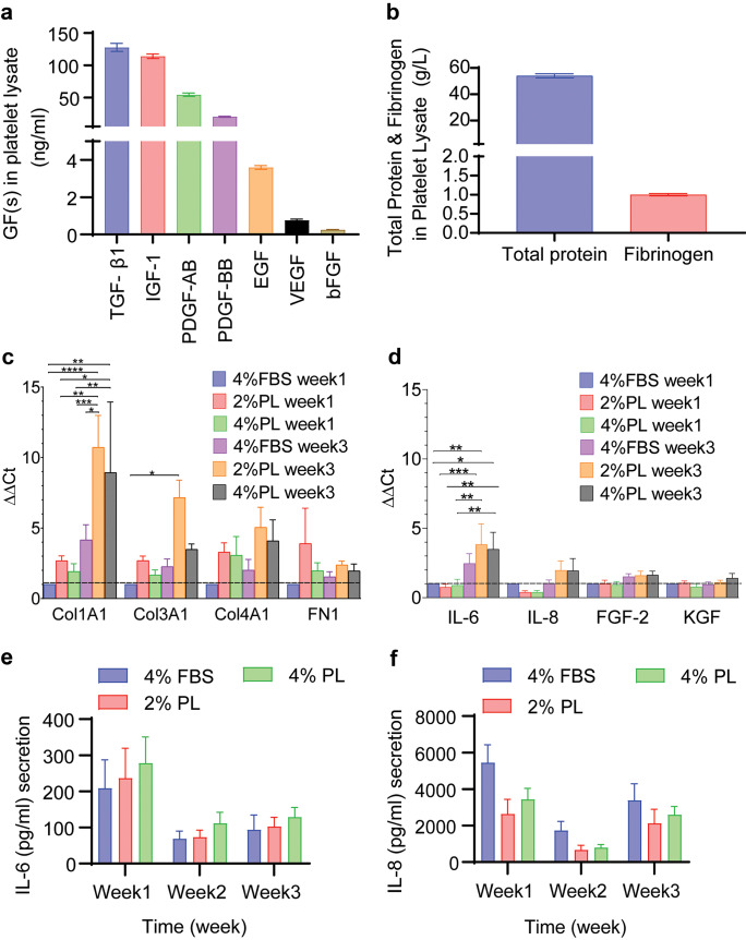
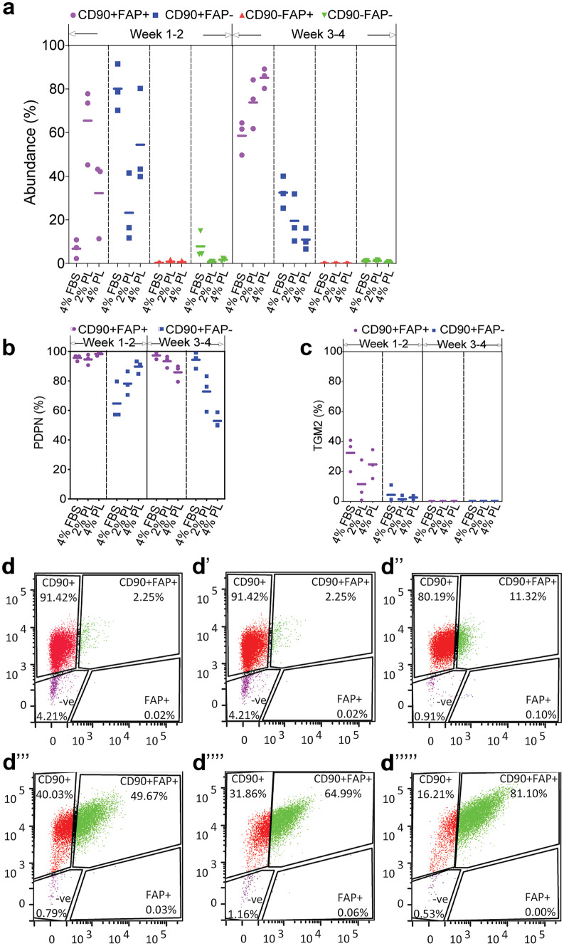
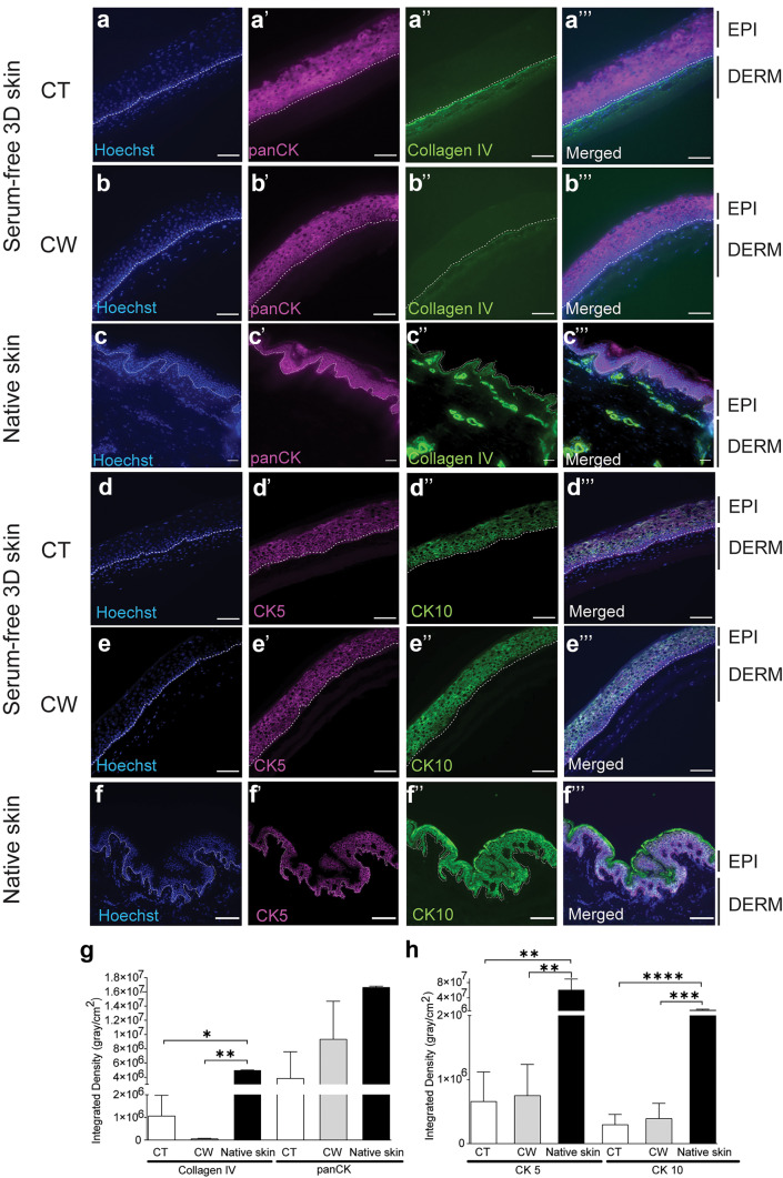
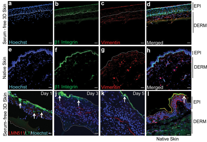
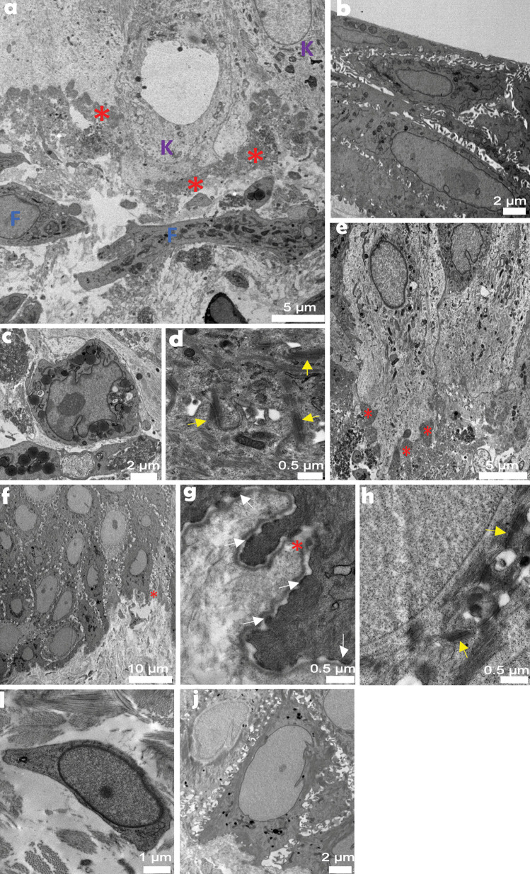
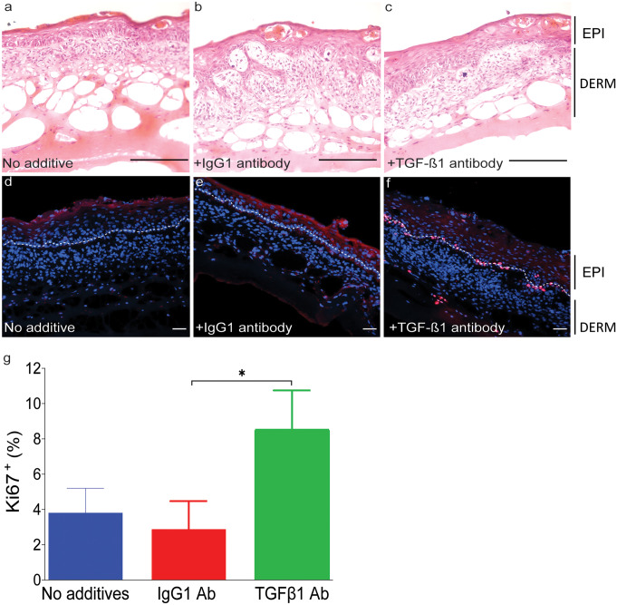
Safety concerns associated with foetal bovine serum (FBS) have restricted its translation into clinics. We hypothesised that platelet lysate (PL) can be utilised as a safe alternative to produce serum-free 3D-engineered skin. PL supported a short-term expansion of fibroblasts, with negligible replication-induced senescence and directed epidermal stratification. PL-expanded fibroblasts were phenotypically separated into three subpopulations of CD90+FAP+, CD90+FAP- and CD90-FAP+, based on CD90 (reticular marker) and FAP (papillary marker) expression profile. PL drove the expansion of the intermediate CD90+ FAP+ subpopulation in expense of reticular CD90+FAP-, which may be less fibrotic once grafted. The 3D-engineered skin cultured in PL was analysed by immunofluorescence using specific markers. Detection of ColIV and LMN-511 confirmed basement membrane. K10 confirmed near native differentiation pattern of neo-epidermis. CD29- and K5-positive interfollicular stem cells were also sustained. Transmission and scanning electron microscopies detailed the ultrastructure of the neo-dermis and neo-epidermis. To elucidate the underlying mechanism of the effect of PL on skin maturation, growth factor contents in PL were measured, and TGF-β1 was identified as one of the most abundant. TGF-β1 neutralising antibody reduced the number of Ki67-positive proliferative cells, suggesting TGF-β1 plays a role in skin maturation. Moreover, the 3D-engineered skin was exposed to lucifer yellow on days 1, 3 and 5. Penetration of lucifer yellow into the skin was used as a semi-quantitative measure of improved barrier function over time. Our findings support the concept of PL as a safe and effective serum alternative for bioengineering skin for cell therapies.

Keywords: Collagen IV; Platelet lysate; Primary keratinocytes culture; Senescence; TGF-β1.


Conflict of interest statement

The authors declare no competing interests.

Figures

Fig. 1
Fig. 1
Platelet lysate (PL) can support expansion of dermal fibroblasts ex vivo. Primary adult fibroblast proliferation in media supplemented with 0.5%, 1%, 2% and 4% PL, compared to 4% FBS were measured by a cell counting and b Alamar Blue on day 2, day 4 and day 6 post-seeding. c Fibroblasts were kept in media supplemented with 0.5% PL, 1% PL, 2% PL, 4% PL or 4% FBS for over 3 months. Cell numbers were determined at each split. Values represent mean + / − SEM in each group (n = 3 per group). Data analysed using an unpaired t-test
Fig. 2
Fig. 2
Replication-induced senescence in fibroblasts expanded in PL-supplemented media. Senescence in fibroblasts expanded in 2% and 4% PL, compared to 4% FBS, was quantified by measuring a absolute telomere length and bf β-galactosidase accumulation in cells. Panels be and b’e’ visualise, weeks 1–2 and weeks 7–8 stained fibroblast cultures, respectively. Panels e’’ and e’’’ present stained fibroblasts from weeks 11 to 12 and weeks 15 to 16 cultures, respectively. Fibroblasts in 4% FBS-supplemented standard media were kept in culture for up to 16 weeks as a reference. Values represent mean values + / − SEM in each group (* = p ≤ 0.05, ** = p ≤ 0.01, *** = p ≤ 0001, n = 5 per group)
Fig. 3
Fig. 3
PL contains a number of growth factors that stimulate fibroblasts. a transforming growth factor β1 (TGF-β1), insulin-like growth factor 1 (IGF-1), platelet-derived growth factors (PDGF-AB, and PDGF-BB), epidermal growth factor (EGF), vascular endothelial growth factor (VEGF) and basic fibroblast growth factor (bFGF or FGF-2) concentrations in PL (n = 8). b Total protein and fibrinogen concentrations in PL (n = 8) c,d collagen 1a1 (Col1A1), collagen 3a1 (Col3A1), collagen 4a1 (Col4A1) and fibronectin-1 (FN1), Interleukin-6 (IL-6), Interleukin-8 (IL-8), fibroblast growth factor-2 (FGF-2) and keratinocyte growth factor (KGF) expression were analysed in fibroblasts expanded in 2% and 4% PL over 3 weeks, compared to 4% FBS. Graphs represent mean values + / − SEM in each group (n = 5 per group) (* = p ≤ 0.05, ** = p ≤ 0.01, *** = p ≤ 0.001). ef IL-6 and IL-8 secretion by fibroblasts isolated and expanded in 2% PL-, 4% PL- or 4% FBS-supplemented media was measured by ELISA after 1, 2 and 3 weeks culture (n = 4)
Fig. 4
Fig. 4
Resolution of fibroblast subpopulations expanded in PL-supplemented media. Freshly isolated dermal fibroblasts were expanded in 2% PL-, 4% PL- or 4% FBS-supplemented media as a control over 4 weeks. a The abundance of CD90 and FAP fibroblast markers were analysed by flow cytometry. The abundance of b PDPN and c TGM2 in CD90/FAP subpopulations were measured (n = 3 per group). d The gating strategy to separate CD90 and FAP subpopulations. Panels dd’’ represent fibroblasts in 4% FBS, 2% PL and 4% PL after 1–2 weeks of expansion, respectively. Panels d’’’d’’’’’ represent fibroblasts in 4% FBS, 2% PL and 4% PL after 3–4 weeks of expansion, respectively
Fig. 5
Fig. 5
Serum-free 3D-engineered skin structure. ac Collagen IV (ColIV, a basement membrane marker) and pan-cytokeratin (panCK) immunofluorescence staining showed higher ColIV deposition in the basement membrane when fibroblasts were seeded on top (CT = cells on top) of the hydrogel. The amount of cytokeratin in the epidermis was similar whether the fibroblasts were seeded on top or within the hydrogel (CW = cells within). df CK5 (a marker for interfollicular stem and progenitor keratinocytes) and CK10 (a differentiation marker) immunofluorescence staining confirmed CK5 expression in basal keratinocytes, when fibroblasts were seeded on top or within the hydrogel, although at a lower level, when compared to native skin. Basal keratinocytes did not express CK10, similar to native skin, regardless of whether fibroblasts were seeded on top or within the hydrogel. EPI, epidermis; DERM, dermis; and white dotted line, dermal/epidermal junction (scale bar 100 µm). e Integrated density quantitation for ColIV and panCK measured using FIJI software (n = 3). f Integrated density quantitation for CK5 and CK10 (* = p ≤ 0.05, ** = p ≤ 0.01, *** = p ≤ 0.001) (n = 4)
Fig. 6
Fig. 6
Functional analysis of serum-free 3D-engineered skin. ad Serum-free 3D skin and eh native skin sections were co-stained for CD29 (a marker for interfollicular stem and progenitor keratinocytes) and Vimentin (a fibroblast marker) on day-5 post keratinocyte seeding (n = 3) (scale bar 100 µm). EPI, epidermis; DERM, dermis; and white dotted line, dermal/epidermal junction. il The barrier function was detected by confocal microscopy showing reduced lucifer yellow (LY) penetration overtime on days 1, 3 and 5 post keratinocytes seeding, and it was compared to lack of LY penetration on native skin. Laminin-511 (LMN-511), in red, marks the basement membrane that divides dermal and epidermal compartments. LY is shown in green and the white arrows show the depth of cells from the surface that have taken up the LY dye. The figure is a representative of two independent experiments (scale bar 100 µm)
Fig. 7
Fig. 7
Transmission electron microscopy analysis of serum-free 3D-engineered skin. Atop basement membrane zone (a), tightly packed basal keratinocytes (e) formed a stratum, distinct to loosely spread fibroblasts below. Corneocytes (b), basal keratinocytes (a, e) and organelle-rich fibroblasts (c) observed across skin construct cross-section were complemented by desmosomes (d) and basal membrane ultrastructures. In between, a still disorganised basement membrane built up as a thick and non-linear entity. Native human skin provided an architecturally robust tissue organisation in the basement membrane zone (f). Fibroblast cytoplasm (i) was less organelle-packed. Basement membrane (g) was a thin, linear structure, desmosomes (h) linked neighbouring keratinocytes (j), while hemidesmosomes (g) adhered basal keratinocytes to the basement membrane. Red asterisk, basement membrane; K, keratinocyte; F, fibroblast; yellow arrow, desmosome; and white arrow, hemidesmosome. Scale bar values are in micrometres
Fig. 8
Fig. 8
Serum-free 3D skin maturation is mediated, at least partly, through TGF-β1 signalling. Serum-free 3D-engineered skin was cultured in the presence of either TGFβ1 neutralising or IgG1 control antibodies. ac The top panel shows haematoxylin and eosin staining on day 5 post keratinocyte seeding, in media alone, media plus isotype control antibody, and TGF-β1 neutralising antibody, respectively. Stratified epidermis was detectable even in the presence of TGF-β1 antibody. df Sections were analysed for the presence of Ki67 (a marker for proliferating cells) by immunofluorescence. EPI, epidermis; DERM, dermis; white dotted line, dermal/epidermal junction (scale bar 100 µm). g Ki67.+ (%) cells were counted in four fields of view per experiment for statistical analysis. There was significantly less proliferation in serum-free 3D skin in presence of TGF-β1 neutralising antibody compared to the isotype control antibody. Graph represents two independent experiments (* = p ≤ 0.05)

References

    1. Akbarzadeh S, McKenzie MB, Rahman MM, Cleland H (2021) Allogeneic platelet-rich plasma: is it safe and effective for wound repair? Eur Surg Res 1–12 - PubMed
    1. Altran SC, Yoshito D, Isaac C, Sufi BS, Herson MR, Conceicao RO, Ferreira MC, Mathor MB. Human platelet lysate as a substitute for fetal bovine serum in human keratinocyte cell cultures conforming epithelia for transplantation. J Cell Tissue Res. 2013;13(2):3603–3609.
    1. Antoniades HN, Galanopoulos T, Neville-Golden J, Kiritsy CP, Lynch SE. Injury induces in vivo expression of platelet-derived growth factor (PDGF) and PDGF receptor mRNAs in skin epithelial cells and PDGF mRNA in connective tissue fibroblasts. Proc Natl Acad Sci U S A. 1991;88(2):565–569. doi: 10.1073/pnas.88.2.565. - DOI - PMC - PubMed
    1. Astori G, Amati E, Bambi F, Bernardi M, Chieregato K, Schafer R, Sella S, Rodeghiero F. Platelet lysate as a substitute for animal serum for the ex-vivo expansion of mesenchymal stem/stromal cells: present and future. Stem Cell Res Ther. 2016;7(1):93. doi: 10.1186/s13287-016-0352-x. - DOI - PMC - PubMed
    1. Banakh I, Cheshire P, Rahman M, Carmichael I, Jagadeesan P, Cameron NR, Cleland H, Akbarzadeh S (2020) A comparative study of engineered dermal templates for skin wound repair in a mouse model. Int J Mol Sci 21(12) - PMC - PubMed

Substances