. 2022 Oct 21;20(1):455.
doi: 10.1186/s12951-022-01657-6.

Exosome-like nanovesicles derived from Phellinus linteus inhibit Mical2 expression through cross-kingdom regulation and inhibit ultraviolet-induced skin aging

Affiliations

Exosome-like nanovesicles derived from Phellinus linteus inhibit Mical2 expression through cross-kingdom regulation and inhibit ultraviolet-induced skin aging

Jingxia Han et al. J Nanobiotechnology. .

Abstract

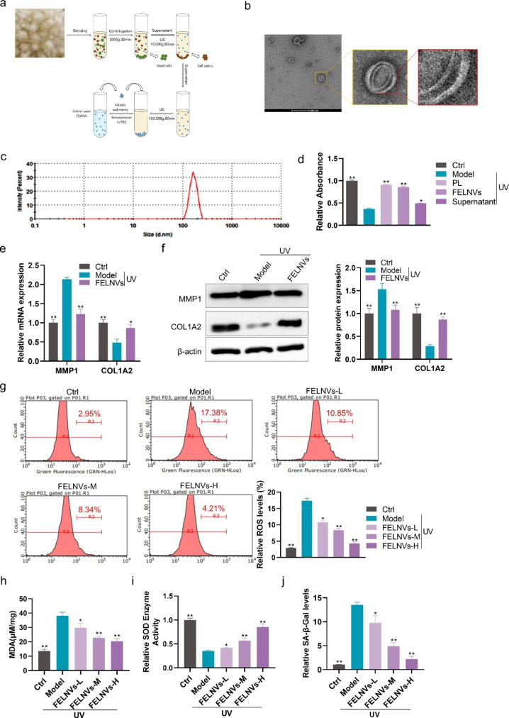
Background: Phellinus linteus (PL), which is a typical medicinal fungus, has been shown to have antitumor and anti-inflammatory activities. However, studies on the effect of anti-photoaging are limited. Studies have shown that exosome-like nanovesicles are functional components of many medicinal plants, and miRNAs in exosome-like nanovesicles play a cross-kingdom regulatory role. At present, research on fungi exosome-like nanovesicles (FELNVs) is few.

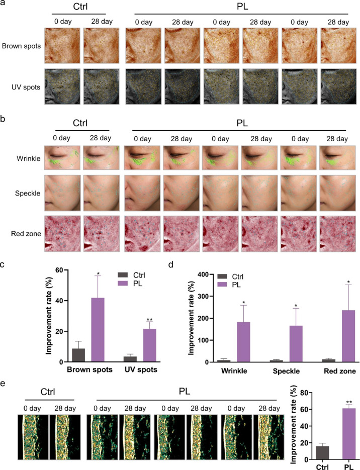
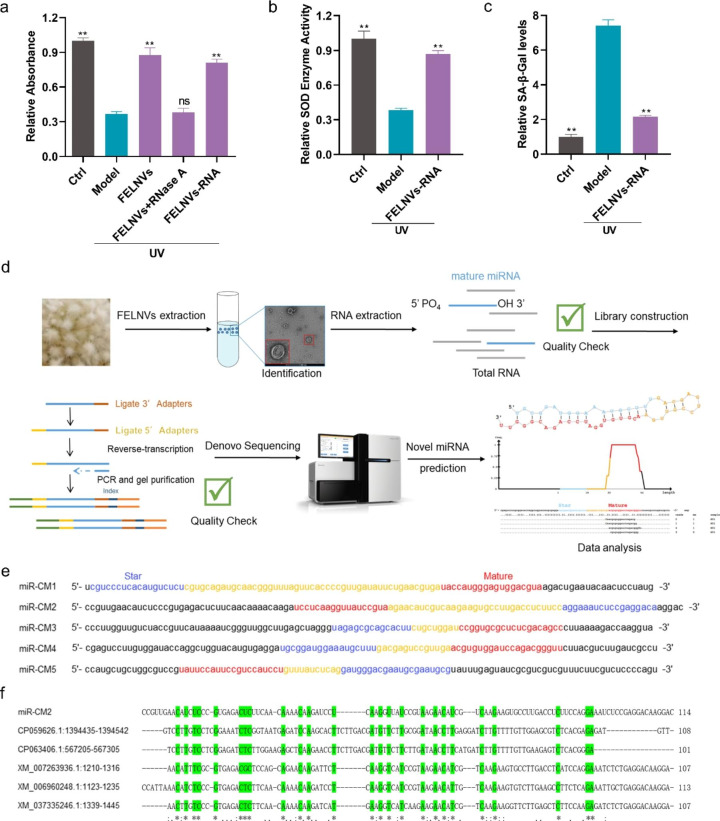
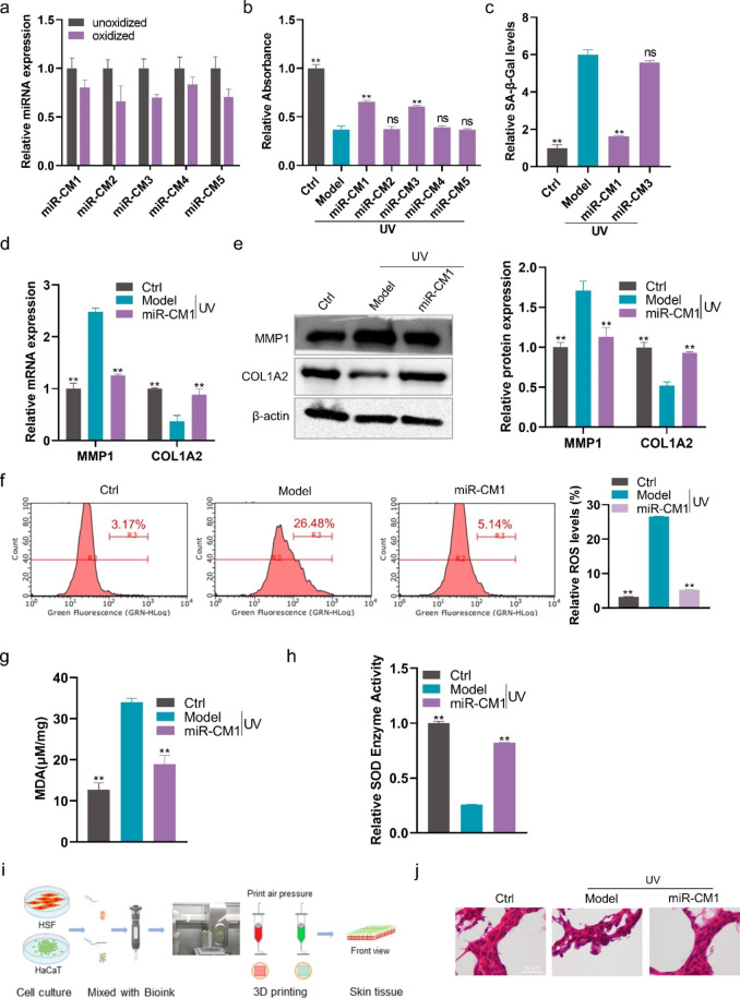
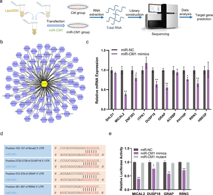
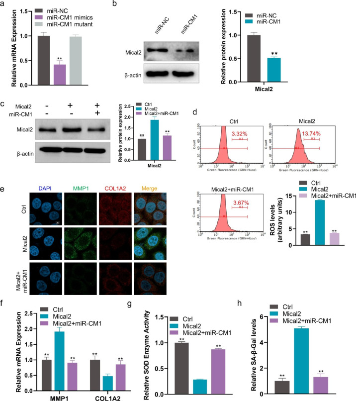
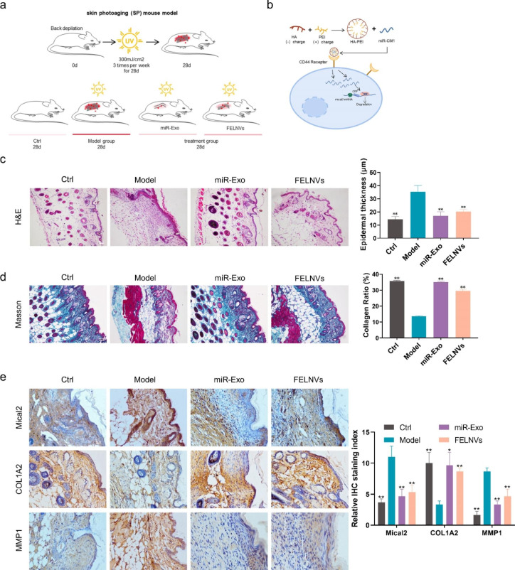
Results: We systematically evaluated the anti-aging effects of PL. FELNVs of PL were isolated, and the functional molecular mechanisms were evaluated. The results of volunteer testing showed that PL had anti-aging activity. The results of component analysis showed that FELNVs were the important components of PL function. FELNVs are nanoparticles (100-260 nm) with a double shell structure. Molecular mechanism research results showed that miR-CM1 in FELNVs could inhibit Mical2 expression in HaCaT cells through cross-kingdom regulation, thereby promoting COL1A2 expression; inhibiting MMP1 expression in skin cells; decreasing the levels of ROS, MDA, and SA-β-Gal; and increasing SOD activity induced by ultraviolet (UV) rays. The above results indicated that miR-CM1 derived from PL inhibited the expression of Mical2 through cross-kingdom regulation and inhibited UV-induced skin aging.

Conclusion: miR-CM1 plays an anti-aging role by inhibiting the expression of Mical2 in human skin cells through cross-species regulation.

Keywords: Anti-aging effects; Cross-kingdom regulations; Fungi exosome-like nanovesicles; Skin aging; miRNAs.


Conflict of interest statement

The authors declare no competing interests.

Figures

Fig. 1
Fig. 1
Effect of PL extract on the skin of volunteers. a, b Skin image detected by VISIA imaging system showing the brown spots, UV spots, wrinkles, speckles, and red zone of volunteers. c, d Statistical analyses of the improvement rate of brown spots, UV spots, wrinkles, speckles, and red zone. e Collagen density in the arm of volunteers detected by the Dermalab instrument. Data are expressed as mean ± SD (*P < 0.05, **P < 0.01)
Fig. 2
Fig. 2
FELNVs can resist UV-induced aging in HaCaT cells. a Flow diagram to show FELNV extraction using differential ultracentrifugation. b Representative TEM image of purified FELNVs from PL. Scale bar, 500 nm. c Average size of FELNVs was characterized by NanoSight analysis. d Effect of PL extract (PL), FELNVs, and supernatant on proliferation of UV-treated HaCaT cells by CCK8 assay. Model: UV-treated. e Effect of FELNVs on mRNA expression levels of MMP1 and COL1A2 in UV-treated HaCaT cells tested by qRT-PCR. f Effect of FELNVs on the expression of MMP1 and COL1A2 in UV-treated HaCaT cells tested by Western blot. g–j Relative ROS levels (g), MDA content (h), relative SOD enzyme activity (i), and relative SA-β-Gal levels (j) in UV-treated HaCaT cells with FELNVs treatment at different concentrations. Data are expressed as mean ± SD (*P < 0.05, **P < 0.01)
Fig. 3
Fig. 3
RNAs in FELNVs exert anti-aging effects. a Effect of FELNVs, RNA free components (FELNVs + RNase A), and RNA components (FELNVs-RNA) on proliferation in UV-treated HaCaT cells by CCK8 assay. b, c Relative SOD enzyme activity (b) and relative SA-β-Gal levels (c) in UV-treated HaCaT cells with RNA treatment of FELNVs. d Flow diagram to show de novo miRNA sequencing and prediction of novel miRNAs. e Sequences of five novel miRNAs (miR-CM1 to CM5) of FELNVs. f Sequence alignment of miR-CM2 with nucleotide collection (nr/nt) of NCBI. Data are expressed as mean ± SD (*P < 0.05, **P < 0.01)
Fig. 4
Fig. 4
miR-CM1 in FELNVs exert anti-aging effects. a Comparison of expression levels of the five novel miRNAs of FELNVs with/without oxidation treatment. b Effect of five novel miRNAs on proliferation in UV-treated HaCaT cells by CCK8 assay. c Relative SA-β-Gal levels in UV-treated HaCaT cells with miR-CM1 or miR-CM3. d Effect of miR-CM1 on mRNA expression levels of MMP1 and COL1A2 in UV-treated HaCaT cells was tested by qRT-PCR. e Effect of miR-CM1 on the expression of MMP1 and COL1A2 proteins in UV-treated HaCaT cells was tested by Western blot. f–h Relative ROS levels (f), MDA content (g) and relative SOD enzyme activity (h) in UV-treated HaCaT cells with miR-CM1 treatment. i Flow diagram to show 3D cell printing. j Representative imaging of H&E stain of skin tissue from 3D printing and cultures. Data are expressed as mean ± SD (*P < 0.05, **P < 0.01)
Fig. 5
Fig. 5
miR-CM1 has cross-kingdom regulatory activity. a Flow diagram to show transcriptome Illumina sequencing in HaCaT cells with miR-CM1 transfection. b The predicted results of miR-CM1 regulating mRNA. c mRNA expression levels of SHLD1, MICAL2, ZNF383, ITPK1, DUSP18, GRAP, ACRBP, PHYHIP, RRN3 and HBEGF in HaCaT cells with miR-CM1 transfection were tested by qRT-PCR. d The matching results between the seed region of miR-CM1 and the 3’UTR region of MICAL2, DUSP18, GRAP and RRN3. e Relative luciferase activity of MICAL2, DUSP18, GRAP and RRN3 with miR-CM1 transfection by dual luciferase reporter gene assay. Data are expressed as mean ± SD (*P < 0.05, **P < 0.01)
Fig. 6
Fig. 6
miR-CM1 resists UV-induced aging by inhibiting Mical2 expression. a mRNA expression levels of Mical2 in HaCaT cells with miR-CM1 transfection tested by qRT-PCR. b Protein expression of Mical2 in HaCaT cells with miR-NC or miR-CM1 transfection were tested by Western blot. c Protein expression of Mical2 in HaCaT cells with Mical2 transfection or co-transfection of Mical2 and miR-CM1 were tested by Western blot. d ROS levels in HaCaT cells with Mical2 transfection and co-transfection of Mical2 and miR-CM1. e Representative imaging of IF in HaCaT cells labeled for MMP1 (green), COL1A2 (red), and nuclear (DAPI: blue) in different treatment groups. f mRNA expression levels of MMP1 and COL1A2 in HaCaT cells with miR-CM1 transfection and co-transfection of Mical2 and miR-CM1 tested by qRT-PCR. g, h SOD enzyme activity (g) and relative SA-β-Gal levels (h) in HaCaT cells with miR-CM1 transfection and co-transfection of Mical2 and miR-CM1. Data are expressed as mean ± SD (*P < 0.05, **P < 0.01)
Fig. 7
Fig. 7
miR-CM1 encapsulated in artificial exosomes can reduce UV-induced aging in mice. a Schematic of the mouse photoaging model. Treatment groups were separately given drug treatment of miR-Exo and FELNVs to the dorsal skin of mice before UVA irradiation. b Schematic demonstrating miR-CM1 encapsulated in artificial exosomes (EXO mimics) into cells to function. c-e Representative images of H&E staining (c), Masson staining (d), and immunohistochemistry (IHC) (e) of skin tissue from mice. Data statistics for epidermal thickness, collagen ratio and relative IHC staining index are shown on the right. Data are expressed as mean ± SD (*P < 0.05, **P < 0.01)

References

    1. Fisher GJ, Kang S, Varani J, Bata-Csorgo Z, Wan Y, Datta S, Voorhees JJ. Mechanisms of Photoaging and Chronological Skin Aging. Arch Dermatol. 2002;138:1462–70. - PubMed
    1. Ansary TM, Hossain MR, Kamiya K, Komine M, Ohtsuki M. Inflammatory Molecules Associated with Ultraviolet Radiation-Mediated Skin Aging. Int J Mol Sci 2021, 22. - PMC - PubMed
    1. Fisher GJ, Wang Z, Datta SC, Varani J, Kang S, Voorhees JJ. Pathophysiology of Premature Skin Aging Induced by Ultraviolet Light. N Engl J Med. 1997;337:1419–29. - PubMed
    1. Kammeyer A, Luiten RM. Oxidation events and skin aging. Ageing Res Rev. 2015;21:16–29. - PubMed
    1. Chen C, Liu X, Qi S, Yan ACPD, Zhang J. Hepatoprotective effect of Phellinus linteus mycelia polysaccharide (PL-N1) against acetaminophen-induced liver injury in mouse. Int J Biol Macromol. 2020;154:1276–84. - PubMed

Supplementary concepts