. 2023 Jan;95(1):e28246.
doi: 10.1002/jmv.28246. Epub 2022 Nov 2.

Development and characterization of a new monoclonal antibody against SARS-CoV-2 NSP12 (RdRp)

Affiliations

Development and characterization of a new monoclonal antibody against SARS-CoV-2 NSP12 (RdRp)

Wen Meng et al. J Med Virol. 2023 Jan.

Abstract

SARS-CoV-2 NSP12, the viral RNA-dependent RNA polymerase (RdRp), is required for viral replication and is a therapeutic target to treat COVID-19. To facilitate research on SARS-CoV-2 NSP12 protein, we developed a rat monoclonal antibody (CM12.1) against the NSP12 N-terminus that can facilitate functional studies. Immunoblotting and immunofluorescence assay (IFA) confirmed the specific detection of NSP12 protein by this antibody for cells overexpressing the protein. Although NSP12 is generated from the ORF1ab polyprotein, IFA of human autopsy COVID-19 lung samples revealed NSP12 expression in only a small fraction of lung cells including goblet, club-like, vascular endothelial cells, and a range of immune cells, despite wide-spread tissue expression of spike protein antigen. Similar studies using in vitro infection also generated scant protein detection in cells with established virus replication. These results suggest that NSP12 may have diminished steady-state expression or extensive posttranslation modifications that limit antibody reactivity during SARS-CoV-2 replication.

Keywords: COVID-19; NSP12; RNA-dependent RNA polymerase, RdRp; SARS-CoV-2; monoclonal antibody.


Conflict of interest statement

The authors declare no conflict of interest.

Figures

Figure 1
Figure 1
Application of CM12.1 in immunoblotting and immunofluorescence assay. (A) Structural demonstration of epitope distributions in SARS‐CoV‐2. NSP12 epitopes (epitope 1: N terminus; epitope 2: linker region) are all exposed on the surface of the predicted protein structure, maximumly covering possible epitope regions. The SARS‐CoV‐2 RNA‐dependent polymerase complex formed by NSP12, NSP7, and NSP8 is shown (PDB ID: 6M71). Figure was created using BioRender. (B) Immunofluorescence assay showing the detection of C‐terminally FLAG‐tagged NSP12 (FLAG‐NSP12) and full‐length NSP12 (NSP12 FL) on paraffin‐embedded formalin‐fixed cell pellet slides by CM12.1. EV, empty vector. (C) Illustration of the fragments’ distributions in SARS‐CoV‐2 NSP12. (D) Immunoblotting showing epitope mapping result of CM12.1. Whole‐cell lysates were harvested from 293 cells transfected with equal amounts of NSP12 fragments. Lanes 1: EV, 2: FLAG‐NSP12, 3: FLAG‐NSP12 1‐249 aa, 4: FLAG‐NSP12 1‐581 aa, 5: FLAG‐NSP12 131‐932 aa, 6: FLAG‐NSP12 582‐932 aa, 7: NSP12 FL. β‐tubulin served as loading control. (E) Vero E6 cells were infected with SARS‐CoV‐2 and lysed 2 days postinfection. Lysates from infected and uninfected cells were immunoblotted with CM12.1, anti‐Spike S2, and β‐tubulin.
Figure 2
Figure 2
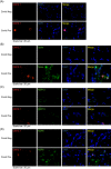
Application of CM12.1 in the detection of NSP12 in COVID‐19 patients. (A) Images of immunofluorescence detection of NSP12 using CM12.1 in COVID‐19 negative and positive lung tissues. 4′,6‐diamidino‐2‐phenylindole (DAPI) stained nuclei. (B) Images of immunofluorescence detection for NSP12 and SARS‐CoV‐2 spike protein S1 subunit (Spike) in COVID‐19 negative and positive lung tissues. DAPI stained nuclei. (C) Images of immunofluorescence detection for NSP12 and SARS‐CoV‐2 NSP13 protein in COVID‐19 negative and positive lung tissues. DAPI stained nuclei. (D) Images of immunofluorescence detection for NSP12 and SARS‐CoV‐2 NSP8 protein in COVID‐19 negative and positive lung tissues. DAPI stained nuclei.
Figure 3
Figure 3
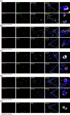
Multicolor immunofluorescence staining of SARS‐CoV‐2 NSP12, Spike and cellular markers in lung tissues from COVID‐19 patients. Images of immunofluorescence detection for (A) SARS‐CoV‐2 NSP12 (CM12.1), spike protein S1 subunit (Spike), and MUC5AC (goblet cells, pseudo color white) or MUC5B (club‐like cells, pseudo color white). Nuclei were stained with 4′,6‐diamidino‐2‐phenylindole (DAPI). (B) SARS‐CoV‐2 NSP12 (CM12.1), spike protein S1 subunit (Spike), and CD34 or CD144 (vascular endothelial cells, pseudo color white). Nuclei were stained with DAPI. (C) SARS‐CoV‐2 NSP12 (CM12.1), spike protein S1 subunit (Spike), and CD68 or CD14 (monocytes and macrophages, pseudo color white). Nuclei were stained with DAPI. (D) SARS‐CoV‐2 NSP12 (CM12.1), spike protein S1 subunit (Spike), and CD56 (NK cells, pseudo color white) or ELA‐2 (neutrophils, pseudo color white). Nuclei were stained with DAPI. (E) SARS‐CoV‐2 NSP12 (CM12.1), spike protein S1 subunit (Spike), and CD8 (CD8+ T cells, pseudo color white). Nuclei were stained with DAPI.

References

    1. Lu R, Zhao X, Li J, et al. Genomic characterisation and epidemiology of 2019 novel coronavirus: implications for virus origins and receptor binding. Lancet. 2020;395(10224):565‐574. 10.1016/S0140-6736(20)30251-8 - DOI - PMC - PubMed
    1. Wu F, Zhao S, Yu B, et al. A new coronavirus associated with human respiratory disease in China. Nature. 2020;579(7798):265‐269. 10.1038/s41586-020-2008-3 - DOI - PMC - PubMed
    1. Zhou P, Yang XL, Wang XG, et al. A pneumonia outbreak associated with a new coronavirus of probable bat origin. Nature. 2020;579(7798):270‐273. 10.1038/s41586-020-2012-7 - DOI - PMC - PubMed
    1. Gao SJ, Guo H, Luo G. Omicron variant (B.1.1.529) of SARS‐CoV‐2, a global urgent public health alert! J Med Virol. 2022;94(4):1255‐1256. 10.1002/jmv.27491 - DOI - PMC - PubMed
    1. Harvey WT, Carabelli AM, Jackson B, et al. SARS‐CoV‐2 variants, spike mutations and immune escape. Nat Rev Microbiol. 2021;19(7):409‐424. 10.1038/s41579-021-00573-0 - DOI - PMC - PubMed

Publication types