. 2022 Dec;600(24):5311-5332.
doi: 10.1113/JP283789. Epub 2022 Dec 2.

Descending pathways from the superior colliculus mediating autonomic and respiratory effects associated with orienting behaviour

Affiliations

Descending pathways from the superior colliculus mediating autonomic and respiratory effects associated with orienting behaviour

Erin Lynch et al. J Physiol. 2022 Dec.

Abstract

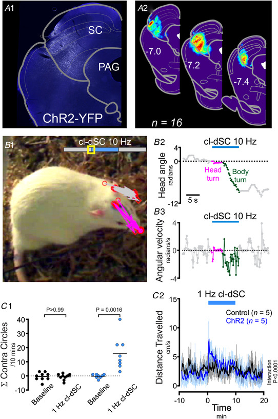
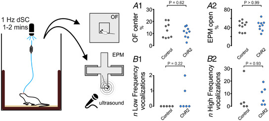
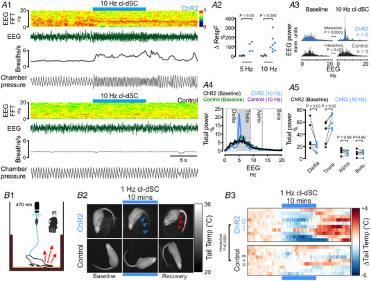
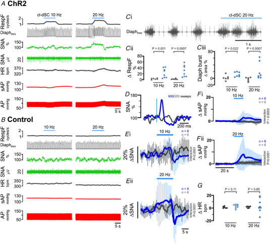
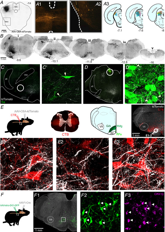
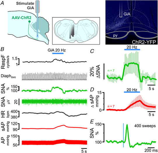
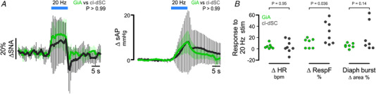
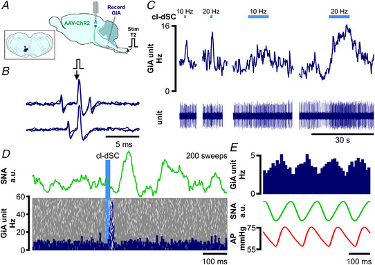
The ability to discriminate competing external stimuli and initiate contextually appropriate behaviours is a key brain function. Neurons in the deep superior colliculus (dSC) integrate multisensory inputs and activate descending projections to premotor pathways responsible for orienting, attention and defence, behaviours which involve adjustments to respiratory and cardiovascular parameters. However, the neural pathways that subserve the physiological components of orienting are poorly understood. We report that orienting responses to optogenetic dSC stimulation are accompanied by short-latency autonomic, respiratory and electroencephalographic effects in awake rats, closely mimicking those evoked by naturalistic alerting stimuli. Physiological responses were not accompanied by detectable aversion or fear, and persisted under urethane anaesthesia, indicating independence from emotional stress. Anterograde and trans-synaptic viral tracing identified a monosynaptic pathway that links the dSC to spinally projecting neurons in the medullary gigantocellular reticular nucleus (GiA), a key hub for the coordination of orienting and locomotor behaviours. In urethane-anaesthetized animals, sympathoexcitatory and cardiovascular, but not respiratory, responses to dSC stimulation were replicated by optogenetic stimulation of the dSC-GiA terminals, suggesting a likely role for this pathway in mediating the autonomic components of dSC-mediated responses. Similarly, extracellular recordings from putative GiA sympathetic premotor neurons confirmed short-latency excitatory inputs from the dSC. This pathway represents a likely substrate for autonomic components of orienting responses that are mediated by dSC neurons and suggests a mechanism through which physiological and motor components of orienting behaviours may be integrated without the involvement of higher centres that mediate affective components of defensive responses. KEY POINTS: Neurons in the deep superior colliculus (dSC) integrate multimodal sensory signals to elicit context-dependent innate behaviours that are accompanied by stereotypical cardiovascular and respiratory activities. The pathways responsible for mediating the physiological components of colliculus-mediated orienting behaviours are unknown. We show that optogenetic dSC stimulation evokes transient orienting, respiratory and autonomic effects in awake rats which persist under urethane anaesthesia. Anterograde tracing from the dSC identified projections to spinally projecting neurons in the medullary gigantocellular reticular nucleus (GiA). Stimulation of this pathway recapitulated autonomic effects evoked by stimulation of dSC neurons. Electrophysiological recordings from putative GiA sympathetic premotor neurons confirmed short latency excitatory input from dSC neurons. This disynaptic dSC-GiA-spinal sympathoexcitatory pathway may underlie autonomic adjustments to salient environmental cues independent of input from higher centres.

Keywords: arousal; cardiovascular; innate behaviours; sensorimotor integration; sympathetic.


Conflict of interest statement

None declared.

Figures

Figure 1
Figure 1. Optogenetic dSC stimulation causes orienting and circling behaviours in awake rats
A1, distribution of ChR2‐YFP fluorescence in a representative experiment and (A2) normalized ChR2‐YFP distribution from 16 animals. Optogenetic stimulation of this region evoked contraversive orienting: Panel B depicts movements evoked by moderate‐intensity cl‐dSC stimulation in a typical experiment. B1, an annotated video frame captured after 1 s of stimulation; head position in preceding frames is indicated by arrows drawn between the optical fibre and tip of the nose in grey (baseline) and magenta (stimulation). B2 and 3, head angle and angular velocity during the same experiment. C, behavioural effects of sustained cl‐dSC stimulation in rats injected with ChR2 and control vectors. Incidence of contraversive circling (C1) and distanced travelled (C2) during 10 min 1 Hz cl‐dSC stimulation in ChR2‐treated and control rats. **P < 0.01 paired t test, ####interaction P < 0.0001, two‐way repeated measures ANOVA. Error bars indicate standard deviation from the mean throughout. [Colour figure can be viewed at wileyonlinelibrary.com]
Figure 2
Figure 2. Optogenetic dSC stimulation is not associated with anxiety‐like behaviours or ultrasonic vocalisation
The presence of anxiety‐like behaviours was assayed immediately after optical cl‐dSC stimulation using the open field (OF, A1) or elevated plus maze (EPM, A2) apparatus: no significant difference was detected between rats treated with ChR2 or control vectors. Similarly, no effects on the incidence of low‐ or high‐frequency ultrasonic vocalizations were detected while exploring the EPM (B1 and B2). [Colour figure can be viewed at wileyonlinelibrary.com]
Figure 3
Figure 3. cl‐dSC stimulation drives arousal, ventilation and tail vasoconstriction in ChR2‐treated but not vector control rats
A1, simultaneous recording of electroencephalogram (EEG) and respiratory rate from ChR2 (upper) and control vector treated (lower panels) animals showing tachypnoea and EEG desynchronization (0.5–4.5 Hz) with increased theta power (4.5–9 Hz). Pooled data shown in A2 (increase in respiratory frequency; *,**Mann‐Whitney P < 0.05, 0.01), A3, pooled EEG (0.5–20 Hz) before and during photostimulation in ChR2 and control rats; frequency bands are defined in A4. A5, changes in Delta and Theta power in ChR2 rats as a percentage of total EEG power, *P < 0.05 (Sidak's post hoc test after two‐way repeated measures ANOVA). B1, tail temperature was measured by infrared thermography before, during and after 10 min of 1 Hz cl‐dSC stimulation. B2, thermal imagery illustrates tail‐cooling in ChR2 experiment during stimulation (blue arrowheads), and tail temperature rebound during the recovery period in a representative experiment (red arrowheads). B3, heat maps illustrate recordings from 10 ChR2 and nine control vector treated rats: each row represents an individual experiment ###, #### two‐way repeated measures ANOVA interaction P < 0.001, <0.0001. [Colour figure can be viewed at wileyonlinelibrary.com]
Figure 4
Figure 4. Respiratory and cardiovascular effects of cl‐dSC stimulation under urethane anaesthesia
Representative recordings from rats treated with ChR2 (A) and control (B) vectors. Boxed region in Panel A denotes expanded trace shown in Panel Ci. Respiratory effects (Panel C) manifested as an increase in diaphragmatic EMG burst frequency (Cii) and amplitude (Ciii). Single‐pulse cl‐dSC stimulation evoked short‐latency monophasic bursts in SNA (D: vertical blue line denotes laser onset); repetitive cl‐dSC stimulation evoked frequency‐dependent recruitment of SNA (E) and systolic blood pressure (sAP, F), with variable effects on heart rate (HR, G). Bold traces in E and F indicate mean; light traces indicate SEM. *,**,***Mann‐Whitney P < 0.05, <0.01, <0.001; ####two‐way repeated measures ANOVA interaction P < 0.0001. [Colour figure can be viewed at wileyonlinelibrary.com]
Figure 5
Figure 5. Anterograde labelling from the cl‐dSC
A, example of AAV‐CBA‐tdTomato injection site in the cl‐dSC; fibres emerging from the injection site projected to the contralateral colliculi via the commissure of the inferior colliculus (CiC, A1) or followed a descending trajectory via the lateral midbrain (desc, A2). A3, composite diagram showing distribution of injection sites in six experiments. B, histological sections from one experiment, illustrating the brainstem distribution of tdTomato labelling. The descending bundle split into two tracts in the pons, a more substantial ventral bundle that surrounded the superior olive on all sides (SO, Bi), and a lesser and exclusively ipsilateral dorsal tract that innervated the dorsomedial spinal trigeminal nucleus (DMSp5, Bii) with large‐calibre fibres. The ventral branch occupied a column extending from the ventral surface to an apex at the locus coeruleus, was sparsely mirrored on the contralateral brainstem, and comprised both large‐ and fine‐calibre fibres and putative terminals. Caudal to the facial nerve (nVII, Bii) the ventral branch was most densely concentrated in the medullary reticular formation (mRF, Biii) dorsal to the pyramidal tract (py) at the level of the caudal pole of the facial nucleus. Labelled fibres were infrequently encountered in the caudal medulla (Biv) and virtually absent from spinal sections (Bv). Coordinates indicate the rostrocaudal position with respect to the bregma. Putative targets of the cl‐dSC outputs included rare tyrosine hydroxylase (TH) immunoreactive neurons in the A5 (C) and A6 (D) cell groups, and a conspicuous terminal field in the gigantocellular alpha region of the reticular formation (GiA, E). Putative cl‐dSC projections to spinally projecting neurons in the GiA were identified in experiments in which anterograde cl‐dSC labelling was combined with retrograde labelling of spinally projecting neurons by microinjection of cholera toxin B (CTB) at the thoracic intermediolateral cell column: top row of panel E shows a schematic of the experimental strategy; brightfield transverse thoracic spinal cord section superimposed with CTB injection sites in red; and an atlas plate highlighting the GiA, midline raphe magnus (RMg) and pallidus regions (RPa) with a low‐power image showing the distribution of CTB‐labelled cell bodies (red) and cl‐dSC terminals (white). E1–3, high‐power confocal images from the circled region show close apposition between cl‐dSC terminals and spinally projecting GiA neurons. F, similar results were obtained using AAV1‐mediated trans‐synaptic tagging: spinally projecting neurons were transduced by injection of Cre‐dependent AAVretro‐hSyn‐DIO‐GFP in the thoracic spinal cord, GFP expression controlled by the trans‐synaptic trafficking of AAV1‐hSyn‐Cre from cl‐dSC. F1, GFP‐immunoreactive neurons were exclusively found in GiA and included CHX10‐immunoreactive V2A neurons (arrowheads) and CHX‐10 negative cells (F2 and 3). [Colour figure can be viewed at wileyonlinelibrary.com]
Figure 6
Figure 6. Respiratory and cardiovascular responses to optogenetic stimulation of cl‐dSC‐GiA terminals under urethane anaesthesia
A, illustration of (L–R) experimental strategy, locations of optical fibres in different experiments and a histological specimen illustrating fibre position and terminal ChR2 labelling. B, physiological recording of respiratory, sympathetic and cardiovascular responses to GiA stimulation; pooled sympathetic and pressor responses from seven experiments are shown in C and D. E, stimulus‐triggered average of sympathetic response to 0.5 Hz stimulation from the experiment shown in B. [Colour figure can be viewed at wileyonlinelibrary.com]
Figure 7
Figure 7. Direct comparison of responses evoked by optogenetic stimulation of cl‐dSC neurons (dark grey) and their GiA terminals (green)
Outputs examined were sympathetic nerve activity and systolic blood pressure (A), and heart rate, respiratory frequency and respiratory burst area (B). Significantly different responses were only detected in respiratory frequency (*P < 0.05, unpaired t test). [Colour figure can be viewed at wileyonlinelibrary.com]
Figure 8
Figure 8. Optogenetic cl‐dSC stimulation activates spinally projecting cardiovascular and non‐ cardiovascular GiA neurons
A, extracellular recordings were made from bulbospinal GiA neurons under urethane anaesthesia after transduction of cl‐dSC neurons with ChR2 and fibre optic implantation; inset shows recording sites of seven neurons. B, spinally projecting neurons exhibited constant‐latency antidromic spikes in response to electrical stimulation of the T2 spinal cord (top trace) which collided with spontaneous orthodromic spikes (lower trace). C, cl‐dSC stimulation evoked frequency‐dependent increases in baseline activity: trace shows typical responses to short (1 s) and long (10 s) trains of stimulation at 10 and 20 Hz (same neuron as B). D, low‐frequency cl‐dSC stimulation evoked short‐latency response in the same neuron (laser‐triggered peristimulus time histogram (dark blue) with overlaid raster (white spikes, grey background, lower panel) that preceded simultaneously recorded splanchnic sympathetic responses (green trace, upper panel)). E, putative sympathetic premotor neurons were identified by covariation of spontaneous neuronal activity (top trace) with SNA (green) and pulsatile arterial pressure (lower trace). [Colour figure can be viewed at wileyonlinelibrary.com]

Similar articles

Cited by

References

    1. Aicher, S. A. , Reis, D. J. , Nicolae, R. , & Milner, T. A. (1995). Monosynaptic projections from the medullary gigantocellular reticular formation to sympathetic preganglionic neurons in the thoracic spinal cord. Journal of Comparative Neurology, 363(4), 563–580. - PubMed
    1. Babic, T. , & Ciriello, J. (2004). Medullary and spinal cord projections from cardiovascular responsive sites in the rostral ventromedial medulla. Journal of Comparative Neurology, 469(3), 391–412. - PubMed
    1. Baudrie, V. , Tulen, J. H. , Blanc, J. , & Elghozi, J. L. (1997). Autonomic components of the cardiovascular responses to an acoustic startle stimulus in rats. Journal of Autonomic Pharmacology, 17(5), 303–309. - PubMed
    1. Boehnke, S. E. , & Munoz, D. P. (2008). On the importance of the transient visual response in the superior colliculus. Current Opinion in Neurobiology, 18(6), 544–551. - PubMed
    1. Bondarenko, E. , Beig, M. I. , Hodgson, D. M. , Braga, V. A. , & Nalivaiko, E. (2015). Blockade of the dorsomedial hypothalamus and the perifornical area inhibits respiratory responses to arousing and stressful stimuli. American Journal of Physiology Regulatory, Integrative and Comparative Physiology, 308(10), R816‐R822. - PubMed

Publication types