Review
. 2023 Apr;57(4):977-991.
doi: 10.1002/jmri.28496. Epub 2022 Oct 22.

MRI of Lymphedema

Affiliations
Review

MRI of Lymphedema

Betsa Parsai Salehi et al. J Magn Reson Imaging. 2023 Apr.

Abstract

Lymphedema is a devastating disease that has no cure. Management of lymphedema has evolved rapidly over the past two decades with the advent of surgeries that can ameliorate symptoms. MRI has played an increasingly important role in the diagnosis and evaluation of lymphedema, as it provides high spatial resolution of the distribution and severity of soft tissue edema, characterizes diseased lymphatic channels, and assesses secondary effects such as fat hypertrophy. Many different MR techniques have been developed for the evaluation of lymphedema, and the modality can be tailored to suit the needs of a lymphatic clinic. In this review article we provide an overview of lymphedema, current management options, and the current role of MRI in lymphedema diagnosis and management. EVIDENCE LEVEL: 5 TECHNICAL EFFICACY: Stage 5.

Keywords: MRI; lymphatic Imaging; lymphedema.


Figures

Figure 1.
Figure 1.
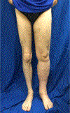
Patient with left lower extremity lymphedema with a 56 % volume difference between right and left leg.
Figure 2:
Figure 2:
Examples of different surgical approaches to lymphedema treatment. (A) Pre and post images of an upper extremity debulking with the collection canister showing total fat (*) removed. (B) Lymph node transplant (arrow) into a lower extremity. (C) Lymphovenous bypass showing the anastomosis (*); each etched square on the blue backplate measures 1 mm.
Figure 3:
Figure 3:
Diagram of lymphatic drainage. Lymphatic channels from the extremities drain into lymph nodes at the axilla and inguinal regions which can resorb lymph and send efferent channels more centrally. Central lymphatics drain primarily into the thoracic duct (*), which directly communicates with the left subclavian vein.
Figure 4:
Figure 4:
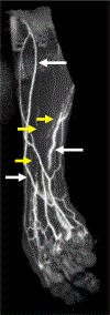
MIP image from an MRL following subcutaneous injection of gadobutrol at three locations along the dorsum of the left hand. While lymphatic channels are demonstrated (yellow arrows), there is substantial contamination from superficial veins (white arrows) which demonstrate relatively greater signal intensity which obscures the lymphatics.
Figure 5:
Figure 5:
DARC MRL in 32-year-old female with right lower extremity lymphedema. A, Coronal 3D SPGR TE 0.9 ms MIP angiographic image shows enhancement of blood vasculature (red arrows) and lymphatic vasculature (yellow arrow). B, Coronal 3D SPGR TE 4.4 ms MIP lymphangiographic image with enhancement of lymphatic vessels (yellow arrow) and suppression of venous signal. C, Cor 3D T2w image with increased subcutaneous thickness and prominent epifascial edema (blue arrow).
Figure 6:
Figure 6:
Axial T2-weighted fat-nulled STIR image across the mid thighs of a 46-year-old female with chronic bilateral lower extremity lymphedema demonstrates three hallmark features: honeycombing (*), dermal thickening (arrows) and epifascial fluid (arrowhead).
Figure 7:
Figure 7:
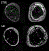
Axial STIR (top) and DIXON-fat (bottom) images of the mid forearms in a 43-year-old male with chronic left lower extremity lymphedema. There is extensive confluent subcutaneous fluid and dermal thickening highlighted on STIR, however, DIXON-fat images reveal a substantial amount of fat hypertrophy relative to the right forearm which is not as conspicuous on STIR.
Figure 8:
Figure 8:
65-year-old-female with history of breast cancer and right arm lymphedema. Cor 3D SPGR lymphangiography demonstrates contrast flowing along prominent collecting vessels (yellow arrows) with retrograde flow of contrast into the dermis (yellow arrow heads). It is unclear what component of this contrast is within a dilated lymphatic system versus extravasated out of the lymphatic vasculature.
Figure 9:
Figure 9:
MR Lymphangiography in 27-year-old male with scrotal and lower extremity lymphedema. A, Coronal 3D SPGR MIP angiographic image shows lymphatic vessels draining from the right inguinal lymph node (not shown) into the scrotum (yellow arrows). B, Cor 3D T2 w image with scrotal wall edema and honeycombing of the left thigh (blue arrows).
Figure 10:
Figure 10:
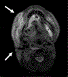
Axial T2-weighted image at the upper neck of a 38-year-old with persistent facial swelling following right hemi-glossectomy and radiation for squamous cell carcinoma. Extensive edema overlies the mandibles bilaterally, worse on the right with extension to the upper right neck (arrows).

References

    1. Cemal Y, Pusic A, Mehrara BJ. Preventative measures for lymphedema: Separating fact from fiction. J Am Coll Surg 2011;213(4):543–551. doi:10.1016/j.jamcollsurg.2011.07.001 - DOI - PMC - PubMed
    1. Suami H, Pan WR, Taylor GI. Changes in the lymph structure of the upper limb after axillary dissection: Radiographic and anatomical study in a human cadaver. Plast Reconstr Surg 2007;120(4):982–991. doi:10.1097/01.prs.0000277995.25009.3e - DOI - PubMed
    1. Hespe G, Nitti M, Mehrara B. Pathophysiology of Lymphedema. In: Lymphedema Cham: Springer International Publishing; 2015:9–18.
    1. Szuba A, Rockson SG. Lymphedema: Anatomy, Physiology and Pathogenesis Vol 2.; 1997. - PubMed
    1. Farzaliyev F, Hamacher R, Steinau Professor HU, Bertram S, Podleska LE. Secondary angiosarcoma: A fatal complication of chronic lymphedema. J Surg Oncol 2020;121(1):85–90. doi:10.1002/jso.25598 - DOI - PubMed

Publication types