. 2023 Jan;145(1):71-82.
doi: 10.1007/s00401-022-02513-5. Epub 2022 Oct 22.

Expanded analysis of high-grade astrocytoma with piloid features identifies an epigenetically and clinically distinct subtype associated with neurofibromatosis type 1

Affiliations

Expanded analysis of high-grade astrocytoma with piloid features identifies an epigenetically and clinically distinct subtype associated with neurofibromatosis type 1

Patrick J Cimino et al. Acta Neuropathol. 2023 Jan.

Abstract

High-grade astrocytoma with piloid features (HGAP) is a recently recognized glioma type whose classification is dependent on its global epigenetic signature. HGAP is characterized by alterations in the mitogen-activated protein kinase (MAPK) pathway, often co-occurring with CDKN2A/B homozygous deletion and/or ATRX mutation. Experience with HGAP is limited and to better understand this tumor type, we evaluated an expanded cohort of patients (n = 144) with these tumors, as defined by DNA methylation array testing, with a subset additionally evaluated by next-generation sequencing (NGS). Among evaluable cases, we confirmed the high prevalence CDKN2A/B homozygous deletion, and/or ATRX mutations/loss in this tumor type, along with a subset showing NF1 alterations. Five of 93 (5.4%) cases sequenced harbored TP53 mutations and RNA fusion analysis identified a single tumor containing an NTRK2 gene fusion, neither of which have been previously reported in HGAP. Clustering analysis revealed the presence of three distinct HGAP subtypes (or groups = g) based on whole-genome DNA methylation patterns, which we provisionally designated as gNF1 (n = 18), g1 (n = 72), and g2 (n = 54) (median ages 43.5 years, 47 years, and 32 years, respectively). Subtype gNF1 is notable for enrichment with patients with Neurofibromatosis Type 1 (33.3%, p = 0.0008), confinement to the posterior fossa, hypermethylation in the NF1 enhancer region, a trend towards decreased progression-free survival (p = 0.0579), RNA processing pathway dysregulation, and elevated non-neoplastic glia and neuron cell content (p < 0.0001 and p < 0.0001, respectively). Overall, our expanded cohort broadens the genetic, epigenetic, and clinical phenotype of HGAP and provides evidence for distinct epigenetic subtypes in this tumor type.

Keywords: DNA Methylation; HGAP; High-grade astrocytoma with piloid features; NF1; Neurofibromatosis type 1.


Conflict of interest statement

Conflict of interest The authors declare that there are no conflicts of interest.

Figures

Fig. 1
Fig. 1
Representative high-grade histologic features frequently present in HGAP. a HGAP tumors often had an infiltrative growth pattern. b, c Tumors repeatedly demonstrated pleomorphism and/or poorly differentiated cytomorphologies. d Increased mitotic activity occurred in a subset of cases. e Palisading necrosis and/or f) microvascular proliferation were present in a majority of HGAP tumors. (Scale bars: a 500 μm, b 100 μm, c 100 μm, d 50 μm, e 500 μm, f 100 μm)
Fig. 2
Fig. 2
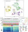
HGAP resolves into three epigenetically determined subtypes. a Uniform Manifold Approximation and Projection (UMAP) dimensionality reduction confirms that HGAP forms a distinct methylation cluster when compared to other CNS glial/glioneuronal tumors. b Unsupervised hierarchical clustering heatmap of whole-genome CpG methylation for HGAP uncovers the presence of three distinct HGAP epigenetic subtypes. c UMAP dimensionality reduction of HGAP only confirms the presence of three HGAP subtypes. IT Infratentorial, ST supratentorial, PF posterior fossa, SC spinal cord)
Fig. 3
Fig. 3
NF1 germline status, somatic alterations, and NF1 methylation define a relevant HGAP subtype. a HGAP subtype gNF1 is enriched for patients having Neurofibromatosis Type 1 (NF1) (gNF1 = 33.3%, g1 = 6.9%, g2 = 3.6%) and b) ATRX alterations (gNF1 = 87.5%, g1 = 59.6%, g2 = 47.5%). b CDKN2A/B (gNF1 = 83.3%, g1 = 83.3%, g2 = 85.5%) and NF1 (gNF1 = 62.5%, g1 = 41.3%, g2 = 32.4%) alterations are common across HGAP subtypes. c There are 5 of 30 (16.7%) CpG probes in the NF1 regulatory region that are differentially methylated in the gNF1 subtype, 3 of which are in the geneenhancer region
Fig. 4
Fig. 4
Clinical features of HGAP methylation subtypes. Median age with interquartile range for a all HGAPs and b HGAP subtypes (p > 0.05 for all pairwise comparisons, Mann–Whitney U tests). Localization for c all HGAP tumors and d HGAP by subtype (STsupratentorial, PFposterior fossa, SC spinal cord). Progression-free survival (PFS) with number at risk for e all HGAP subtypes and f gNF1 subtype versus the others. Overall survival (OS) with number at risk for g all HGAP subtypes and h gNF1 subtype versus the others
Fig. 5
Fig. 5
Gene Ontology (GO) pathway analysis of HGAP subtypes. a Schematic for differentially methylated CpG probes between HGAP subtypes and downstream GO analysis. b Top 10 GO pathways that are dysregulated in gNF1 compared to other HGAP subtypes when utilizing all gene structure methylation sites for CpG sites that are either hypermethylated or hypomethylated. c General gene structure of evaluated regions. d RNA processing dysregulation in the gNF1 subtype is a function of hypermethylation or hypomethylation in the gene body region, not promoter region of the associated genes
Fig. 6
Fig. 6
Deconvolution of the tumor microenvironment in HGAP. a Correlation matrix and b principal component analysis of neoplastic and non-neoplastic cell components within HGAP as a single tumor type. c Neoplastic cell contents as a function of HGAP subtype, which is highest in the g2 subtype (error bars as median values with 95% CI). d Non-neoplastic cell contents as a function of HGAP subtype, indicating that glia and neurons are enriched in the gNF1 subtype while monocytes and possibly B cells are enriched in the g1 subtype. e Heatmap summarizing the relative non-neoplastic cell contents in each HGAP subtype

References

    1. Alexander BM, Trippa L, Gaffey S, Arrillaga-Romany IC, Lee EQ, Rinne ML et al. (2019) Individualized Screening Trial of Innovative Glioblastoma Therapy (INSIGhT): a Bayesian adaptive platform trial to develop precision medicines for patients with glioblastoma. JCO Precis Oncol. 10.1200/PO.18.00071 - DOI - PMC - PubMed
    1. Bates D, Machler M, Bolker B, Walker S (2015) Fitting linear mixe-effects models using lme4. J Stat Softw 67:1–48. 10.18637/jss.v067.i01 - DOI
    1. Bender K, Perez E, Chirica M, Onken J, Kahn J, Brenner W et al. (2021) High-grade astrocytoma with piloid features (HGAP): the Charite experience with a new central nervous system tumor entity. J Neurooncol 153:109–120. 10.1007/s11060-021-03749-z - DOI - PMC - PubMed
    1. Capper D, Jones DTW, Sill M, Hovestadt V, Schrimpf D, Sturm D et al. (2018) DNA methylation-based classification of central nervous system tumours. Nature 555:469–474. 10.1038/nature26000 - DOI - PMC - PubMed
    1. Cimino PJ, McFerrin L, Wirsching HG, Arora S, Bolouri H, Rabadan R et al. (2018) Copy number profiling across glioblastoma populations has implications for clinical trial design. Neuro Oncol 20:1368–1373. 10.1093/neuonc/noy108 - DOI - PMC - PubMed

Publication types