. 2023:2562:109-122.
doi: 10.1007/978-1-0716-2659-7_6.

Hybridization Chain Reaction Fluorescence In Situ Hybridization (HCR-FISH) in Ambystoma mexicanum Tissue

Affiliations

Hybridization Chain Reaction Fluorescence In Situ Hybridization (HCR-FISH) in Ambystoma mexicanum Tissue

Alex M Lovely et al. Methods Mol Biol. 2023.

Abstract

In situ hybridization is a standard procedure for visualizing mRNA transcripts in tissues. The recent adoption of fluorescent probes and new signal amplification methods have facilitated multiplexed RNA imaging in tissue sections and whole tissues. Here we present protocols for multiplexed hybridization chain reaction fluorescence in situ hybridization (HCR-FISH) staining, imaging, cell segmentation, and mRNA quantification in regenerating axolotl tissue sections. We also present a protocol for whole-mount staining and imaging of developing axolotl limbs.

Keywords: Axolotl; Hybridization chain reaction; In situ hybridization; Limb development; Limb regeneration.


Conflict of interest statement

Competing Interests Authors declare no competing interests.

Figures

Fig. 1
Fig. 1
HCR schematic diagram. Schematic of v3.HCR-FISH on tissue sections. Transcripts of interest are hybridized with pools of split initiator probes containing unique initiators, B1–B5 (a). Following hybridization, initiator-specific fluorescently labeled hairpins are applied to the samples and a hybridization chain reaction is initiated (b). Sections are imaged for each gene of interest (c). Probes with hairpins are removed from the sections with a DNase treatment and probe wash (d). Subsequent rounds of hybridization are performed using new probe pools. (Adapted from “Loop-Mediated Isothermal Amplification (LAMP),” by BioRender.com (2021). Retrieved from https://app.biorender.com/biorender-templates)
Fig. 2
Fig. 2
Hybridization chamber-well locations. Areas on the slide correlating to the hybridization chambers are marked with a sharpie. Sections are collected onto a functionalized coverslip placed on top of the marked slide. This ensures proper placement of the sections for the hybridization chamber
Fig. 3
Fig. 3
Multiplexed FISH. Two sequential rounds of v3.HCR-FISH performed on a regenerating axolotl limb tissue section. The DAPI image is shown for reference (a) along with four genes stained in round 1: Midkine (Mdk) (b), Keratin 5 (Krt5) (c), Axolotl anterior gradient (Aag) (d), Thrombospondin 1 (Thbs1) (e) and an overlay of all four genes (f). Four genes stained in round 2: Hyaluronan And Proteoglycan Link Protein 3 (g), Keratin 17 (h), Methyltransferase-like (i), Laminin beta 1 (j) and an overlay of all 4 genes (k). 50-μm scale bar is included on DAPI image and a dotted line represents the epithelium/mesenchyme boundary
Fig. 4
Fig. 4
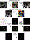
Graphical depiction of data analysis workflow. Major steps and outputs from HCR analysis pipeline. First, nuclei/cells are segmented using Cellpose (a). This mask is saved and then imported into CellProfiler where it is converted into regions of interest (b). HCR-FISH puncta are then identified in CellProfiler and assigned to a parent cell (c). The data can be outputted into a spreadsheet for further analysis
Fig. 5
Fig. 5
Whole-mount FISH. Whole-mount, 3D visualization of Shh (magenta), Prrx1 (yellow), and Fgf8 (cyan) in a stage 46 axolotl limb bud (a). A single z-slice of the limb bud (b). Scale bar = 100 μm

References

    1. Gall JG, Pardue ML (1969) Formation and detection of RNA-DNA hybrid molecules in cytological preparations. Proc Natl Acad Sci U S A 63(2):378–383. 10.1073/pnas.63.2.378 - DOI - PMC - PubMed
    1. John HA, Birnstiel ML, Jones KW (1969) RNA-DNA hybrids at the cytological level. Nature 223(5206):582–587. 10.1038/223582a0 - DOI - PubMed
    1. Herrington CS, Burns J, Graham AK, Evans M, McGee JO (1989) Interphase cytogenetics using biotin and digoxigenin labelled probes I: relative sensitivity of both reporter molecules for detection of HPV16 in CaSki cells. J Clin Pathol 42(6):592–600. 10.1136/jcp.42.6.592 - DOI - PMC - PubMed
    1. Tautz D, Pfeifle C (1989) A non-radioactive in situ hybridization method for the localization of specific RNAs in Drosophila embryos reveals translational control of the segmentation gene hunchback. Chromosoma 98(2):81–85. 10.1007/bf00291041 - DOI - PubMed
    1. Aigner S, Pette D (1990) In situ hybridization of slow myosin heavy chain mRNA in normal and transforming rabbit muscles with the use of a nonradioactively labeled cRNA. Histochemistry 95(1):11–18. 10.1007/bf00737222 - DOI - PubMed

Publication types

LinkOut - more resources