Review
. 2022 Dec:191:114583.
doi: 10.1016/j.addr.2022.114583. Epub 2022 Oct 19.

Applications of focused ultrasound-mediated blood-brain barrier opening

Affiliations
Review

Applications of focused ultrasound-mediated blood-brain barrier opening

Catherine M Gorick et al. Adv Drug Deliv Rev. 2022 Dec.

Abstract

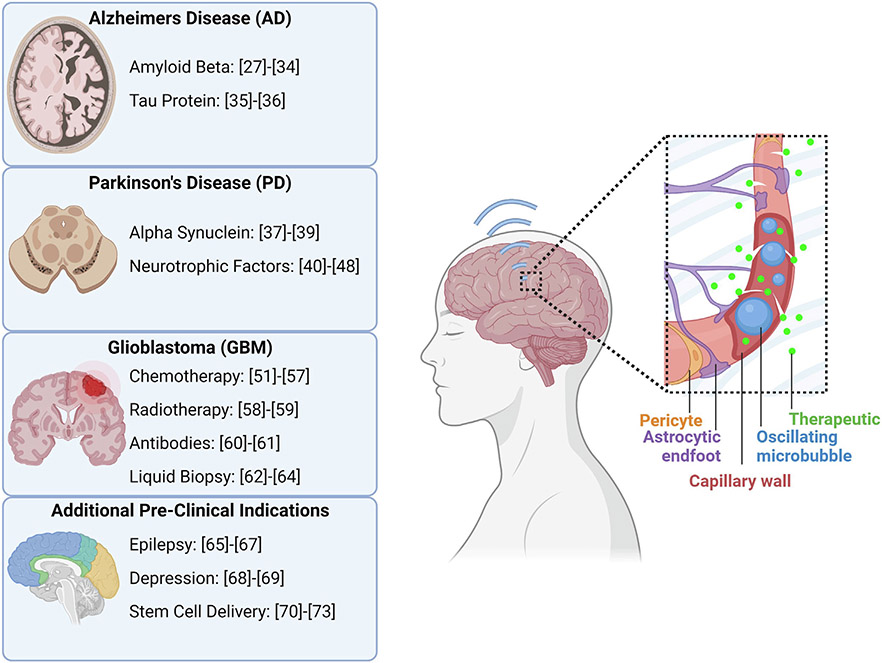
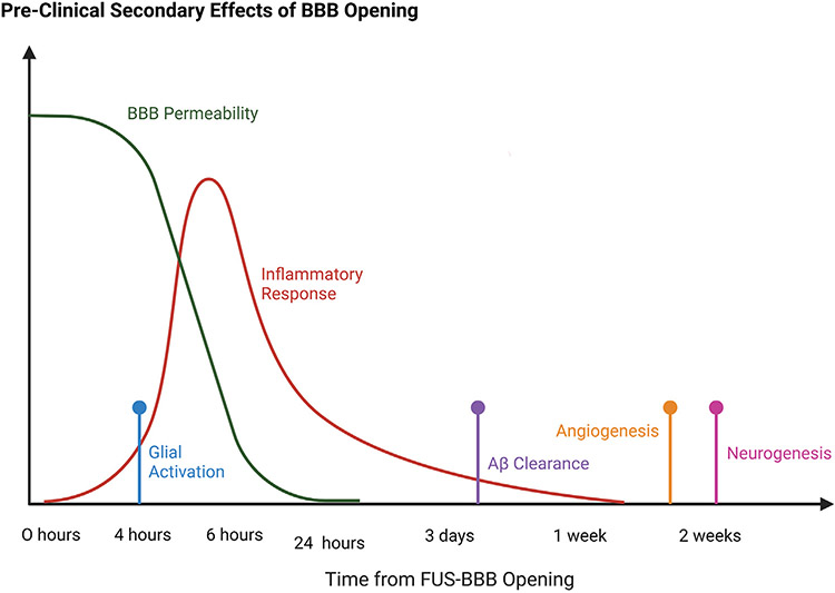
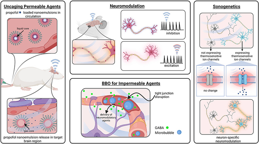
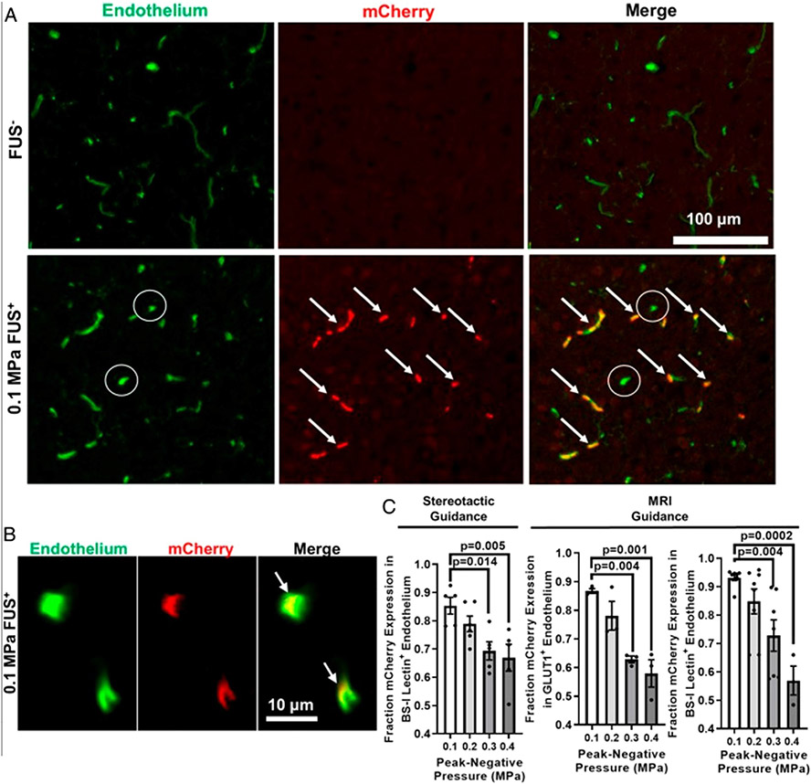
The blood brain barrier (BBB) plays a critically important role in the regulation of central nervous system (CNS) homeostasis, but also represents a major limitation to treatments of brain pathologies. In recent years, focused ultrasound (FUS) in conjunction with gas-filled microbubble contrast agents has emerged as a powerful tool for transiently and non-invasively disrupting the BBB in a targeted and image-guided manner, allowing for localized delivery of drugs, genes, or other therapeutic agents. Beyond the delivery of known therapeutics, FUS-mediated BBB opening also demonstrates the potential for use in neuromodulation and the stimulation of a range of cell- and tissue-level physiological responses that may prove beneficial in disease contexts. Clinical trials investigating the safety and efficacy of FUS-mediated BBB opening are well underway, and offer promising non-surgical approaches to treatment of devastating pathologies. This article reviews a range of pre-clinical and clinical studies demonstrating the tremendous potential of FUS to fundamentally change the paradigm of treatment for CNS diseases.


Conflict of interest statement

Declaration of Competing Interest The authors declare that they have no known competing financial interests or personal relationships that could have appeared to influence the work reported in this paper.

Figures

Fig. 1.
Fig. 1.
The physiology of the blood brain barrier, and the permeabilization of the blood brain barrier with microbubbles and focused ultrasound. (A) Focused ultrasound induced volumetric oscillations of microbubbles. (B) Transcytosis of a therapeutic through an endothelial cell. (C) Sonoporation of an endothelial cell. (D) Disruption of tight junctions between adjacent endothelial cells.
Fig. 2.
Fig. 2.
A summary of the pre-clinical therapeutic targets for which drugs and genes are being delivered with FUS to the brain for various disease applications. References correspond to studies investigating that therapeutic target with FUS BBBO. Created with Biorender.com.
Fig. 3.
Fig. 3.
A summary of the timeline of various secondary physiological effects of FUS-mediated BBBO. Adapted from a figure in Todd et al., 2020 [91]. Made with Biorender.com.
Fig. 4.
Fig. 4.
A summary of the neuroscience tools being studied at the pre-clinical level which utilize FUS for various neurological applications. Created with Biorender.com.
Fig. 5.
Fig. 5.
FUS peak-negative acoustic pressure (PNP) may be tuned to yield sonoselective cerebrovascular endothelial transfection. (A and B) Confocal images of FUS+ (0.1 MPa) and contralateral FUS brain tissue showing expression of mCherry reporter gene (red) with respect to endothelial cells (BS-I lectin, green). Arrows denote mCherry colocalization with endothelium. (C) Bar graphs of fraction of mCherry expression in cerebrovascular endothelium as a function of PNP. Highly selective endothelial transfection is observed at low PNPs (i.e., 0.1 MPa and 0.2 MPa). One-way ANOVAs followed by Dunnett’s multiple comparisons tests. [140].

References

    1. Daneman R, Prat A, The Blood-Brain Barrier, Cold Spring Harb. Perspect. Biol 7 (1) (2015) a020412. - PMC - PubMed
    1. Fu BM, Transport Across the Blood-Brain Barrier, in: 2018: pp. 235–259. 10.1007/978-3-319-96445-4_13. - DOI - PubMed
    1. Wolburg H, Lippoldt A, Tight junctions of the blood–brain barrier, Vascul. Pharmacol 38 (2002) 323–337, 10.1016/S1537-1891(02)00200-8. - DOI - PubMed
    1. Segarra M, Aburto MR, Acker-Palmer A, Blood-Brain Barrier Dynamics to Maintain Brain Homeostasis, Trends Neurosci. 44 (2021) 393–405, 10.1016/j.tins.2020.12.002. - DOI - PubMed
    1. Abbott NJ, Patabendige AAK, Dolman DEM, Yusof SR, Begley DJ, Structure and function of the blood–brain barrier, Neurobiol. Dis 37 (2010) 13–25, 10.1016/j.nbd.2009.07.030. - DOI - PubMed

Publication types