

Understanding the aging hypothalamus, one cell at a time
- PMID: 36272823
- PMCID: PMC9671837
- DOI: 10.1016/j.tins.2022.10.004
Understanding the aging hypothalamus, one cell at a time
Abstract
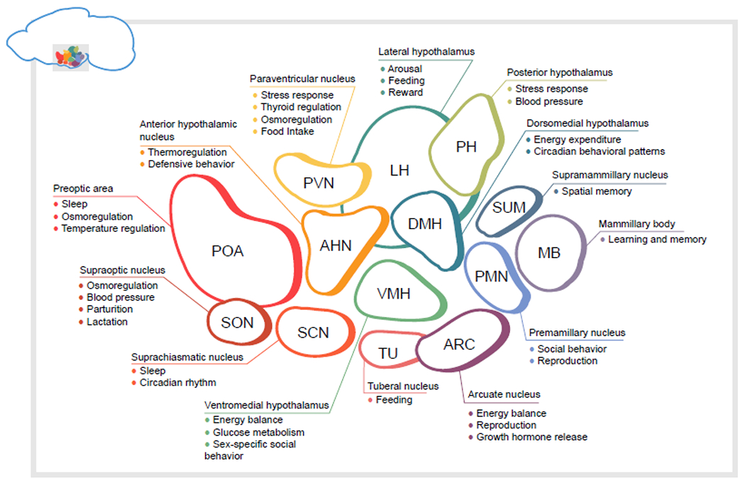
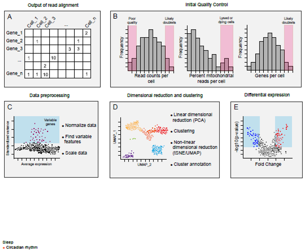
The hypothalamus is a brain region that integrates signals from the periphery and the environment to maintain organismal homeostasis. To do so, specialized hypothalamic neuropeptidergic neurons control a range of processes, such as sleep, feeding, the stress response, and hormone release. These processes are altered with age, which can affect longevity and contribute to disease status. Technological advances, such as single-cell RNA sequencing, are upending assumptions about the transcriptional identity of cell types in the hypothalamus and revealing how distinct cell types change with age. In this review, we summarize current knowledge about the contribution of hypothalamic functions to aging. We highlight recent single-cell studies interrogating distinct cell types of the mouse hypothalamus and suggest ways in which single-cell 'omics technologies can be used to further understand the aging hypothalamus and its role in longevity.
Keywords: homeostasis; longevity; metabolism; single-cell RNA-seq.
Copyright © 2022 The Author(s). Published by Elsevier Ltd.. All rights reserved.
Conflict of interest statement
Declaration of interests The authors declare no conflicts of interest in relation to this work.
Figures
Similar articles
-
Single-cell analysis of the aging female mouse hypothalamus.Nat Aging. 2022 Jul;2(7):662-678. doi: 10.1038/s43587-022-00246-4. Epub 2022 Jul 4. Nat Aging. 2022. PMID: 36285248 Free PMC article.
-
Hypothalamic redox balance and leptin signaling - Emerging role of selenoproteins.Free Radic Biol Med. 2018 Nov 1;127:172-181. doi: 10.1016/j.freeradbiomed.2018.02.038. Epub 2018 Mar 5. Free Radic Biol Med. 2018. PMID: 29518483 Free PMC article. Review.
-
Role of the central nervous system in cell non-autonomous signaling mechanisms of aging and longevity in mammals.J Physiol Sci. 2024 Aug 31;74(1):40. doi: 10.1186/s12576-024-00934-3. J Physiol Sci. 2024. PMID: 39217308 Free PMC article. Review.
-
Hypothalamic carnitine metabolism integrates nutrient and hormonal feedback to regulate energy homeostasis.Mol Cell Endocrinol. 2015 Dec 15;418 Pt 1:9-16. doi: 10.1016/j.mce.2015.08.002. Epub 2015 Aug 8. Mol Cell Endocrinol. 2015. PMID: 26261054 Review.
-
Single-Cell RNA-Seq Reveals Hypothalamic Cell Diversity.Cell Rep. 2017 Mar 28;18(13):3227-3241. doi: 10.1016/j.celrep.2017.03.004. Cell Rep. 2017. PMID: 28355573 Free PMC article.
Cited by
-
Interaction of GPER-1 with the endocrine signaling axis in breast cancer.Front Endocrinol (Lausanne). 2025 Jan 24;16:1494411. doi: 10.3389/fendo.2025.1494411. eCollection 2025. Front Endocrinol (Lausanne). 2025. PMID: 39936103 Free PMC article. Review.
-
Function of brain-derived neurotrophic factor in the hypothalamus: Implications for depression pathology.Front Mol Neurosci. 2022 Nov 16;15:1028223. doi: 10.3389/fnmol.2022.1028223. eCollection 2022. Front Mol Neurosci. 2022. PMID: 36466807 Free PMC article. Review.
-
Recent advances in understanding neuronal diversity and neural circuit complexity across different brain regions using single-cell sequencing.Front Neural Circuits. 2023 Mar 30;17:1007755. doi: 10.3389/fncir.2023.1007755. eCollection 2023. Front Neural Circuits. 2023. PMID: 37063385 Free PMC article. Review.
-
Hypertension: Causes and Consequences of Circadian Rhythms in Blood Pressure.Circ Res. 2024 Mar 15;134(6):810-832. doi: 10.1161/CIRCRESAHA.124.323515. Epub 2024 Mar 14. Circ Res. 2024. PMID: 38484034 Free PMC article. Review.
-
Single-cell and spatial omics: exploring hypothalamic heterogeneity.Neural Regen Res. 2025 Jun 1;20(6):1525-1540. doi: 10.4103/NRR.NRR-D-24-00231. Epub 2024 Jul 10. Neural Regen Res. 2025. PMID: 38993130 Free PMC article.
References
-
- Deorah S et al. (2006) Trends in brain cancer incidence and survival in the United States: Surveillance, Epidemiology, and End Results Program, 1973 to 2001. Neurosurg. Focus 20, E1 - PubMed
-
- Hou Y et al. (2019) Ageing as a risk factor for neurodegenerative disease. Nat. Rev. Neurol 15, 565–581 - PubMed
Publication types
MeSH terms
Grants and funding
LinkOut - more resources
Full Text Sources
