. 2022 Oct 22;12(1):17778.
doi: 10.1038/s41598-022-22307-9.

Vitamin D enhances type I IFN signaling in COVID-19 patients

Affiliations

Vitamin D enhances type I IFN signaling in COVID-19 patients

Shirin Hafezi et al. Sci Rep. .

Abstract

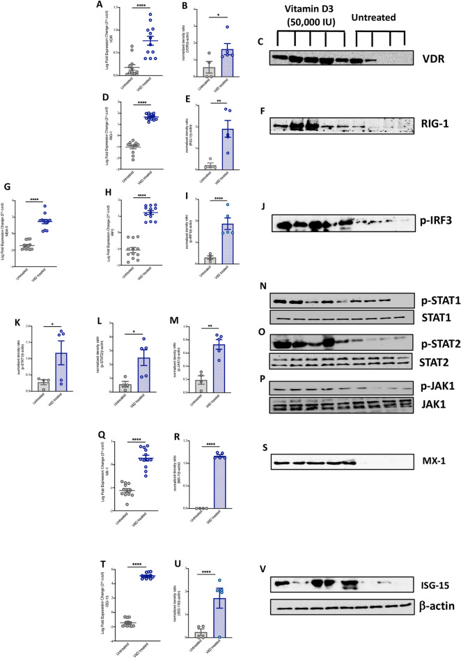
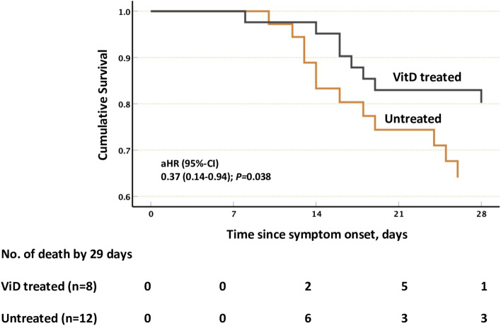
The ability of Vitamin D (VitD) to modulate antiviral responses through induction of antimicrobial peptide is well established. However, the effect of VitD on host responses to SARS-CoV-2 is not well investigated. We here report the ability of VitD to enhance host IFN-alpha/beta (a/β) signaling both in vitro and among severe COVID-19 patients treated with VitD. Blood and saliva specimens were obtained from severe COVID-19 patients treated (43 patients), or not (37 patients), with vitD, during their stay in intensive care unit. Patients were followed up to 29 days following admission, and patient survival outcomes were collected. Higher activity levels of RIG-1/MDA-5 and JAK-STAT signaling pathways were observed with significantly higher gene and protein levels of antiviral interferon stimulating genes (ISGs) such as MX-1 and ISG-15; both in vitro, following treatment of PBMCs with vitD, and in whole blood and saliva specimens of VitD treated patients. Moreover, VitD treated patients had lower risk of all-cause mortality by day 29 compared to untreated patients (adjusted hazard ratio, 0.37, 95% confidence interval of 0.14-0.94; P = 0.038). The herein uncovered regulatory role of VitD on type I IFNs suggests the importance of insuring a normal level of VitD for the prevention and probably treatment of SARS-CoV-2 infection. Additional mechanistic studies, however, are needed to fully elucidate the antiviral effects of VitD particularly in the setting of COVID-19 infection.


Conflict of interest statement

The authors declare no competing interests.

Figures

Figure 1
Figure 1
Increased IFNα/β pathway signaling and higher expression of ISGs in human airway epithelial cells and PBMCs following VitD treatment. (A) Enrichment of IFNα/β pathway in IFNα, poly I:C, SARS-CoV-2, and VitD treated human airway epithelial cells (HAECs). Normalized enrichment Score (NES) of IFNα (1.41), poly I:C (1.00), SARS-CoV-2 (1.40), and VitD (1.005) treated HAECs. (B) The gene expression levels of RIG-1, MDA-5, IRF3, IRF9, MX-1, and ISG-15 in IFNα, poly I:C, SARS-CoV-2, and VitD treated HAECs treated cells. (C) Overlays of genes in Reactome Interferon α/β signaling pathway (R-HSA-909733) and VitD target gene lists. For IFNα, the RNAseq data was obtained from 3 replicates of IFNα-treated BEAS-2B cells using GEO: GSE148829 dataset. For poly I:C or VitD, the RNAseq data was obtained from 3 replicates of poly I:C or calcitriol (VitD) treated HAECs using GEO: GSE106885 dataset, while for SARS-CoV-2, the RNAseq data was obtained from 3 replicates of SARS-CoV-2 infected HAECs were extracted from GEO: GSE147507 dataset. Results are presented as log fold change ± SE of gene expression between cases and controls. (DF) The mRNA and protein levels of VDR in VitD (50 nM of calcitriol for 8 h) treated PBMCs. (GL) Protein levels of p-STAT1, p-STAT2 and p-JAK1 in VitD and/or IFNα (50 nM of calcitriol and/or 1 μg/ml of IFNα for 8 h) treated PBMCs. (MO) The mRNA and protein levels of ISG-15 in VitD and/or IFNα treated PBMCs. PBMCs were isolated from peripheral blood of healthy donors (n = 3). Statistic test: comparison was done using unpaired t-test or Mann–Whitney U test, depending on the skewness of the data. *P < 0.05, **P < 0.01, ***P < 0.001, ****P < 0.0001.
Figure 2
Figure 2
Increased IFNα/β signaling in blood of VitD treated COVID-19 patients. (AC) The mRNA and protein levels of VDR in whole blood of VitD treated (cholecalciferol was administrated as 50,000 IU weekly) and untreated COVID-19 patients. (DJ) The mRNA and/or protein levels of RNA cytosolic sensing proteins, such as RIG-1, MDA5, and p-IRF3 in whole blood of VitD treated and untreated COVID-19 patients. (K) The plasma levels of IFNα in VitD treated and VitD untreated COVID-19 patients. (LQ) Protein levels of p-STAT1, p-STAT2, and p-JAK1 in blood of VitD treated and VitD untreated COVID-19 patients. (RX) The mRNA and/or protein levels of IRF3 and ISGs such as MX-1 and ISG-15 in blood of VitD treated and VitD untreated COVID-19 patients. The whole blood of VitD treated (n = 9; 6 males and 3 females) and untreated COVID-19 patients (n = 9; 6 males and 3 females) were randomly chosen from cohorts of VitD treated (n = 43; 36 males and 7 females) or untreated (n = 37; 27 males and 10 females) COVID-19 patients. Statistic test: comparison was done using unpaired t-test or Mann–Whitney U test, depending on the skewness of the data. *P < 0.05, **P < 0.01.
Figure 3
Figure 3
Increased IFNα/β signaling in saliva of VitD treated COVID-19 patients. (AC) The mRNA and protein levels of VDR in saliva of VitD treated (cholecalciferol was administrated as 50,000 IU weekly) and untreated COVID-19 patients. (DJ) The mRNA levels of RNA cytosolic sensing proteins, such as RIG-1, MDA5, and p-IRF3 in saliva of VitD treated and untreated COVID-19 patients. (KP) Protein levels of p-STAT1, p-STAT2, and p-JAK1 in saliva of VitD treated and VitD untreated COVID-19 patients. (QV) The mRNA levels of ISGs such as MX-1 and ISG-15 in saliva of VitD treated and VitD untreated COVID-19 patients. The saliva of VitD treated (n = 5; 3 males and 2 females) and untreated COVID-19 patients (n = 4; 2 males and 2 females) were randomly chosen from cohorts of VitD treated (n = 43; 36 males and 7 females) or untreated (n = 37; 27 males and 10 females) COVID-19 patients. Statistic test: comparison was done using unpaired t-test or Mann–Whitney U test, depending on the skewness of the data. *P < 0.05, **P < 0.01.
Figure 4
Figure 4
Lower 29-day all-cause mortality of VitD treated COVID-19 patients. The Kaplan–Meier curve of VitD treated (n = 43, 8 death events) and untreated COVID-19 patients (n = 37, 12 death events). The Cox proportional hazards regression model was adjusted for patient’s demographics factors (age, gender, and body mass index), comorbidities (diabetes mellitus), and COVID-19 related severity serum marker (D-dimer and C-reactive protein).
Figure 5
Figure 5
Schematic overview of the study findings. VitD enhances signaling of RIG-1/MDA-5 and JAK-STAT pathways and the resultant production of MX-1 and ISG-15 antiviral ISGs.

References

    1. Casanova J-L, et al. A global effort to define the human genetics of protective immunity to SARS-CoV-2 infection. Cell. 2020;181:1194–1199. doi: 10.1016/j.cell.2020.05.016. - DOI - PMC - PubMed
    1. Lee JS, Shin E-C. The type I interferon response in COVID-19: Implications for treatment. Nat. Rev. Immunol. 2020;20:585–586. doi: 10.1038/s41577-020-00429-3. - DOI - PMC - PubMed
    1. Mdkhana B, et al. Nucleic acid-sensing pathways during SARS-CoV-2 infection: Expectations versus reality. J. Inflamm. Res. 2021;14:199–216. doi: 10.2147/JIR.S277716. - DOI - PMC - PubMed
    1. Yamada T, et al. RIG-I triggers a signaling-abortive anti-SARS-CoV-2 defense in human lung cells. Nat. Immunol. 2021;22:820–828. doi: 10.1038/s41590-021-00942-0. - DOI - PubMed
    1. Rehwinkel J, Gack MU. RIG-I-like receptors: Their regulation and roles in RNA sensing. Nat. Rev. Immunol. 2020;20:537–551. doi: 10.1038/s41577-020-0288-3. - DOI - PMC - PubMed

Publication types