. 2022 Oct 22;13(10):893.
doi: 10.1038/s41419-022-05322-6.

Acinar ATP8b1/LPC pathway promotes macrophage efferocytosis and clearance of inflammation during chronic pancreatitis development

Affiliations

Acinar ATP8b1/LPC pathway promotes macrophage efferocytosis and clearance of inflammation during chronic pancreatitis development

Wan-Jun Yang et al. Cell Death Dis. .

Erratum in

Abstract

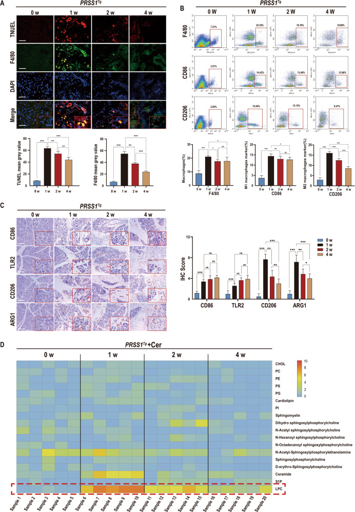
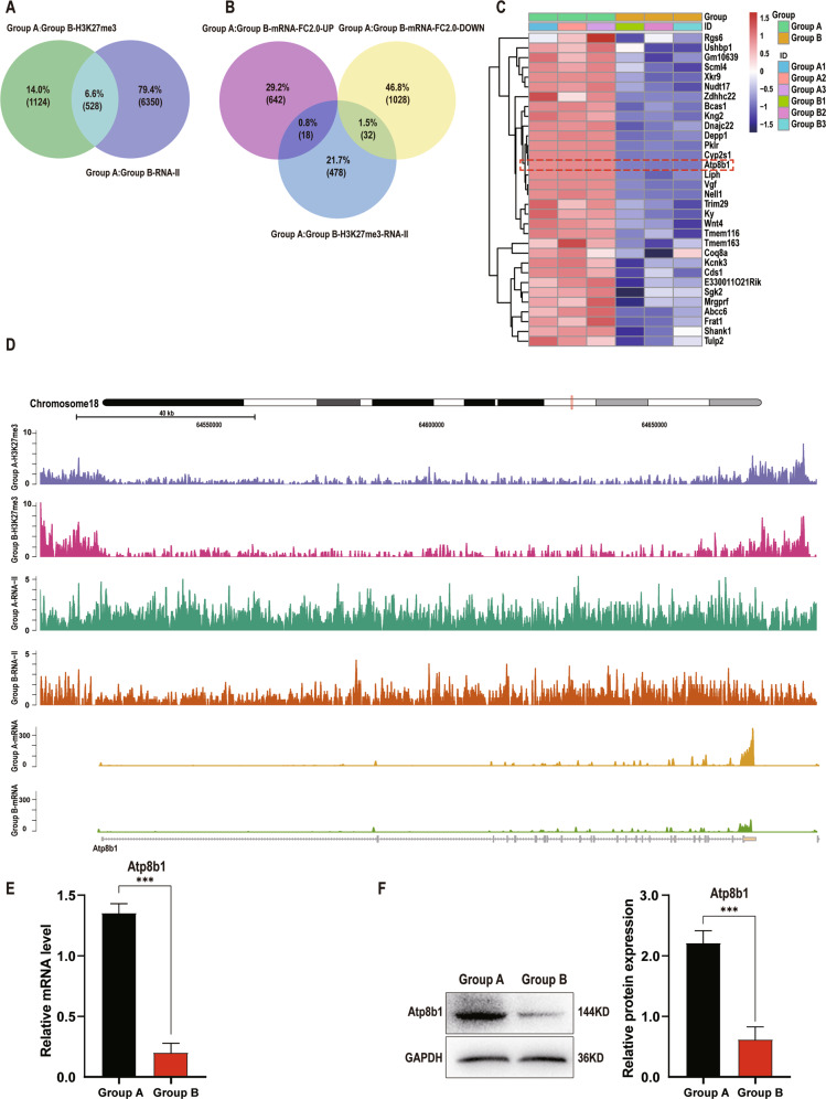
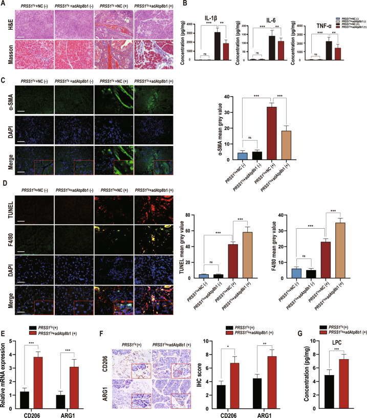
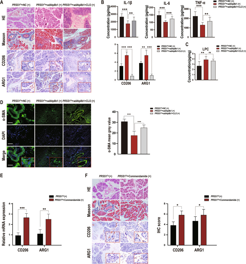
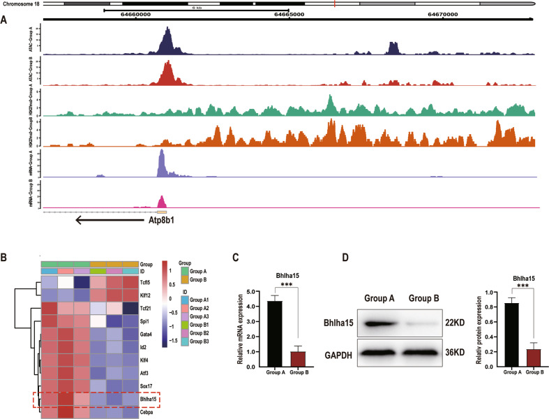
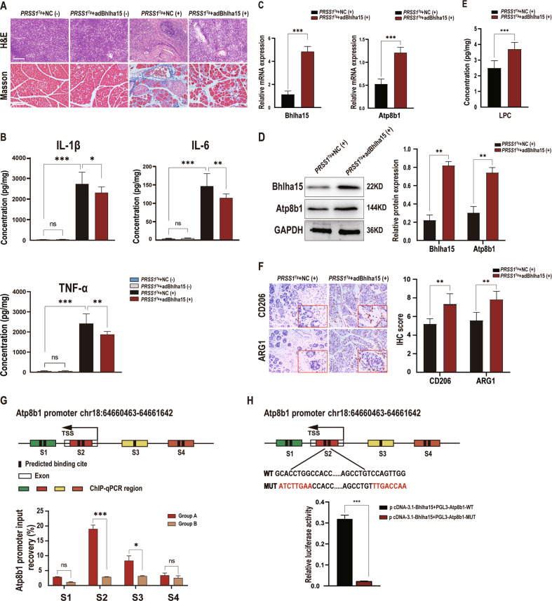
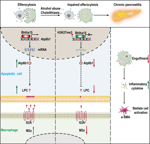
Noninflammatory clearance of dying cells by professional phagocytes, termed efferocytosis, is fundamental in both homeostasis and inflammatory fibrosis disease but has not been confirmed to occur in chronic pancreatitis (CP). Here, we investigated whether efferocytosis constitutes a novel regulatory target in CP and its mechanisms. PRSS1 transgenic (PRSS1Tg) mice were treated with caerulein to mimic CP development. Phospholipid metabolite profiling and epigenetic assays were performed with PRSS1Tg CP models. The potential functions of Atp8b1 in CP model were clarified using Atp8b1-overexpressing adeno-associated virus, immunofluorescence, enzyme-linked immunosorbent assay(ELISA), and lipid metabolomic approaches. ATAC-seq combined with RNA-seq was then used to identify transcription factors binding to the Atp8b1 promoter, and ChIP-qPCR and luciferase assays were used to confirm that the identified transcription factor bound to the Atp8b1 promoter, and to identify the specific binding site. Flow cytometry was performed to analyze the proportion of pancreatic macrophages. Decreased efferocytosis with aggravated inflammation was identified in CP. The lysophosphatidylcholine (LPC) pathway was the most obviously dysregulated phospholipid pathway, and LPC and Atp8b1 expression gradually decreased during CP development. H3K27me3 ChIP-seq showed that increased Atp8b1 promoter methylation led to transcriptional inhibition. Atp8b1 complementation substantially increased the LPC concentration and improved CP outcomes. Bhlha15 was identified as a transcription factor that binds to the Atp8b1 promoter and regulates phospholipid metabolism. Our study indicates that the acinar Atp8b1/LPC pathway acts as an important "find-me" signal for macrophages and plays a protective role in CP, with Atp8b1 transcription promoted by the acinar cell-specific transcription factor Bhlha15. Bhlha15, Atp8b1, and LPC could be clinically translated into valuable therapeutic targets to overcome the limitations of current CP therapies.


Conflict of interest statement

The authors declare no competing interests.

Figures

Fig. 1
Fig. 1. Identification of impaired efferocytosis and dysregulated phospholipid metabolism in chronic pancreatitis(CP).
To establish a caerulein-induced CP model, PRSS1Tg mice were intraperitoneally injected with 15 μg/ml caerulein dissolved in phosphate-buffered saline at a dose of 50 μg/kg each hour for 8 hours, twice per week, for a total of four weeks. A TUNEL staining to show apoptosis were performed and immunofluorescence staining was to detect the expression of F4/80 in pancreatic tissues from caerulein-treated PRSS1Tg mice at weeks 0, 1, 2, and 4. DAPI staining to show nucleus was performed. The mean gray values were used to quantify the expression of F4/80 and the degree of apoptosis. B Flow cytometry was performed to analyze the proportion of macrophages maker(F4/80+PE-CY7), M1-like macrophages marker(CD86 + APC), and M2-like macrophages marker(CD206 + PE) in the pancreatic tissues of caerulein-treated PRSS1Tg mice at weeks 0, 1, 2, and 4. C The expression levels of M1 macrophage markers (CD86 and TRL2) and M2a macrophage markers (CD206 and ARG1) in pancreatic tissues from caerulein-treated PRSS1Tg mice were measured by immunohistochemistry, and immunohistochemical scoring was performed. D The levels of phospholipid metabolites were assessed by liquid chromatography-tandem mass spectrometry (LC–MS/MS), orange represented high expression, blue mean low expression. The data are presented as the means ± SDs. Three biological replicates were performed. Significant differences between two groups were analyzed by Student’s t test, and one-way analysis of variation was performed to investigate the differences among more than two groups. ns no significant difference; *p ≤ 0.05; **p ≤ 0.01; ***p ≤ 0.001. Scale bars = 100 µm.
Fig. 2
Fig. 2. Atp8b1 is downregulated in CP tissues from PRSS1Tg mice.
H3K27me3 ChIP-seq, RNA pol II ChIP-seq and mRNA-seq analyses of pancreatic tissues from Group A (treated with caerulein for 1 week) and Group B (treated with caerulein for 4 weeks) were applied to identify genes that potentially regulate phospholipid metabolism. A Venn diagram analysis of the overlap between the H3K27me3 ChIP-seq and RNA pol II ChIP-seq. B Venn diagram analysis of the overlap between the above 528 genes and differentially expressed mRNAs in Group A and Group B separately. C Heatmap of the 32 downregulated genes. D Signal tracks showing the binding and co-occupancy of H3K27me3 and RNA pol II at the Atp8b1 gene locus and mRNA-seq signals for genes in pancreatic acinar cells in Groups A and B. Purple, Group A-H3K27me3; Red, Group B-H3K27me3, Green, Group A- RNA pol II; Orange, Group B- RNA pol II; Yellow, Group A-mRNA; Cyan-blue, Group B- mRNA. E The total Atp8b1 mRNA and F protein expression levels in pancreatic tissue in Group A and B were measured by quantitative RT-PCR and western blotting, respectively. The results were measured by densitometry and expressed by expression relative to GAPDH as the reference protein. The data are presented as the means ± SDs; n = 3 biological replicates. ***p ≤ 0.001.
Fig. 3
Fig. 3. Atp8b1 reverses the impairment of efferocytosis and dysregulation of phospholipid metabolism in acinar cells during CP development.
AdAtp8b1 was used to induce Atp8b1 overexpression in the pancreas of PRSS1Tg mice. The pancreases of PRSS1Tg mice were infected with adenoviral vectors harbouring a scrambled adRNA for the negative control (NC). A Structural alterations and fibrosis in pancreatic tissues from adAtp8b1 and NC groups PRSS1Tg mice treated (+) or untreated (−) with caerulein were evaluated by H&E staining and Masson’s trichrome staining, respectively. B The levels of IL-1β, IL-6, and TNF-α in pancreatic tissues from caerulein-treated and untreated PRSS1Tg mice were quantitatively analyzed by ELISA. C α-SMA expression in pancreatic tissues from caerulein-treated and untreated PRSS1Tg mice was detected by immunofluorescence staining, the mean gray values calculated by ImageJ were used to quantitatively analyze the degree of fibrosis. The mean gray values was calculated using integrated density divided by area. D Apoptosis and F4/80 expression were detected by a TUNEL assay and immunofluorescence staining, respectively, in pancreatic tissues from caerulein-treated and untreated PRSS1Tg mice. The mean gray values calculated by ImageJ were used to quantify these parameters. E The mRNA expression of CD206 and ARG1 in the pancreatic tissues from adAtp8b1-treated and untreated PRSS1Tg CP mice was measured by RT-PCR. F Immunohistochemical staining was used to detect CD206 and ARG1 expression in pancreatic tissues from adAtp8b1-treated and untreated PRSS1Tg CP mice. G The concentration of LPC in pancreatic tissues from adAtp8b1-treated and untreated PRSS1Tg CP mice was assessed by LC–MS/MS. PRSS1Tg (+) and PRSS1Tg (−) represented mice treated and untreated with caerulein, respectively. The data are presented as the means ± SDs. ns no significant difference; *p ≤ 0.05; **p ≤ 0.01; ***p ≤ 0.001. Scale bars = 100 µm.
Fig. 4
Fig. 4. LPC plays a role in promoting efferocytosis by binding to its receptor G2A.
CLO was used for macrophage knockout in adAtp8b1 PRSS1Tg mice treated with careulein. A Structural alterations, fibrosis, and the expression of CD206 and ARG1 were evaluated by H&E staining, Masson’s trichrome staining, and immunohistochemical staining, respectively, of pancreatic tissues from PRSS1Tg mice treated with caerulein in the adAtp8b1 and macrophage knockout groups. B Quantitative expression of IL-1β, IL-6, and TNF-α in pancreatic tissues from caerulein-treated PRSS1Tg mice. C The concentration of LPC in pancreatic tissues from caerulein-treated PRSS1Tg mice was measured by LC–MS/MS. D α-SMA expression detected by immunofluorescence staining was used to analyze the degree of fibrosis from PRSS1Tg mice treated with caerulein in the adAtp8b1 and macrophage knockout groups. The mean gray values were used to quantitatively analyze the degree of fibrosis. E The mRNA expression of CD206 and ARG1 in the pancreatic tissues from adAtp8b1-treated and untreated PRSS1Tg CP mice was measured by RT-PCR. F Structural alterations, fibrosis, and the expression of CD206 and ARG1 were evaluated by H&E staining, Masson’s trichrome staining, and immunohistochemical staining, respectively, of pancreatic tissues from CP model mice treated with or without commendamide (a G2A agonist). The data are presented as the means ± SDs. ns no significant difference; *p ≤ 0.05; **p ≤ 0.01; ***p ≤ 0.001. Scale bars = 100 µm.
Fig. 5
Fig. 5. Bhlha15 binds to the promoter of Atp8b1 in acinar cells.
A Signal tracks showing the open chromatin region in the Atp8b1 promoter, as determined by ATAC-seq, in Group A and B. The differential peaks in the two groups are shown above the tracks. The binding and co-occupancy of H3K27me3 and mRNA-seq signals of genes in pancreatic acinar cells from Groups A and B w ere visualized with Integrative Genomics Viewer. B Heatmap shown 11 genes were identified by predicting the binding sites of transcription factor to the open chromatin regions around the TSS of Atp8b1 using Jaspar (threshold score, > 80). The mRNA (C) and protein (D) expression levels of Bhlha15 in pancreatic tissue were determined by quantitative RT–PCR and western blotting, respectively. ***p ≤ 0.001.
Fig. 6
Fig. 6. Overexpression of Bhlha15 ameliorates apoptosis and CP progression.
AdBhlha15 was used to overexpress Bhlha15 in the pancreas of caerulein-treated or untreated PRSS1Tg mice. The pancreases of PRSS1Tg mice were infected with adenoviral vectors harbouring a scrambled adRNA for the negative control (NC). A Structural alterations and fibrosis were evaluated by HE staining and Masson’s trichrome staining, respectively, of pancreatic tissues from PRSS1Tg mice. B Quantitative analysis of IL-1β, IL-6, and TNF-α levels in pancreatic tissues from caerulein-treated or untreated PRSS1Tg mice. C The Bhlha15 and Atp8b1 mRNA expression levels in pancreatic tissue from the Bhlha15 overexpression group and negative control group (NC) treated with caerulein were measured by quantitative RT–PCR. D The protein expression of Bhlha15 and Atp8b1 in pancreatic tissues from adBhlha15-treated or negative control PRSS1Tg mice treated with caerulein was detected by western blotting and quantified. E The concentration of LPC in pancreatic tissues from adBhlha15-treated or untreated PRSS1Tg CP mice was measured by LC–MS/MS. F Immunohistochemical staining was performed to detect CD206 and ARG1 expression in pancreatic tissues from PRSS1Tg mice. G Binding of Bhlha15 to the Atp8b1 promoter region was assessed in vitro in pancreatic acinar cells from mice in Group B (treated with caerulein for 4 weeks) or Group A (treated with caerulein for 1 week) by ChIP using anti-Bhlha15 antibodies or IgG. Input and immunoprecipitated DNA purified by ChIP were analyzed using quantitative RT–PCR, relative mRNA levels were calculated using the comparative ΔΔCt method and presented as a percentage of the controls. mRNA levels of GAPDH genes were used as controls for normalization. The predicted Bhlha15 binding sites are marked S1, S2, S3, and S4. H The luciferase assay indicated that the S2 region of the Atp8b1 promoter was the target binding site for Bhlha15. A relative firefly luciferase/ Renilla luciferase of each samples were calculated, then mean firefly luciferase/Renilla luciferase ratio of biological replicates were subsequently calculated. To obtain fold enrichment of each group compared to NC group, NC group was designed as normalization, The red text indicates the mutant binding sites. The data are presented as the means ± SDs. ns no significant difference; *p ≤ 0.05; **p ≤ 0.01; ***p ≤ 0.001.
Fig. 7
Fig. 7. Graphical abstract.
Mechanistic diagram showing how efferocytosis effect CP process through regulating Bhlha15/Atp8b1/LPC pathway to alter the regulation of phospholipid metabolism. Under normal conditions on the left, macrophages engulf apoptotic acinar cells through efferocytosis to resolve inflammation. Alcohol abuse and cholelithiasis can injury pancreas and impaire the efficiency of efferocytosis. Impaired efferocytosis decreases the efficiency of apoptotic cell clearance. H3K27me3 of histones suppresses Atp8b1 transcription, and decrease LPC concentration. This reduction means that less LPC is available to interact with G2A and fewer macrophages are attracted to engulf apoptotic cells, leading to further inflammation.

References

    1. Singh VK, Yadav D, Garg PK. Diagnosis and management of chronic pancreatitis: A review. JAMA. 2019;322:2422–34. - PubMed
    1. Machicado JD, Dudekula A, Tang G, Xu H, Wu BU, Forsmark CE, et al. Period prevalence of chronic pancreatitis diagnosis from 2001–2013 in the commercially insured population of the United States. Pancreatology. 2019;19:813–8. - PMC - PubMed
    1. Gardner TB, Adler DG, Forsmark CE, Sauer BG, Taylor JR, Whitcomb DC. ACG clinical guideline: Chronic pancreatitis. AM J Gastroenterol. 2020;115:322–39. - PubMed
    1. Shelton CA, Umapathy C, Stello K, Yadav D, Whitcomb DC. Hereditary pancreatitis in the United States: Survival and rates of pancreatic cancer. AM J Gastroenterol. 2018;113:1376. - PMC - PubMed
    1. Beyer G, Habtezion A, Werner J, Lerch MM, Mayerle J. Chronic pancreatitis. Lancet. 2020;396:499–512. - PubMed

Publication types

MeSH terms